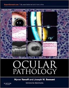
Ocular Pathology, 7e (Expert Consult Title: Online + Print) 7th Edition

[amazon template=iframe image2&asin=1455728748]

2015 BMA Medical Book Awards Highly Commended in Pathology Category! Comprehensive, yet concise and clinically oriented, the new edition of Ocular Pathology brings you the very latest advances of every aspect of ocular pathology. From updated information on today’s imaging techniques, to the implementation of genetic data to better understand disease, this esteemed medical reference book promises to keep you at the forefront of your field

“This seventh edition of Ocular Pathology by Myron Yanoff and Joseph Sassani is a superb update of what has become the single best ophthalmic pathology reference text for ophthalmologists, pathologists and researchers.” Foreword by: J. Douglas Cameron, Ophthalmology and Visual Neurosciences, University of Minnesota School of Medicine, June 2015

  • Take advantage of clinical “pearls” that offer you the benefits of proven strategies.
  • Quickly reference information with help from a convenient outline format, ideal for today’s busy physician.
  • Visualize every concept by viewing 1,900 illustrations, 1,600 of which are in full color, from the collections of internationally renowned leaders in ocular pathology.

  • Understand the role of VEGF and other factors in the pathobiology of diabetic complications, as well as the pathobiology of myocilin and the TIGR gene in the development of glaucoma.
  • Review the latest features related to the pathobiology of central corneal thickness.
  • Stay abreast of the latest in ocular pathology with coverage of the classification system for retinoblastoma; immunopathology of herpes keratitis; and genetic features of persistent hyperplastic primary vitreous.
  • Access the entire text online at Expert Consult, and test your visual recognition and understanding of disease with a new online image review/testing feature.

DOWNLOAD THIS BOOK FREE HERE

http://upsto.re/A8UokFf

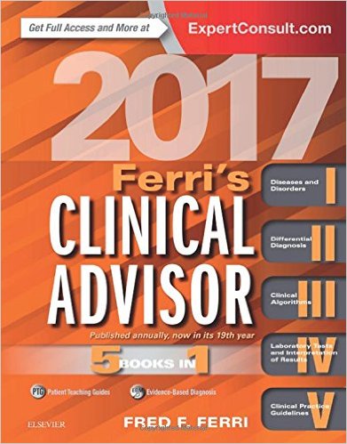
Ferri’s Clinical Advisor 2017: 5 Books in 1, 1e (Ferri’s Medical Solutions) 1 Har/Psc Edition

[amazon template=iframe image2&asin=032328048X]

Access up-to-date diagnostic and treatment information on more than 700 common medical conditions with Ferri’s Clinical Advisor 2017, which boasts the popular “5 books in 1” format! Published annually and now in its 19th year, it provides quick guidance on diseases and disorders, differential diagnoses, medical algorithms, laboratory tests, and clinical practice guidelines, while additional electronic content equips you with e-only topics, images, tables, and much more.

  • Updated content by experts in key clinical fields helps you keep pace with the speed of modern medicine.
  • Popular “5 books in 1” format includes cross-references, outlines, bullets, tables, boxes, and algorithms to help expedite search.


  • Expert Consult eBook version included with purchase. This enhanced eBook experience gives you access to all of the text, more than 90 e-only topics, algorithms, additional images and tables, EBM boxes, and fully updated patient teaching guides. Links between each section connect you from one topic to the relevant material associated with it.
  • Diseases and Disorders section features more than 300 new figures and tables, as well as 20 new topics including: cyclic vomiting syndrome, traveler’s diarrhea, chronic pruritus, post-herpetic neuralgia, enteropathic arthritis, and hoarding disorder.
  • Differential Diagnosis section highlights 50 new topics, including: alcohol-related seizures, dysentery and inflammatory enterocolitis, hair loss, cystic and solid pancreatic lesions, and COPD decompensation.
  • New algorithms offer important diagnostic information on 19 added conditions, including allergic reaction to vaccines, cardiac arrest, occupational asthma, urinary tract infection, and vertigo and dizziness.
  • Current ICD-10 insurance billing codes help expedite insurance reimbursements.

DOWNLOAD THIS BOOK FREE HERE

http://upsto.re/A8UWp7y

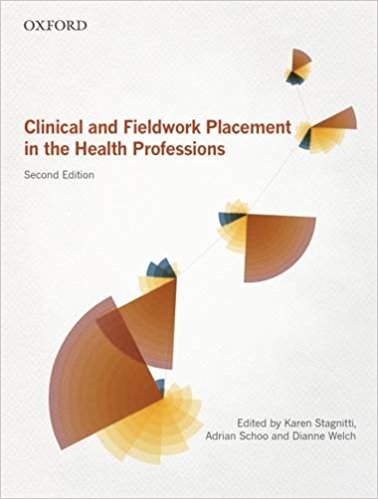
Clinical and Fieldwork Placement in the Health Profession 2nd Edition

[amazon template=iframe image2&asin=0195519604]

In all health professions, students are required to spend a certain number of hours in a healthcare setting working within their discipline-specific profession. This requirement is essential to becoming a competent health professional and practitioner. Clinical and Fieldwork Placement in the Health Professions, second edition, helps to prepare students for their work in clinical and fieldwork settings.

The text is divided into three parts. Part 1 Issues for Practice presents information that is important for students and supervisors to know-regardless of the clinical setting. This includes how to get the most out of your fieldwork, working in teams and how to prepare for placement. Part 2 Contexts of Practice explores various clinical settings (i.e. working with mothers and babies, working in acute care, working in mental health) and guides the student through what to prepare for, what to expect and issues that would be helpful to be aware of during placement. Part 3 Transition to Practice provides information on the transition from student to fieldwork educator and on some of the issues involved in supervision of a student.

This second edition continues to encourage multidisciplinary practice, reinforcing that the key to good health practice is to have all health professionals working together successfully with shared skills in reflective practice, engaging clients in care and ethical practice. New to this edition are chapters on ‘Working in Palliative Care’ and ‘Becoming a Reflective Practitioner’, more detail on the competency standards for students on placement, and new pedagogical features-‘Supervisor Profiles’ to help students prepare for clinical placements in various settings, and ‘Portfolio Development Exercises’ to help shape a students’ professional practice portfolio.

DOWNLOAD THIS BOOK FREE HERE

http://upsto.re/A8UW4a9

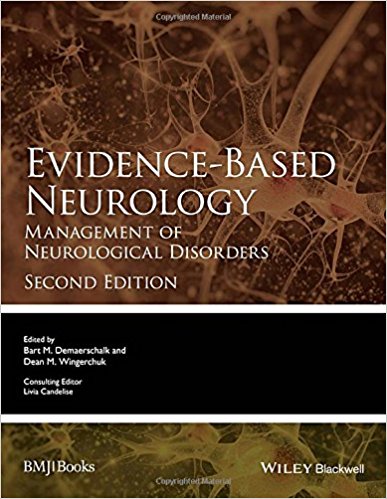
Evidence-Based Neurology: Management of Neurological Disorders (Evidence-Based Medicine) 2nd Edition

[amazon template=iframe image2&asin=0470657782]

From an expert editor team drawn from the Cochrane Neurological Network, Evidence-Based Neurology provides specialists and those in training with the skills and knowledge to apply evidence-based practice in the clinical setting.

  • fills the gap between guidelines and primary studies as well as between primary and secondary scientific medical literature
  • summarizes the most recent and important findings on treatments for neurological patients
  • measures the benefit and, when applicable, the risk of harm inherent in specific neurological interventions
  • now includes new non-clinical topics of interest to neurologists such as education and research

DOWNLOAD THIS BOOK FREE HERE

http://upsto.re/A8UVZ78

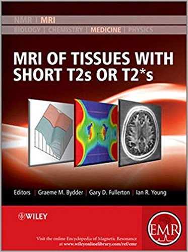
MRI of Tissues with Short T2s or T2*s 1st Edition

[amazon template=iframe image2&asin=0470688351]

The content of this volume has been added to eMagRes (formerly Encyclopedia of Magnetic Resonance) – the ultimate online resource for NMR and MRI.

Up to now MRI could not be used clinically for imaging fine structures of bones or muscles. Since the late 1990s however, the scene has changed dramatically. In particular, Graeme Bydder and his many collaborators have demonstrated the possibility – and importance – of imaging structures in the body that were previously regarded as being “MR Invisible”. The images obtained with a variety of these newly developed methods exhibit complex contrast, resulting in a new quality of images for a wide range of new applications.

This Handbook is designed to enable the radiology community to begin their assessment of how best to exploit these new capabilities. It is organised in four major sections – the first of which, after an Introduction, deals with the basic science underlying the rest of the contents of the Handbook. The second, larger, section describes the techniques which are used in recovering the short T2 and T2* data from which the images are reconstructed. The third and fourth sections present a range of applications of the methods described earlier. The third section deals with pre-clinical uses and studies, while the final section describes a range of clinical applications. It is this last section that will surely have the biggest impact on the development in the next few years as the huge promise of Short T2 and T2* Imaging will be exploited to the benefit of patients.

In many instances, the authors of an article are the only research group who have published on the topic they describe. This demonstrates that this Handbook presents a range of methods and applications with a huge potential for future developments.

About EMR Handbooks / eMagRes Handbooks 

The Encyclopedia of Magnetic Resonance (up to 2012) and eMagRes (from 2013 onward) publish a wide range of online articles on all aspects of magnetic resonance in physics, chemistry, biology and medicine. The existence of this large number of articles, written by experts in various fields, is enabling the publication of a series of EMR Handbooks / eMagRes Handbooks on specific areas of NMR and MRI. The chapters of each of these handbooks will comprise a carefully chosen selection of articles from eMagRes. In consultation with the eMagRes Editorial Board, the EMR Handbooks / eMagRes Handbooks  are coherently planned in advance by specially-selected Editors, and new articles are written (together with updates of some already existing articles) to give appropriate complete coverage. The handbooks are intended to be of value and interest to research students, postdoctoral fellows and other researchers learning about the scientific area in question and undertaking relevant experiments, whether in academia or industry.

DOWNLOAD THIS BOOK FREE HERE

http://upsto.re/A8UVe59

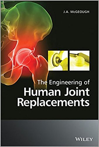
The Engineering of Human Joint Replacements 1st Edition

[amazon template=iframe image2&asin=0470740272]

Since the major pioneering of joint replacement surgery more than fifty years ago, much research and progress has been made in the field of arthroplasty with new insights into better materials, types of cement and bone-cell compatible coatings, and a better understanding of the causes of implant failure. With an increasingly ageing population the requirement for arthroplastic surgery is manifest; over 800,000 hips worldwide are replaced each year, and replacement surgery is performed for almost every joint of the body.

The Engineering of Human Joint Replacements covers the design, engineering, production and manufacture of human joint replacements, as well as associated engineering concerns such as surface coatings, orthopedic bone cement, the causes and effects of wear and tear, and rapid prototyping for clinical evaluation. Materials evaluation and selection is discussed, as well as production processes and insertion methods. The author provides an overview of skeletal anatomy and the effects of pain and deterioration in order to put the engineering principles into a medical context. Examples of joint replacements for the most common regions of the body are included, and aspects of clinical studies of these cases are discussed.

Key Features:

• Provides an overview of the engineering materials and processes involved in the manufacture of    human joint replacements
• Sets the scene for engineers and clinicians embarking on research into joint replacements
• Includes clinical and industrial examples and points the way to future developments
• Provides information on medical device companies with an engineering guide to the requirements for joint replacement

The Engineering of Human Joint Replacements bridges the divide between engineering and orthopaedic surgery, offering an introductory text to young engineers entering the field, as well as a reference for medical staff who will benefit from an understanding of the materials and methods used in their design, engineering and manufacture.

DOWNLOAD THIS BOOK FREE HERE

http://upsto.re/A8pQRVF

Tumors of the Jugular Foramen 1st ed. 2017 Edition

[amazon template=iframe image2&asin=3319433660]

This book covers all aspects of the diagnosis and treatment of tumors of the jugular foramen – cranial base lesions that present particular diagnostic and management difficulties and remain challenging to remove despite advances in surgery and interventional neuroradiology. The authors have developed novel concepts and surgical techniques relating to management of the facial nerve, reconstruction of the cranial base, and resection of large tumors with intra- and extracranial extension. These techniques, as well as anatomic studies in cadavers, are presented by means of surgical photographs, illustrations, and videos. Tumors of the Jugular Foramen will be of interest to all specialists involved in the management of these challenging tumors, including neurosurgeons, ENT surgeons, neuroradiologists, neurologists, neurointensivists, radiotherapists, oncologists, and physiotherapists.

About the Author

Ricardo Ramina, MD, PhD, studied medicine at the Pontifical Catholic University in Curitiba, Brazil, before completing residencies in neurosurgery in Curitiba, Frankfurt, and Hannover.  In 1986 he was appointed Chairman of the Neurosurgical Department at Nações Hospital in Curitiba, Brazil and since 2004 he has been Chairman of the Neurosurgical Department of the Neurological Institute of Curitiba [WFNS (World Federation of Neurosurgical Societies) Post-Graduate Training Center]. Since 2006 he has been Professor of the Postgraduate Course in Surgery at the Pontifical Catholic University of Curitiba, having previously been an Associated Professor of Neurosurgery at George Washington University, Washington and State University of Campinas, São Paulo, Brazil. He has published more than 160 papers in peer-reviewed journals and has given more than 600 lectures and presentations on various topics in neurosurgery as an invited guest or guest of honor at national and international congresses.

Marcos S. Tatagiba, MD, PhD, studied medicine at the University of the State of Rio de Janeiro, Brazil, before undertaking neurosurgical training from 1987 to 1993 in the Department of Neurosurgery, Nordstadt Hospital, Hannover, Germany, under the chairmanship of Prof. Madjid Samii. He presented his doctoral and post-doctoral theses at Hannover Medical School in 1992 and 1998. Since 2000 he has been Vice Director of the International Neuroscience Institute, Hannover; Vice Director of the Department of Neurosurgery, University of Freiburg; Director and Chairman of the Department of Neurosurgery, University of Tübingen; and Chairman and Director, Department of Neurosurgery, University of Zurich. Professor Tatagiba is the author of 237 articles in peer-reviewed journals and the recipient of several prizes, including the Neurosurgical Research Foundation Prize from the German Society of Neurosurgery (2002) and the Innovationspreis Medizintechnik – BMBF (2007). Professor Tatagiba served as President of the German Academy of Neurosurgery in 2010-11 and is President-elect of the German Society of the Skull Base.

DOWNLOAD THIS BOOK FREE HERE

http://upsto.re/A87fD3m

Recognizing Child Abuse in Radiology 1st ed. 2017 Edition

[amazon template=iframe image2&asin=3319443224]

This book is both a personal review of experience in the diagnosis of child abuse and its differential diagnosis, intended to instruct beginners, and a reliable source of information for the more experienced health care provider. Emphasis is placed on normal findings that simulate abuse, true and false diagnoses of metabolic bone disease as the cause of fractures, abusive head trauma, and the problem of zealous abuse-denying “experts.” Plain radiography is emphasized, with appropriate mention of, ultrasound, CT, MRI, – in detecting childhood abuse is clearly explained, and individual chapters focus on neuroradiological evaluation and the radiological assessment of abdominal, thoracic, and subcutaneous abnormalities. Guidance is provided on how to exclude a range of alternative diagnoses, on the documentation of concerns over potential abuse, and on the needs of the clinician in relation to the radiologist. Recognizing Child Abuse in Radiology will be of value for radiologists in training and practice, for health care workers, and for other interested professionals.

About the Author

Alan E. Oestreich, MD, is a pediatric radiologist since 1980 at the Cincinnati Children’s Hospital Medical Center in Cincinnati, Ohio, and is Emeritus Professor of radiology at the University of Cincinnati College of Medicine. From 1972 – 1979 he was pediatric radiologist at the University of Missouri-Columbia. He a Fellow of the American College of Radiology, Honorary Member of the European Society of Paediatric Radiology, emeritus member of the Society for Pediatric Radiology, and past chairman of the Radiology Section of the National Medical Association. He is a member of the Child Abuse Team at Cincinnati Children’s Hospital. He has written and lectured extensively on the subject of radiology of child abuse. Among his previous books are An Atlas of Pediatric Orthopedic Radiology, with co-author Alvin Crawford, MD. He received his MD from the Johns Hopkins Medical School in 1965.

Marguerite M. Caré, MD is an assistant professor in the Department of Radiology, Division of Neuroradiology, at Cincinnati Children’s Hospital Medical Center. Caré joined the CCHMC faculty after completing a pediatric radiology fellowship in 1998 and a pediatric neuroradiology fellowship in 2000 at Cincinnati Children’s Hospital Medical Center. She has been the radiology representative to the multidisciplinary Child Abuse Team at the CCHMC Mayerson Center since 1998 and has recently become the radiology representation to the CCHMC Trauma Surgical Program. Caré has a major interest in the neuroimaging findings in child abuse and has presented at both local and national meetings on this topic. She also has research interests in Tuberous Sclerosis with her neurology colleagues. Caré serves on the CCHMC Radiology Department education committee with a primary role in third year medical student education for the department.

DOWNLOAD THIS BOOK FREE HERE

http://upsto.re/A87fj56

Managing Gastrointestinal Complications of Diabetes 1st ed. 2017 Edition

[amazon template=iframe image2&asin=3319486616]

An ideal clinical handbook for medical professionals that regularly treat patients with diabetes and comorbid gastrointestinal complications. The concise format provides evidence-based content with a targeted emphasis on diagnosis, as up to 75% of patients visiting diabetes clinics report significant gastrointestinal symptoms. Includes guidelines for complication prevention, treatment, and long-term disease management.

DOWNLOAD THIS BOOK FREE HERE

http://upsto.re/A87fWxM

Neuropharmacology of New Psychoactive Substances (NPS): The Science Behind the Headlines (Current Topics in Behavioral Neurosciences) 1st ed. 2017 Edition

[amazon template=iframe image2&asin=3319524429]

Designer drugs, or new psychoactive substances (NPS), are synthetic chemicals that mimic the effects of classic drugs of abuse. There has been an alarming worldwide increase in the abuse of NPS in recent years. NPS are cheap, easy to obtain, and often legally available. In this volume, leading experts summarize the latest studies regarding the molecular mechanisms of action, behavioral effects, and adverse consequences of popular NPS. Specific chapters clarify the differences between various types of NPS, namely: stimulants, cannabinoids and hallucinogens. Thus, this volume broadens our understanding of NPS and provides insight into the rapidly evolving “new drug” phenomenon.

DOWNLOAD THIS BOOK FREE HERE

http://upsto.re/A875quq

1 646 647 648 649 650 907