Clinical Anatomy by Regions Ninth Edition
[amazon template=iframe image2&asin=1451110324]
Medical Books Library for Doctors, Physicians, Surgeons, Dentists, Intensivists, Physician Assistants, Nurses, Medical Technicians and Medical Students
Medical books library


[amazon template=iframe image2&asin=1451110324]
[amazon template=iframe image2&asin=1119055520]
Fundamentals of Anatomy and Physiology for Nursing and Healthcare Students is a succinct but complete overview of the structure and function of the human body, with clinical applications throughout. Designed specifically for nursing and healthcare students, the new edition of this best-selling textbook provides a user-friendly, straightforward, jargon-free introduction to the subject.
Key features:
This edition is now supported by an accompanying study guide to facilitate the learning and revision of the content within this book: ‘Fundamentals of Anatomy and Physiology Workbook: A Study Guide for Nurses and Healthcare Students’
Download this book free here
[amazon template=iframe image2&asin=0323377564]
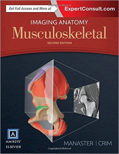
Now in its second edition, Imaging Anatomy: Musculoskeletal is a complete anatomic atlas of the musculoskeletal system, boasting an improved organization with easily accessible information that is standardized for each body region. Brand new chapters, updated anatomical coverage, and highly detailed imagescombine to make this quick yet in-depth resource ideal for day-to-day reference.
Download this book free here


[amazon template=iframe image2&asin=9400988168]
AFTER ten years’ preparation the first edition of our Atlas of Human Anatomy was published between 1946 and 1951. Our experience enabled us to improve each of the subsequent editions and the present one has also been thoroughly revised and enlarged to allow the inclusion of more instructive illus trations. Throughout we have adhered to our original intention that this work should be a well propor tioned Atlas of life-like illustrations primarily for medical students but also useful to the practising physician and surgeon. The introduction of topographical illustrations in the third volume has been welcomed by readers and, while not embarking on histology, semi-microscopic figures have been introduced into some chapters for a better understanding of function. We did not deviate without reason from the currently accepted methods of illustrating the elements of the different systems such as bones, joints, muscles, vessels and nerves and we were at pains to base our illustrations on original dissections and to include in them only essential details. The use of colour in the illustrations, introduced by the Italian anatomist Aselli (1627), was with didactic intent. The legends to the illustrations of this edition use the nomenclature of the “Nomina Anatomica”, Paris 1955 (PNA) , as revised in New York in 1960.
Download this book free here
[amazon template=iframe image2&asin=0702033952]
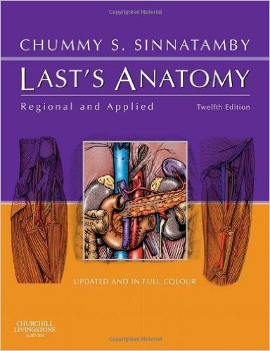
This regional textbook of anatomy is aimed at trainee surgeons and medical students. Throughout it is rich in applied clinical content, knowledge of which is essential for both clinical examination and surgical procedures. Although regional in approach each chapter is structured to clearly explain the structure and function of the component systems. The author brings his continuing experience of teaching anatomy to trainee surgeons to ensure the contents reflects the changing emphasis of anatomical knowledge now required.
Download this book free here
[amazon template=iframe image2&asin=0323077226]
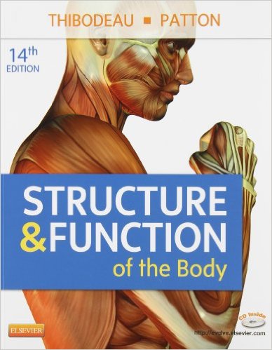
Simple and straightforward, Thibodeau and Patton’s Structure & Function of the Body, 14th Edition makes the difficult concepts of anatomy and physiology clear and easier to understand. Focusing on the normal structure and function of the human body and what the body does to maintain homeostasis, this introductory text provides more than 400 vibrantly detailed illustrations and a variety of interactive learning tools to help you establish an essential foundation for success in the care of the human body.
Download this book free here
[amazon template=iframe image2&asin=1626231710]
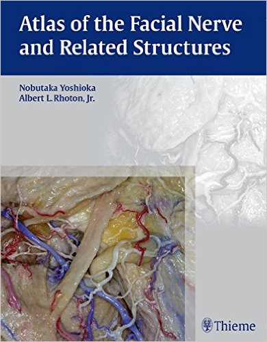
Nobutaka Yoshioka, MD, PhD and Albert L. Rhoton Jr., MD
have created an anatomical atlas of astounding precision. An unparalleled
teaching tool, this atlas opens a unique window into the anatomical intricacies
of complex facial nerves and related structures.
An internationally renowned author, educator, brain anatomist, and
neurosurgeon, Dr. Rhoton is regarded by colleagues as one of the fathers of
modern microscopic neurosurgery. Dr. Yoshioka, an esteemed craniofacial
reconstructive surgeon in Japan, mastered this precise dissection technique
while undertaking a fellowship at Dr. Rhoton’s microanatomy lab, writing in the
preface that within such precision images lies potential for surgical
innovation.
Download this book free here
[amazon template=iframe image2&asin=0443066841]
Download this book free here