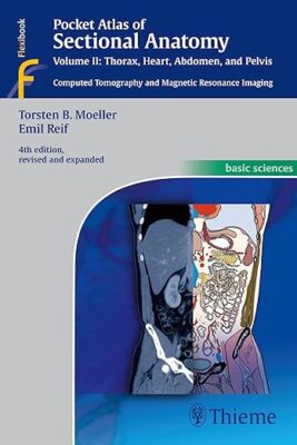
Pocket Atlas of Sectional Anatomy, Vol. II: Thorax, Heart, Abdomen and Pelvis: Computed Tomography and Magnetic Resonance Imaging 4th edition

Pocket Atlas of Sectional Anatomy, Vol. II: Thorax, Heart, Abdomen and Pelvis: Computed Tomography and Magnetic Resonance Imaging 4th edition

This comprehensive, easy-to-consult pocket atlas is renowned for its superb illustrations and ability to depict sectional anatomy in every plane. Together with its two companion volumes, it provides a highly specialized navigational tool for all clinicians who need to master radiologic anatomy and accurately interpret CT and MR images.

Special features of Pocket Atlas of Sectional Anatomy:

  • Didactic organization in two-page units, with high-quality radiographs on one side and brilliant, full-color diagrams on the other
  • Hundreds of high-resolution CT and MR images made with the latest generation of scanners (e.g., 3T MRI, 64-slice CT)
  • Color-coded schematic drawings that indicate the level of each section
  • Consistent color coding, making it easy to identify similar structures across several slices

Updates for the 4th edition of Volume II:

  • CT imaging of the chest and abdomen in all 3 planes: axial, sagittal, and coronal
  • New back-cover foldout featuring pulmonary and hepatic segments and lymph node stations
  • Follows standard international classifications of the American Heart Association for cardiac vessels and the AJCC/UICC for mediastinal lymph nodes

Compact, easy-to-use, highly visual, and designed for quick recall, this book is ideal for use in both the clinical and study settings.

DOWNLOAD THIS MEDICAL BOOK

Normal Findings in Radiography 1st Edition

Normal Findings in Radiography 1st Edition

This book deals with a subject that is seemingly trivial: normal findings in radiology. However, the normal is also the frequent, but not always the simple. Every radiologist has experienced difficulties in the systematics of imaging and with phrasing the diagnosis. This book is intended to answer three questions:

  • How should the diagnosis be phrased?
  • Which general scheme can I use to evaluate an image; how can I check for the degree of normality?
  • Which data allow me to evidence normality, and what measurements should be taken?

In spite of the introducion of digital X-ray imaging, the interpretation of conventional X-rays has not basically changed during the past ten years, which is evidenced by the development of this book. Since its first publication in 1987, it has gone through several unchanged reprints. In the 2nd German edition the text has hardly been changed; however, several new images have been added.

The method of making findings and the systematic method of interpreting images are still relevant today. Consequently, the strict and systematic organization of the book was left unchanged. The new layout leads to a clearer accessibility of the material for the reader.

DOWNLOAD THIS MEDICAL BOOK

Photo Acoustic and Optical Coherence Tomography Imaging: Angiography: – An Application in Vessel Imaging (Volume 3)

Photo Acoustic and Optical Coherence Tomography Imaging: Angiography: – An Application in Vessel Imaging (Volume 3)

This book covers the state-of-the-art techniques of optical coherence tomography angiography (OCTA) imaging for the diagnosis of retinal diseases.

It is part of a three-volume work that describes the latest imaging techniques in which to bring optical coherence tomography (OCT), Fundus Imaging and optical coherence tomography angiography (OCTA) to accurately facilitate the diagnosis of retinal diseases. Clinical disorders of the retina have been attracting the attention of researchers, aiming at reducing the blindness rate. This includes uveitis, diabetic retinopathy, macular edema, endophthalmitis, proliferative retinopathy, age-related macular degeneration and glaucoma. Treatment is significantly dependent on having early and accurate diagnosis, which can be significantly improved by employing the techniques described in the book.


Key Features:


  • Provides a comprehensive overview of all pertinent topics related to optical coherence tomography angiography (OCTA) imaging techniques, applicable to diagnosis of eye disorders.
  • Offers a unique coverage of Neural Networks in distinguishing eye diseases.
  • Machine learning techniques are presented in detail throughout.
  • Many of the chapter contributors are world-class researchers.
  • Extensive references will be provided at the end of each chapter to enhance further study.

DOWNLOAD THIS MEDICAL BOOK 

ABC of Emergency Radiology (ABC Series) 3rd Edition

ABC of Emergency Radiology (ABC Series) 3rd Edition

Rapid acquisition and interpretation of radiographs, portable ultrasound (US) and computed tomography (CT) are now the mainstay of initial successful management of sick and traumatized patients presenting to Accident and Emergency Departments.

The ABC of Emergency Radiology is a simple and logical step-by-step guide on how to interpret radiographs, US and CT. It incorporates all the latest technological advances, including replacing plain radiographs with digital radiographs, changes in imaging protocols and the role of portable US and multidetector CT.

With over 400 illustrations and annotated radiographs, this thoroughly revised third edition provides more images, new illustrations, and new chapters on emergency US and CT that reflect current practice. Each chapter starts with radiological anatomy, standard and then additional views, a systematic approach to interpretation (ABC approach) and followed by a review of common abnormalities.

The ABC of Emergency Radiology is an invaluable resource for accident and emergency staff, trainee radiologists, medical students, nurses, radiographers and all medical personnel involved in the immediate care of trauma patients.

This title is also available as a mobile App from MedHand Mobile Libraries. Buy it now from iTunesGoogle Play or the MedHand Store.

DOWNLOAD THIS MEDICAL BOOK

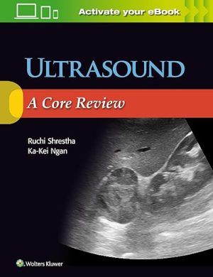
Ultrasound: A Core Review First Edition

Ultrasound: A Core Review First Edition

Uniquely designed for the Core Exam, Ultrasound: A Core Review covers all key aspects of ultrasound, mimicking the image-rich, multiple-choice format of the actual test. Ideal for residents getting ready for the Core Examination, as well as practitioners taking recertification exams, this one-of-a-kind review follows the structure and content of what you’ll encounter on the test, effectively preparing you for Core Exam success!Key Features:

  • Contains 300 image-rich ultrasound questions with answers, explanations, and references.
  • Features high-quality black-and-white, color, and spectral Doppler images throughout.
  • Covers organ-specific ultrasound as well as gynecologic, pregnancy, vascular, musculoskeletal, peritoneal space, retroperitoneum, abdominal wall, chest, and ultrasound-guided procedures.
  • Includes questions on noninterpretive skills such as ultrasound physics and quality and safety.
  • Provides answers and rationales as to why an answer is correct and other choices are incorrect.

Your book purchase includes a complimentary download of the enhanced eBook for iOS, Android, PC & Mac.

Take advantage of these practical features that will improve your eBook experience:

  • The ability to download the eBook on multiple devices at one time — providing a seamless reading experience online or offline
  • Powerful search tools and smart navigation cross-links that allow you to search within this book, or across your entire library of VitalSource eBooks
  • Multiple viewing options that enable you to scale images and text to any size without losing page clarity as well as responsive design
  • The ability to highlight text and add notes with one click

DOWNLOAD THIS MEDICAL BOOK 

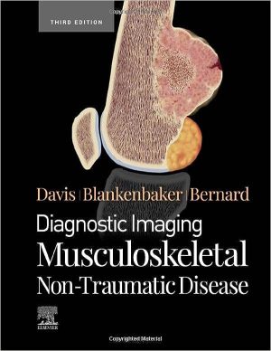
Diagnostic Imaging: Musculoskeletal Non-Traumatic Disease 3rd Edition

Diagnostic Imaging: Musculoskeletal Non-Traumatic Disease 3rd Edition

Covering the entire spectrum of this fast-changing field, Diagnostic Imaging: Musculoskeletal Non-Traumatic Disease, third edition, is an invaluable resource for musculoskeletal radiologists, general radiologists, and trainees―anyone who requires an easily accessible, highly visual reference in this complex area of imaging. Drs. Kirkland W. Davis, Donna G. Blankenbaker, Stephanie A. Bernard, and their team of highly regarded experts provide up-to-date information on recent advances in technology and the understanding of musculoskeletal diseases and disorders to help you make informed decisions at the point of care. The text is lavishly illustrated, delineated, and referenced, making it a useful learning tool as well as a handy reference for daily practice.
Guides readers through the complexities of the full range of non-traumatic musculoskeletal disorders, including arthritis, collagen vascular diseases, bone tumors, soft tissue tumors, infections, systemic diseases, developmental and congenital abnormalities, and metabolic diseases
Contains multiple new chapters on topics such as musculoskeletal genetics, neurinomas, and rapidly progressive osteoarthritis, among others, as well as updates throughout on reclassified lesions, tumors, and neoplasms; musculoskeletal infection details, including image-guided aspirations and biopsies for infections; and evolving medical and surgical treatments for many musculoskeletal conditions
Reflects recent changes in the World Health Organization’s classification of tumors and tumor-like conditions regarding terminology and diagnostic criteria
Covers evolving imaging techniques such as ultrasound in non-traumatic disease imaging, contrast-enhanced ultrasound use in tumor biopsies, enhanced MR of musculoskeletal tumors, and diffusion-weighted MR, and PET/CT and PET/MR use for rapidly progressive osteoarthritis
Provides up-to-date discussions of enhancements in bone and soft tissue tumor pathology and imaging of orthopedic implants and related hardware
Features more than 3,750 annotated images (with an additional 2,100+ digital-only examples), including radiologic images, full-color medical illustrations, clinical and histologic photographs, and gross pathology images
Uses bulleted, succinct text and highly templated chapters for quick comprehension of essential information at the point of care

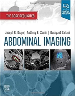
Abdominal Imaging: The Core Requisites 1st Edition

Abdominal Imaging: The bdominal Imaging: The Core Requisites 1st Edition

Taking a high-yield, “just the essentials” approach, Abdominal Imaging: The Core Requisites helps you establish a foundational understanding of both gastrointestinal and genitourinary imaging during rotations, prepare for the core and certifying exams, and refresh your knowledge of key concepts. This new title solves the “information overload” problem often faced by trainee and practicing radiologists by emphasizing the essential knowledge you need in an easy-to-ready hybrid format of traditional text and bullet points.

  • Emphasizes a “just the essentials” approach to foundational abdominal imaging content presented in an easy-to-read, quick reference format, with templated content that includes numerous outlines, tables, pearls, boxed material, and bulleted text for easy reading and efficient recall.

  • Helps you build and solidify core knowledge to prepare you for clinical practice with critical, up-to-date information on GI/GU topics, including relevant anatomy, lesion characterization, tumor staging, indication-based protocols and techniques, and more.

  • Prioritizes high-yield topics and explains key information to help you efficiently and effectively prepare for board exams.

    • Contains problem-based and disease-focused chapters such as right upper quadrant pain, chronic liver disease, colorectal cancer and screening, postoperative imaging, and abdominal/pelvic trauma.

    • Includes reporting tips and recommendations with sample structured reports.

    • Features more than 500 high-quality images spanning a variety of critical abdominal and pelvic disease processes, including discussions of advanced imaging techniques such as multiparametric MRI, dual energy CT, and elastography.

    • Published as part of the newly reimagined Core Requisites series, an update to the popular Requisites seriesfor today’s busy clinician.
    • Enhanced eBook version included with purchase. Your enhanced eBook allows you to access all of the text, figures, and references from the book on a variety of devices.


DOWNLOAD THIS MEDICAL BOOK


Taking a high-yield, “just the essentials” approach, Abdominal Imaging: The Core Requisites helps you establish a foundational understanding of both gastrointestinal and genitourinary imaging during rotations, prepare for the core and certifying exams, and refresh your knowledge of key concepts. This new title solves the “information overload” problem often faced by trainee and practicing radiologists by emphasizing the essential knowledge you need in an easy-to-ready hybrid format of traditional text and bullet points.

  • Emphasizes a “just the essentials” approach to foundational abdominal imaging content presented in an easy-to-read, quick reference format, with templated content that includes numerous outlines, tables, pearls, boxed material, and bulleted text for easy reading and efficient recall.

  • Helps you build and solidify core knowledge to prepare you for clinical practice with critical, up-to-date information on GI/GU topics, including relevant anatomy, lesion characterization, tumor staging, indication-based protocols and techniques, and more.

  • Prioritizes high-yield topics and explains key information to help you efficiently and effectively prepare for board exams.

    • Contains problem-based and disease-focused chapters such as right upper quadrant pain, chronic liver disease, colorectal cancer and screening, postoperative imaging, and abdominal/pelvic trauma.

    • Includes reporting tips and recommendations with sample structured reports.

    • Features more than 500 high-quality images spanning a variety of critical abdominal and pelvic disease processes, including discussions of advanced imaging techniques such as multiparametric MRI, dual energy CT, and elastography.

    • Published as part of the newly reimagined Core Requisites series, an update to the popular Requisites seriesfor today’s busy clinician.
    • Enhanced eBook version included with purchase. Your enhanced eBook allows you to access all of the text, figures, and references from the book on a variety of devices.

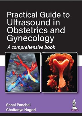
Practical Guide to Ultrasound in Obstetrics and Gynecology: A comprehensive book 1st Edition

Practical Guide to Ultrasound in Obstetrics and Gynecology: A comprehensive book 1st Edition

This book is one more addition in the popular series of Practical Guide to Ultrasound in Obstetrics and Gynecology—A Comprehensive Book by the authors and editors Chaitanya Nagori and Sonal Panchal. They are known for their ‘to-the-point’ writing and crystal clear explanations.

DOWNLOAD THIS MEDICAL BOOK

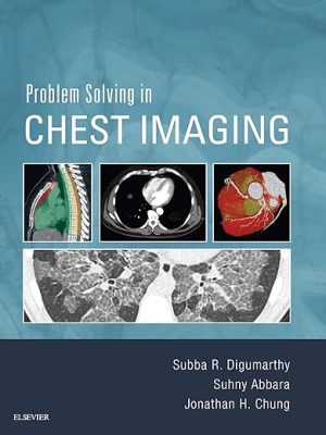
Problem Solving in Chest Imaging 1st Edition


Problem Solving in Chest Imaging 1st Edition

Optimize diagnostic accuracy with Problem Solving in Chest Imaging, a new volume in the Problem Solving in Radiology series. This concise title offers quick, authoritative guidance from experienced radiologists who focus on the problematic conditions you’re likely to see—and how to reach an accurate diagnosis in an efficient manner.

  • Addresses the practical aspects of chest imaging—perfect for practitioners, fellows, and senior level residents who may or may not specialize in chest radiology, but need to use and understand it.

  • Helps you make optimal use of the latest imaging techniques and achieve confident diagnoses.

  • Presents content by organ system and commonly encountered problems, with problem solving techniques integrated throughout.

  • Features more than 1,500 high-quality images that provide a clear picture of what to look for when interpreting studies.

  • Focuses on the core knowledge needed for successful results, covering anatomy, imaging techniques, imaging approach, entities by pathologic disease and anatomic region, and special situations. Key topics include Diffuse Lung Disease, Neoplasms of the Lung and Airways, Interstitial Lung Disease, Smoking-Related Lung Diseases, and Cardiovascular Disease.
  • Shows how to avoid common problems that can lead to an incorrect diagnosis. Tables and boxes with tips, pitfalls, and other teaching points show you what to look for, while problem-solving advice helps you make sound clinical decisions.

DOWNLOAD THIS MEDICAL BOOK

Webb, Müller and Naidich’s High-Resolution CT of the Lung Sixth Edition

Webb, Müller and Naidich’s High-Resolution CT of the Lung Sixth Edition

Selected as a Doody’s Core Title for 2022 and 2023!

Over the past 30 years high-resolution CT (HRCT) has matured to become an integral part of the multidisciplinary evaluation in diffuse lung disease. In this regard, Webb, Muller and Naidich’s High-Resolution CT of the Lung, 6th Edition, is a ‘gold standard’ reference that aims to keep radiologists and pulmonologists alike at the cutting edge of the ever-evolving field of thoracic imaging. The new US-European author team continues the tradition of excellence which readers have come to expect while the underlying layout and ethos — established by the ‘founding’ author team — remain. The new edition aims to brings readers up to date not only with recent advances but also with the important conceptual changes in thinking in various fields of thoracic imaging. Also featured in this updated edition is authoritative guidance on HRCT findings and differential diagnosis, as well as the characteristics of the common lung diseases assessed using HRCT, all enhanced by a multitude of new images and updated content throughout.

  • Covers the physical and anatomic principles governing HRCT imaging and the importance of the ‘pattern-based’ approach to HRCT interpretation.
  • Updates to chapters on interstitial lung diseases and airways diseases & connective tissue diseases reflect the dramatic changes in thinking in these fields.
  • Includes new and improved features including key HRCT findings in separate tables in relevant sections, where relevant, and new and exciting topics including artificial intelligence and machine learning. [Reword to: Includes improved features such as key HRCT findings in separate tables, and new topics including artificial intelligence and machine learning. ?]
  • Contains full-color illustrations of histologic findings in lung disease linked with HRCT manifestations.
  • Provides diagnostic algorithms, an illustrated quick reference guide at the front of the text, and helpful tables and line drawings.

Enrich Your eBook Reading Experience 

  • Read directly on your preferred device(s), such as computer, tablet, or smartphone.
  • Easily convert to audiobook, powering your content with natural language text-to-speech.

DOWNLOAD THIS MEDICAL BOOK

1 2 3 4 5 7