Diagnostic Imaging of the Foot and Ankle 1st Edition

Diagnostic Imaging of the Foot and Ankle 1st Edition

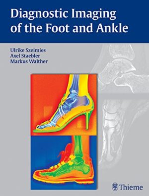
The foot has a special place in musculoskeletal diagnosis due to its complex anatomy and because many similar symptoms can have different causes, each requiring a different approach to treatment. The evaluation of foot disorders and diseases requires close clinical–radiological correlation and communication with foot experts. Foot disorders and injuries increase with age, due in part to the rising popularity of recreational sports in all age groups. Diagnostic Imaging of the Foot and Ankle will help you train your eye to recognize disorders and diseases of the foot and ankle, including those that are often misdiagnosed or overlooked.

Key Features:

  • By practitioners for practitioners: First-hand knowledge from leading surgical and orthopedic foot experts and radiologists
  • Clear and concise: A textbook and reference in a user-friendly layout focused on the foot and ankle
  • Uniform format: Entities are described by definition, clinical presentation, imaging modalities, typical imaging features, differential diagnosis, treatment options, course, and pitfalls
  • Clinical aspects and treatment: Clinical–radiological correlation plus a concise review of treatment options
  • The new standard: This information on the foot and ankle is available nowhere else in such a condensed form
  • Highest quality images: More than 500 superb illustrations including high-resolution images acquired with high-field MRI and multi-channel coils

DOWNLOAD THIS MEDICAL BOOK

Fundamentals of Body MRI (Fundamentals of Radiology) 2nd Edition

Fundamentals of Body MRI (Fundamentals of Radiology) 2nd Edition

Effectively perform and interpret MR body imaging with this concise, highly illustrated resource! Fundamentals of Body MRI, 2nd Edition, by Drs. Christopher Roth and Sandeep Deshmukh, covers the essential concepts residents, fellows, and practitioners need to know, laying a solid foundation for understanding the basics and making accurate diagnoses. This easy-to-use title in the Fundamentals of Radiology series covers all common body MR imaging indications and conditions, while providing new content on physics and noninterpretive skills with an emphasis on quality and safety.

  • More than 1,400 detailed MRI images and 100 algorithms and diagrams highlight key findings and help you grasp visual nuances of images you’re likely to encounter.

  • All common body MR imaging content is covered, along with discussion of how physics, techniques, hardware, and artifacts affect results.

  • Expert Consult™ eBook version included with purchase. This enhanced eBook experience allows you to search all of the text, figures, and references from the book on a variety of devices.
  • Newly streamlined format helps you retrieve important information more quickly.

  • Extensively revised content on the liver, including new MRI contrast agents; new coverage of the spleen; and new safety tips and guidelines keep you up to date.

  • New chapters on GI imaging, the prostate, and the male genitourinary system make this a one-stop reference to address the full range of body MRI.


DOWNLOAD THIS MEDICAL BOOK


Pediatric Emergency Radiology (WHAT DO I DO NOW EMERGENCY MEDICINE)

Pediatric Emergency Radiology (WHAT DO I DO NOW EMERGENCY MEDICINE) 

Part of the “What Do I Do Now?: Emergency Medicine” series, Pediatric Emergency Radiology uses a case-based approach to cover common and important topics in radiology imaging for pediatric emergency care. Each chapter provides a discussion of the diagnosis, key points to remember, and selected references for further reading. Areas of controversy are clearly delineated with a discussion regarding evidence-based options and a balanced view of treatment and disposition decisions. The book addresses a wide range of topics including neonatal respiratory distress, foreign body ingestion, Bronchiolitis, and related radiology issues faced by emergency medicine providers and pediatricians.

Pediatric Emergency Radiology is an engaging collection of thought-provoking cases which clinicians can utilize for effective imaging of pediatric patients. The volume is also a self-assessment tool that tests the reader’s ability to answer the question, “What do I do now?”

DOWNLOAD THIS MEDICAL BOOK

Emergency Imaging of At-Risk Patients – E-Book

Emergency Imaging of At-Risk Patients – E-Book

Radiologists in emergency department settings are uniquely positioned to identify and provide effective, appropriate care to vulnerable patient populations. Emergency Imaging of At-Risk Patients fills a void in the literature by illustrating challenges in emergency and trauma imaging of vulnerable patients using a head-to-toe approach. Drawing on the vast clinical experience of emergency and trauma radiologists from the largest academic medical centers across North America, this reference presents basic and advanced emergency imaging concepts, relevant case studies, current controversies and protocols, and subtle imaging findings that help guide clinicians to efficient and accurate diagnoses and treatments.

  • Provides a comprehensive, evidence-based approach to imaging of non-traumatic and traumatic emergencies in at-risk patients.
  • Covers acute brain, thoracic, abdominal, and musculoskeletal conditions, including the unique challenges of imaging pregnant, geriatric, bariatric, cancer, immunocompromised, and pediatric patients, as well as patients using recreational drugs.
  • Describes specific applications of ultrasound, MRI, radiography, and multidetector computed tomography (MDCT).
  • Reviews recent imaging-related clinical literature and appropriate criteria and guidelines.
  • A valuable tool for emergency and general radiologists, as well as pediatricians, obstetricians, gynecologists, orthopedic, trauma surgeons, and trainees in all specialties.

DOWNLOAD THIS MEDICAL BOOK

ExpertDDx: Chest 2nd Edition

ExpertDDx: Chest 2nd Edition

Now fully revised and up-to-date, Expert DDx: Chest, second edition,quickly guides you to the most likely differential diagnoses based on key imaging findings and clinical information. Expert radiologists Melissa L. Rosado-de-Christenson, Brett W. Carter, and John P. Lichtenberger III present more than 120 cases across a broad cardiothoracic spectrum, classified by general imaging features, modality-specific findings, and clinically-based indications. Readers will find authoritative, superbly illustrated guidance for defining and reporting useful, actionable differential diagnoses that lead to definitive findings for the entire gamut of chest disorders.

  • Presents several clear, sharp, succinctly annotated images for each diagnosis (more than 1,800 annotated images in all); a list of diagnostic possibilities sorted as common, less common, and rare but significant; and brief, bulleted text offering helpful diagnostic clues


  • Shows both typical and variant manifestations of each possible diagnosis


  • Includes new cases, expanded differential considerations, new terminology, and updated imaging throughout


  • Features all relevant imaging modalities, including chest radiography, the latest generation of multi-planar advanced cross-sectional CT and MR imaging, and molecular imaging with FDG PET/CT
  • Covers new and evolving areas such as lung cancer screening and the localization and classification of mediastinal lesions, and contains expanded content on the heart and pericardium


DOWNLOAD THIS MEDICAL BOOK

Nuclear Cardiology and Multimodal Cardiovascular Imaging: A Companion to Braunwald’s Heart Disease 1st Edition

Nuclear Cardiology and Multimodal Cardiovascular Imaging: A Companion to Braunwald’s Heart Disease

1st Edition

Recent years have seen numerous advances in cardiovascular nuclear medicine technology, leading to more precise diagnoses and treatment and an expanded understanding of the molecular basis for cardiac disease. Nuclear Cardiology and Multimodal Cardiovascular Imaging is a one-stop, comprehensive guide to the diagnostic and clinical implications of this complex and increasingly important technology. Part of the Braunwald family of renowned cardiology references, it provides cutting-edge coverage of multimodal cardiac imaging along with case vignettes and integrated teaching content―ideal for cardiologists, cardiology fellows, radiologists, and nuclear medicine physicians.

Features all the latest cardiovascular nuclear medicine studies with practical, evidence-based implications for personalized patient evaluation and treatment.


Presents a consistent, patient-centered approach using integrated case vignettes correlated with specific nuclear medicine imaging findings. Discusses patient assessment criteria, risk factor criteria, pathology, evaluation criteria, outcomes, and other clinical implications.


Covers a full range of imaging technologies, including SPECT/CT, PET/CT, and CT/MR hybrid radionuclide cardiovascular imaging studies.


Addresses emerging clinical applications of nuclear imaging techniques for precision-based medicine, including targeted molecular imaging and cell therapies.


Includes sections on instrumentation/principles of imaging; protocols and interpretation; applications in coronary artery disease, special populations, and heart failure; artificial intelligence, and more.


Contains guidelines and appropriate use documents to provide appropriate context for clinicians.


Features hundreds of high-quality figures including multimodal cardiac imaging studies, anatomic illustrations, and graphs.


Provides Key Point summaries, 50 procedural videos, and 100 multiple-choice questions and answers to reinforce understanding and facilitate review.


Enhanced eBook version included with purchase, which allows you to access all of the text, figures, and references from the book on a variety of devices

DOWNLOAD THIS MEDICAL BOOK

Chest Radiology: Patterns and Differential Diagnoses

[amazon_link asins=’0323498310′ template=’ProductAd’ store=’aishabano-20′ marketplace=’US’ link_id=’982e18e2-07ba-11e9-8e71-5bdbcffd66b6′]

Chest Radiology: Patterns and Differential Diagnoses

DOWNLOAD THIS BOOK FREE HERE

https://upstore.net/DbBRy6z

Sharpen your skills in chest x-ray interpretation using this trusted clinical resource! Chest Radiology: Patterns and Differential Diagnoses, 7th Edition, by Dr. James Reed, walks you through a logical, sequential thought process for the differential diagnoses of 23 radiologic patterns of common chest diseases, using 150 superbly illustrated patient cases. You’ll gain a solid and thorough understanding of how to read and interpret chest x-rays with expert guidance on common disease patterns, differential diagnoses, narrowing down the diagnoses, and further studies (from additional radiographic exams to CT or to biopsy). Each chapter follows a consistent format: Presenting Case, Questions, Discussion, Top 5 Diagnoses, Summary, and Answer Guide.

  • Heavily illustrated with chest radiographs and additional CT, HRCT, and MR correlative images; plus bulleted summary boxes of comprehensive diagnoses lists and interpretation points.
  • An ideal resource for mastering this lower-cost modality before considering more complicated and costly procedures.
    • Illustrated case studies and quizzes feature new and improved questions that address the challenges seen in practice today.

    • Significant updates on carcinoid tumors and pulmonary nodules, including pathologic descriptions, detection, management, and new terminology; new lung cancer screening guidelines and benefits of CT vs. x-ray for screening; and CT description of nodules, detection of pulmonary nodules with chest x-rays, anatomic and perception problems with x-rays in identifying nodules.

    • New content on diffuse disease, including new pathologic terms and the latest impact of high resolution CT (HRCT).

    • Updates on radiation dosing and emphasis on lower dosing, and an expanded emphasis on how to interpret x-rays and CT scans.

  • Expert Consult™ eBook version included with purchase. This enhanced eBook experience allows you to search all of the text, figures, and references from the book on a variety of devices.


Introduction to Medical Imaging: Physics, Engineering and Clinical Applications (Cambridge Texts in Biomedical Engineering)

[amazon_link asins=’0521190657′ template=’ProductAd’ store=’aishabano-20′ marketplace=’US’ link_id=’ecce45af-fd8c-11e8-8783-1f61fbae02df’]

Introduction to Medical Imaging: Physics, Engineering and Clinical Applications (Cambridge Texts in Biomedical Engineering)

DOWNLOAD THIS BOOK FREE HERE

https://upstore.net/DfCKXgj

Covering the basics of X-rays, CT, PET, nuclear medicine, ultrasound, and MRI, this textbook provides senior undergraduate and beginning graduate students with a broad introduction to medical imaging. Over 130 end-of-chapter exercises are included, in addition to solved example problems, which enable students to master the theory as well as providing them with the tools needed to solve more difficult problems. The basic theory, instrumentation and state-of-the-art techniques and applications are covered, bringing students immediately up-to-date with recent developments, such as combined computed tomography/positron emission tomography, multi-slice CT, four-dimensional ultrasound, and parallel imaging MR technology. Clinical examples provide practical applications of physics and engineering knowledge to medicine. Finally, helpful references to specialised texts, recent review articles, and relevant scientific journals are provided at the end of each chapter, making this an ideal textbook for a one-semester course in medical imaging.

Color Atlas of Ultrasound Anatomy

[amazon_link asins=’3131390522′ template=’ProductAd’ store=’aishabano-20′ marketplace=’US’ link_id=’89812f44-fb8f-11e8-bf69-e77a2a205d65′]

Color Atlas of Ultrasound Anatomy

DOWNLOAD THIS BOOK FREE HERE

https://upstore.net/DrEWTd6

Color Atlas of Ultrasound Anatomy, Second Edition presents a systematic, step-by-step introduction to normal sectional anatomy of the abdominal and pelvic organs and thyroid gland, essential for recognizing the anatomic landmarks and variations seen on ultrasound. Its convenient, double-page format, with more than 250 image quartets showing ultrasound images on the left and explanatory drawings on the right, is ideal for rapid comprehension. In addition, each image is accompanied by a line drawing indicating the position of the transducer on the body and a 3-D diagram demonstrating the location of the scanning plane in each organ.

Special features:

More than 60 new ultrasound images in the second edition that were obtained with state-of-the-art equipment for the highest quality resolution A helpful foundation on standard sectional planes for abdominal scanning, with full-color photographs demonstrating probe placement on the body and diagrams of organs shown Front and back cover flaps displaying normal sonographic dimensions of organs for easy reference

Covering all relevant anatomic markers, measurable parameters, and normal values, and including both transverse and longitudinal scans, this pocket-sized reference is an essential learning tool for medical students, radiology residents, ultrasound technicians, and medical sonographers.

1 2 3 4 5