Fundamentals of Body MRI

Fundamentals of Body MRI 

Effectively perform and interpret MR body imaging with this concise, highly illustrated resource! Fundamentals of Body MRI, 2nd Edition, by Drs. Christopher Roth and Sandeep Deshmukh, covers the essential concepts residents, fellows, and practitioners need to know, laying a solid foundation for understanding the basics and making accurate diagnoses. This easy-to-use title in the Fundamentals of Radiology series covers all common body MR imaging indications and conditions, while providing new content on physics and noninterpretive skills with an emphasis on quality and safety.

  • More than 1,400 detailed MRI images and 100 algorithms and diagrams highlight key findings and help you grasp visual nuances of images you’re likely to encounter.

  • All common body MR imaging content is covered, along with discussion of how physics, techniques, hardware, and artifacts affect results.

  • Expert Consult™ eBook version included with purchase. This enhanced eBook experience allows you to search all of the text, figures, and references from the book on a variety of devices.
  • Newly streamlined format helps you retrieve important information more quickly.

  • Extensively revised content on the liver, including new MRI contrast agents; new coverage of the spleen; and new safety tips and guidelines keep you up to date.

  • New chapters on GI imaging, the prostate, and the male genitourinary system make this a one-stop reference to address the full range of body MRI.

DOWNLOAD THIS MEDICAL BOOK 

Workbook for Merrill’s Atlas of Radiographic Positioning and Procedures 15th Edition

Workbook for Merrill’s Atlas of Radiographic Positioning and Procedures 15th Edition

Reinforce your understanding of radiographic positioning and procedures with this practical workbook! Corresponding to the content in Merrill’s Atlas of Radiographic Positioning & Procedures, 15th Edition, this workbook helps you review and practice essential radiographic terminology, anatomy, and positioning concepts. Exercises include multiple-choice and matching questions, imaging evaluation, and more. Written by the textbook’s authors, this workbook prepares you for success on the ARRT certification exam and in clinical radiography practice.

  • Anatomy exercises reinforce your understanding with multiple-choice, matching, and short-answer questions, labeling and identification diagrams, and crossword puzzles.
  • Positioning exercises include multiple-choice, matching, short-answer, true/false, and fill-in-the blank questions, labeling and identification diagrams, and comparisons of standard radiographic projections.
  • Exercises on identifying errors in radiographs prepare you to evaluate radiographs in clinical situations.
  • Pathology exercises help you understand which projections will best demonstrate various pathologies.
  • Self-tests at the end of each chapter include review questions enabling you to assess your comprehension and measure your own progress.
  • NEW! Updated drawings and radiographic images reflect the content updates in the Merrill’s Atlas of Radiographic Positioning & Procedures, 15th Edition text.
  • NEW exercises reflect the latest changes to the American Registry of Radiologic Technologists (ARRT) examination.

DOWNLOAD THIS MEDICAL BOOK

Diagnostic Ultrasound: Abdomen and Pelvis 2nd Edition

Diagnostic Ultrasound: Abdomen and Pelvis 2nd Edition

Develop a solid understanding of ultrasound of the abdomen and pelvis with this practical, point-of-care reference in the popular Diagnostic Ultrasound series. Written by leading experts in the field, the second edition of Diagnostic Ultrasound: Abdomen and Pelvis offers detailed, clinically oriented coverage of ultrasound imaging of this complex area and includes illustrated and written correlation between ultrasound findings and other modalities. The most comprehensive reference in its field, this image-rich resource helps you achieve an accurate ultrasound diagnosis for every patient.

  • Features nearly 15 new chapters that detail updated diagnoses, new terminology, new methodology, new criteria and guidelines, a new generation of scanners, and more
  • Includes 2,500 high-quality images including grayscale, color, power, and spectral (pulsed) Doppler imaging in each chapter and, when applicable, contrast-enhanced ultrasound; plus new videos and animations online
  • Discusses new polycystic ovary syndrome (PCOS) criteria, updated pancreatic cyst guidelines, new ovarian cysts recommendations, shear wave elastography for liver fibrosis, and more
  • Correlates ultrasound findings with CT and MR for improved understanding of disease processes and how ultrasound complements other modalities for a given disease
  • Covers cutting-edge ultrasound techniques, including microbubble contrast and contrast-enhanced US (CEUS) for liver imaging
  • Contains time-saving reference features such as succinct and bulleted text, a variety of test data tables, key facts in each chapter, annotated images, and an extensive index
  • Enhanced eBook version included with purchase. Your enhanced eBook allows you to access all of the text, figures, and references from the book on a variety of devices.

DOWNLOAD THIS MEDICAL BOOK

Ferri’s Best Test: A Practical Guide to Clinical Laboratory Medicine and Diagnostic Imaging (Ferri’s Medical Solutions) 5th Edition

Ferri’s Best Test: A Practical Guide to Clinical Laboratory Medicine and Diagnostic Imaging (Ferri’s Medical Solutions) 5th Edition

Clinicians at all levels must consider a myriad of tests in the diagnostic process, requiring an up-to-date understanding of changing technology and the demands of time- and cost-efficiency. Ferri’s Best Test, 5th Edition, is a unique, easy-to-use guide that simplifies complex information and helps you choose the best test to supplement your clinical diagnostic skills. It includes both lab and imaging tests for concise, convenient access to all diagnostic test options for more than 200 common diseases and disorders.

• Practical, concise, spiral-bound, and pocket-sized―designed from cover to cover for quick, on-the-go reference.
• Three convenient sections provide quick access to key information on clinical laboratory testing, diagnostic imaging, and diagnostic algorithms.
• New and updated content on cardiac computed tomography angiography, computed tomography perfusion imaging, COVID-19, and much more.
• Coverage of 23 new laboratory tests and 14 new diseases and disorders.
• Essential information on indications, advantages, disadvantages, approximate costs, normal ranges, typical abnormalities, likeliest causes, and more.
• Experienced author Dr. Fred Ferri uses a unique, easy-to-follow format to simplify complex information and help you choose the best test for every patient.
• New appendices on electrocardiography and respiratory testing and function.
• Comparison tables and illustrations help improve your test selection.
• Enhanced eBook version included with purchase. Your enhanced eBook includes an image collection with access on a variety of devices.

DOWNLOAD THIS MEDICAL BOOK


Clark’s Essential Guide to Clinical Ultrasound (Clark’s Companion Essential Guides) 1st Edition

Clark’s Essential Guide to Clinical Ultrasound (Clark’s Companion Essential Guides) 1st Edition

This easy-to-understand pocketbook in the highly respected Clark’s stable of diagnostic imaging texts is an invaluable tool for students, sonographers and other ultrasound practitioners, providing practical guidance on clinical ultrasound procedures, summarising current relevant literature and professional body guidelines.

The content is arranged by anatomical system and organ for ease of reference, with each section comprising a short introduction, the indications for the procedure, patient preparation, the imaging procedure itself, along with an image analysis, supported throughout by positioning photographs and clinical images.

Clark’s Essential Guide to Clinical Ultrasound is ideal for all users of clinical ultrasound at both undergraduate and postgraduate level and will also provide a convenient distillation of the latest best practice and guidelines for sonographers, midwives, vascular technologists, ECG technologists, medical doctors, sports injury specialists, paramedics, and other health professionals.

DOWNLOAD THIS MEDICAL BOOK

Diseases of the Heart, Chest & Breast 2011-2014: Diagnostic Imaging and Interventional Techniques 2011th Edition

Diseases of the Heart, Chest & Breast 2011-2014: Diagnostic Imaging and Interventional Techniques 2011th Edition

Written by internationally renowned experts, this volume deals with imaging of diseases of heart, chest and breast. The different topics are disease-oriented and cover all the relevant imaging modalities, including standard radiography, CT, nuclear medicine with PET, ultrasound and magnetic resonance imaging, as well as imaging-guided interventions. This book presents a comprehensive review of current knowledge in imaging of the heart and chest , as well as thoracic interventions and a selection of “hot topics” of breast imaging. It will be particularly relevant for residents in radiology, but also very useful for experienced radiologists and clinicians specializing in thoracic disease and wishing to update their knowledge of this rapidly developing field.

DOWNLOAD THIS MEDICAL BOOK

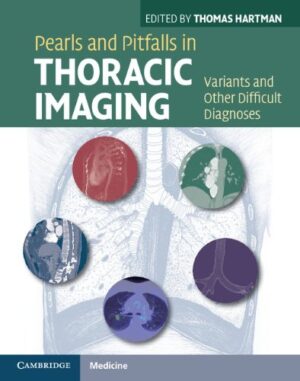
Pearls and Pitfalls in Thoracic Imaging: Variants and Other Difficult Diagnoses 1st Edition

Pearls and Pitfalls in Thoracic Imaging: Variants and Other Difficult Diagnoses 1st Edition

How often have you been confronted with an image on a thoracic CT exam where you knew it didn’t look ‘normal’, but you weren’t sure whether it was ‘abnormal’ either? And if it is abnormal, is there a specific diagnosis you should be able to make directly off the images? Pearls and Pitfalls in Thoracic Imaging is your one-stop resource to answer questions such as: Is this a normal variant or a disease-related abnormality? Are these findings specific for an uncommon disease and if so what is the diagnosis? Is this set of findings strongly suggestive of a diagnosis? Which additional imaging test will allow me to be confident in that diagnosis? Could the ‘abnormality’ be due to an artifact mimicking disease? Written by leading thoracic radiologists and with concise, image-rich descriptions, Pearls and Pitfalls in Thoracic Imaging is an invaluable diagnostic tool for every radiologist.

DOWNLOAD THIS MEDICAL BOOK 

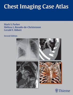
Chest Imaging Case Atlas 2nd Edition

Chest Imaging Case Atlas 2nd Edition

A comprehensive atlas covering the breadth and depth of chest imaging

Written by renowned experts in chest imaging, Chest Imaging Case Atlas, Second Edition enables radiology residents, fellows, and practitioners to hone their diagnostic skills by teaching them how to interpret a large number of radiologic cases. This atlas contains over 200 cases on conditions ranging from Adenoid Cystic Carcinoma to Wegener Granulomatosis. Each case is supported by a discussion of the disease, its underlying pathology, typical and unusual imaging findings, management, and prognosis, providing a comprehensive overview of each disorder.

Special Features of the Second Edition:

  • Over 1500 high-quality images demonstrating normal and pathologic findings and their variations
  • More multiplanar, CT angiographic (CTA), MRI, and 3D imaging is incorporated into the text, helping readers stay current with this rapidly changing technology
  • 40 new cases and updated images in cases from the previous edition
  • A new post-thoracotomy chest section addresses normal post-operative findings and complications associated with common thoracic interventional procedures
  • The neoplastic diseases section includes the new TNM staging system for lung cancer
  • The adult cardiovascular disease section now contains a discussion on univentricular and biventricular or end-stage heart failure including various ventricular assist devices and the Total Artificial Heart, their imaging features, and complications associated with their use
  • The diffuse lung disease section has been expanded to include an approach to HRCT interpretation
  • Case discussions are based on up-to-datereviews of current literature as well as classic landmark articles
  • Pearls are provided to describe the features that may strongly support a specific diagnosis, enabling readers to sharpen their clinical diagnostic skills

This book is an invaluable illustrated reference that all physicians in radiology and chest imaging in particular, including pulmonary medicine physicians and thoracic surgeons, should have on their bookshelf.

DOWNLOAD THIS MEDICAL BOOK


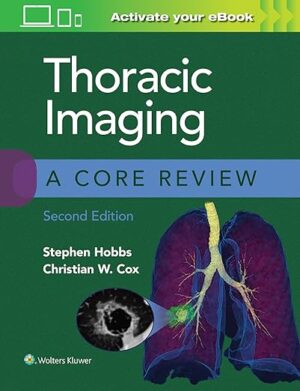
Thoracic Imaging: A Core Review 2nd Edition

Thoracic Imaging: A Core Review 2nd Edition

Prepare for success on the thoracic imaging component of the radiology Core Exam! Thoracic Imaging: A Core Review, 2nd Edition, is an up-to-date, practical review tool written specifically for the Core Exam. This helpful resource contains 300 image-rich, multiple-choice questions with detailed explanations of right and wrong answers, fully revised content, and additional eBook questions to ensure you’re ready for the Core Exam or recertification exam.

  • Features questions in all exam areas, including imaging basics, normal anatomy, ICU radiographs, pulmonary pathology, pleural disease, vascular diseases relevant to thoracic imaging, and trauma.
  • Questions accompanied with 1 or 2 high-quality radiographs, CT, MR, or ultrasound images.
  • Provides concise answers with explanations of each choice followed by relevant, up-to-date references.
  • Follows the structure and content of what you’ll encounter on the test, conveniently organized by topic. 

Enrich Your eBook Reading Experience

  • Read directly on your preferred device(s), such as computer, tablet, or smartphone.
  • Easily convert to audiobook, powering your content with natural language text-to-speech.

DOWNLOAD THIS MEDICAL BOOK

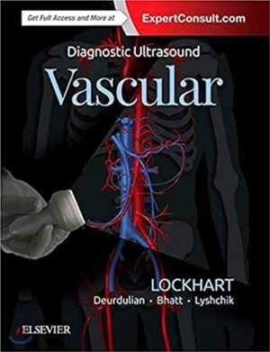
Diagnostic Ultrasound: Vascular 1st Edition

Diagnostic Ultrasound: Vascular 1st Edition

Develop a solid understanding of vascular ultrasound with this practical, point-of-care reference in the popular Diagnostic Ultrasound series. Written by leading experts in the field, Diagnostic Ultrasound: Vascular offers detailed, clinically oriented coverage of ultrasound anatomy, pathology, technique, and diagnosis. This wealth of up-to-date information helps you achieve an accurate vascular ultrasound diagnosis for every patient.

  • Ensures that you stay on top of rapidly evolving vascular ultrasound practice and its expanding applications for everyday clinical use
  • Includes extensively illustrated coverage of sonographic anatomy that depicts pertinent vascular structures of the head and neck, chest and abdomen, and extremities
  • Features image-rich chapters on vascular ultrasound techniques, covering grayscale, color, power, and spectral (pulsed) Doppler imaging, as well as imaging artifacts
  • Provides detailed sonographic descriptions for the vascular diseases and anomalies encountered in clinical practice, including lesions of head and neck, chest and abdomen (including transplants), and extremities, including tips, tricks, and pitfalls
  • Contains a gallery of typical and atypical ultrasound appearances covering a wide spectrum of disease, correlated with CT and MR imaging where appropriate, and detailed artistic renderings
  • Discusses key vascular ultrasound intervention techniques for both diagnosis and treatment
  • Uses a bulleted, templated format that helps you quickly find and understand complex information, as well as thousands of high-quality images and illustrations
  • An ideal reference for radiologists, sonographers, vascular surgeons, and those who are training in these fields
  • Expert Consult™ eBook version included with purchase, which allows you to search all of the text, figures, and references from the book on a variety of devices.

DOWNLOAD THIS MEDICAL BOOK

1 2 3 4 7