Atlas of Image-Guided Intervention in Regional Anesthesia and Pain Medicine Second Edition
1608317048

[amazon template=iframe image2&asin=1608317048]

The Atlas of Image-Guided Intervention in Regional Anesthesia and Pain Medicine is a practical guide for practitioners who perform interventional procedures with radiographic guidance to alleviate acute or chronic pain. The author provides an overview of each technique, with detailed full-color illustrations of the relevant anatomy, technical aspects of each treatment, and a description of potential complications. For this revised and expanded Second Edition, the author also discusses indications for each technique, as well as medical evidence on the technique’s applicability. The new edition features original drawings by a noted medical artist and for the first time includes three-dimensional CT images that correlate with the radiographic images and illustrations for a fuller understanding of the relevant anatomy.

download this book free here

http://upsto.re/s48FfCV

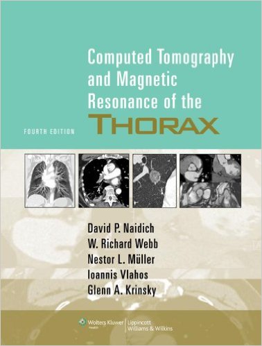
Computed Tomography and Magnetic Resonance of the Thorax Fourth Edition

0781757657

[amazon template=iframe image2&asin=0781757657]

The thoroughly revised, updated Fourth Edition of this classic reference provides authoritative, current guidelines on chest imaging using state-of-the-art technologies, including multidetector CT, MRI, PET, and integrated CT-PET scanning. This edition features a brand-new chapter on cardiac imaging. Extensive descriptions of the use of PET have been added to the chapters on lung cancer, focal lung disease, and the pleura, chest wall, and diaphragm. Also included are recent PIOPED II findings on the role of CT angiography and CT venography in detecting pulmonary embolism. Complementing the text are 2,300 CT, MR, and PET scans made on the latest-generation scanners.

Download this book free here

http://upsto.re/s48Wrvx

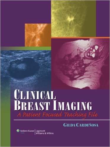
Clinical Breast Imaging: A Patient Focused Teaching File (LWW Teaching File Series) 1st Edition

0781762677

[amazon template=iframe image2&asin=0781762677]

Prepared by a preeminent breast imaging expert, this case-based teaching file atlas presents a clinically oriented approach to screening, diagnostic evaluation, and management of patients with breast conditions encountered by radiologists. Dr. Cardeñosa takes the reader through more than 170 actual patient cases, from classic “Aunt Minnies” to more complex and controversial problems in screening, diagnostic evaluation, and patient management. Cases are thoroughly illustrated with clear, sharp images—over 800 images total—and include multiple imaging studies, pathology studies, and pathologic correlations where appropriate. Emphasis is on determining the clinical significance of abnormalities or potential abnormalities detected on images.

Download this book free here

http://upsto.re/suMdRqa

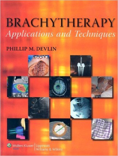
Brachytherapy: Applications and Techniques 1st Edition

0781762774

[amazon template=iframe image2&asin=0781762774]

Written by the foremost experts in the field, this volume is a comprehensive text and practical reference on contemporary brachytherapy. The book provides detailed, site-specific information on applications and techniques of brachytherapy in the head and neck, central nervous system, breast, thorax, gastrointestinal tract, and genitourinary tract, as well as on gynecologic brachytherapy, low dose rate and high dose rate sarcoma brachytherapy, vascular brachytherapy, and pediatric applications.

The book thoroughly describes and compares the four major techniques used in brachytherapy—intracavity, interstitial, surface-dose or mold therapy, and transluminal. Chapters detail particular techniques that are appropriate in specific clinical situations.

Download this book free here

http://upsto.re/suUsHVM

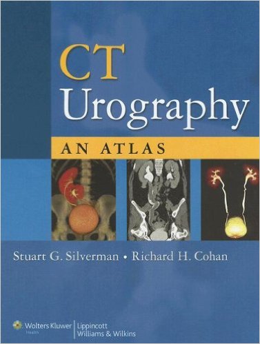
CT Urography: An Atlas 1st Edition

0781787548

[amazon template=iframe image2&asin=0781787548]

Featuring over 500 images, this atlas is the first text on performing and interpreting CT urography. Chapters detail the indications and techniques for CT urography, review the risks of radiation exposure, show how normal urinary tract anatomy and variants appear on CT scans, and demonstrate a wide range of urinary tract abnormalities as they appear on thin-section CT. The final chapter illustrates artifacts and diagnostic pitfalls.

Chapters on abnormalities follow a case-based teaching file format. Each case is presented on a two-page spread, with images and succinct discussion of the entity and how CT urography was used to diagnose it.

download this book free here
http://upsto.re/s9Aupb2

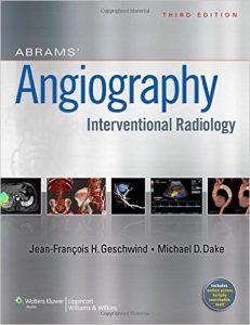
Abrams Angiography: Interventional Radiology 3rd edition

1609137922

[amazon template=iframe image2&asin=1609137922]

Reflecting recent interventions of startling impact and clinical benefit and honoring the field’s legacy of innovation, this Third Edition of Abrams’ Angiography: Interventional Radiology has been completely revamped to reflect the fast-moving landscape of interventional radiology. To provide a fresh take on leading-edge subjects, new series editors, Drs. Jean-Francois H. Geschwind and Michael D. Dake put together a team of 60 of today’s most renowned authorities to focus on the 100 topics essential to mastering contemporary interventional radiology. Now in vibrant full-color, this Third Edition begins with an all-new section on interventional oncology, which includes expert coverage of organ-specific cancers and specialized interventional techniques in cancer care. Many of the themes considered in the vascular sections are rooted in the original stock of the initial landmark Abrams’ volume, updated to focus on current understanding. Features: comprehensive, step-by-step coverage of all contemporary vascular and non-vascular interventional procedures; new Oncology section includes 21 expert chapters that walk through all aspects of interventional oncology, from evaluation of the cancer patient and principles of chemotherapy to management of organ-specific cancers and specialized interventional techniques in cancer care; new detailed coverage complemented by more than 1,100 full-color illustrations; increased focus on surgical anatomy and the details of each technique; expert chapters that capture the groundbreaking achievements of the field from the foremost authorities; and new online solution site provides fully searchable access to the book-anytime, anywhere!

Download this book free here
http://upsto.re/sa8xp6D

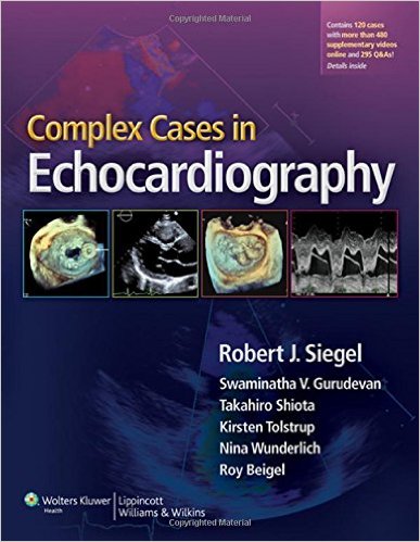
Complex Cases in Echocardiography

1451176465

[amazon template=iframe image2&asin=1451176465]

Complex Cases in Echocardiography is a unique text written by a team of top cardiologists at Cedars Sinai Heart Institute. Drawing from their extensive library of echocardiograms, the authors feature 75 cases demonstrating uncommon and puzzling echo findings. Each case begins with a brief clinical presentation, and related images, followed by multiple-choice questions. Detailed answers include patient outcomes and follow-up recommendations. The cases include m-mode, 2-D, 3-D echo, TEE, and Doppler ultrasound. In addition, three fold-out matching quizzes present a particular physical finding or electrocardiogram to be matched with the patient’s corresponding echocardiogram. With the inclusion of cases seen in emergency situations as well as routine readings in an echo laboratory, this text not only introduces the reader to unusual cases, but also reinforces relevant techniques and applications. A companion website includes the fully searchable text and features additional cases, along with actual case video clips. Features: case-based format featuring 75 unusual and bizarre diagnostic dilemmas; questions and answers associated with each case 3 unique fold-out quizzes; and a companion website with fully searchable text and 45 additional cases.

Download this book free here
http://upsto.re/sov4XeM

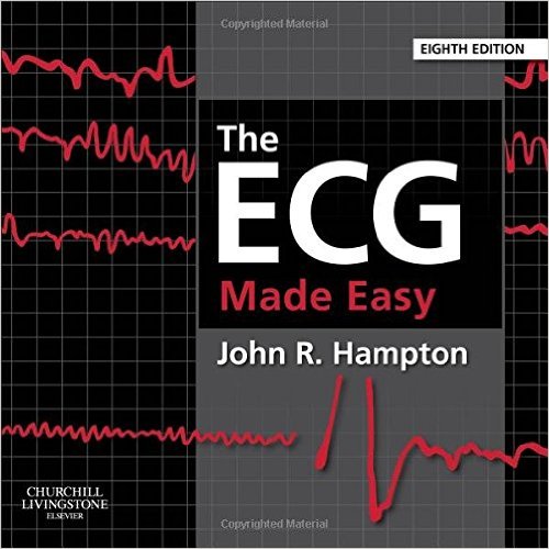
The ECG Made Easy, 8th edition

0702046418

[amazon template=iframe image2&asin=0702046418]

For forty years The ECG Made Easy has been regarded as the best introductory guide to the ECG, with sales of over half a million copies as well as being translated into a dozen languages. Hailed by the British Medical Journal as a “medical classic”, it has been a favourite of generations of medical and health care staff who require clear, basic knowledge about the ECG. This famous book encourages the reader to accept that the ECG is easy to understand and that its use is just a natural extension of taking the patient’s history and performing a physical examination. It directs users of the electrocardiogram to straightforward and accurate identification of normal and abnormal ECG patterns.

Download this book free here
http://upsto.re/sod35kz

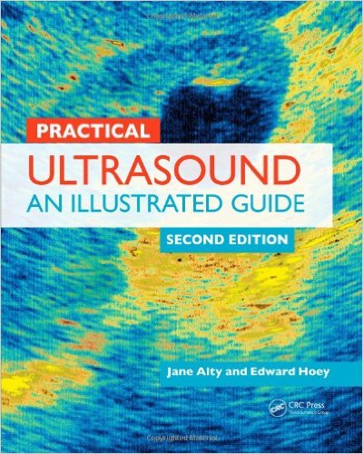
Practical Ultrasound: An Illustrated Guide, Second Edition

1444168290

[amazon template=iframe image2&asin=1444168290]

In the hands of a skilled operator, ultrasound scanning is a simple and easy procedure. However, reaching that level of proficiency can be a long and tedious process. Commended by the British Medical Association, Practical Ultrasound, Second Edition focuses on the scans regularly encountered in a busy ultrasound department and provides everything practitioners need to know to become competent and skilled in scanning.

See What’s New in the Second Edition:


    • New chapters on breast, musculoskeletal, and FAST (focused assessment with sonography in trauma) ultrasonography
    • Revisions to original chapters incorporating up-to-date techniques and protocols

Beginning with the general principles of ultrasound scanning and a guide to using the ultrasound machine, the book provides step-by-step instructions on how to perform scans supplemented by high-quality images and handy tips.

Organized according to anatomical site, the chapters include a review section on useful anatomy, scan protocol presented step by step, and a section on common pathology. Maintaining the popular format of the previous edition, each chapter contains examples of common and clinically relevant pathologies and notes on the salient features of these conditions.

The authors’ precise approach puts an immense amount of knowledge within easy reach, making it an ideal aid for learning the practicalities of ultrasound.

Download this book free here
http://upsto.re/soe6fyE

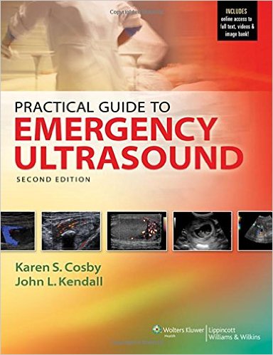
Practical Guide to Emergency Ultrasound 2nd edition

1451175558

[amazon template=iframe image2&asin=1451175558]

Keep pace with the rapid changes in Emergency Ultrasound knowledge. This 2nd edition of the Practical Guide to Emergency Ultrasound addresses your need for a practical, comprehensive, how-to book on ultrasound techniques in the emergency department, including new and expanded applications. Organized in an easy-to-navigate problem-based and symptom-based approach, chapters cover many uses for ultrasound in the ED and proper ultrasound technique. Over 900 images, many in full color, illustrate key concepts and diagnoses, including the use of echocardiography in the ED and newer applications for imaging ocular, musculoskeletal injuries and the use of ultrasound in the management of undifferentiated hypotension and dyspnea. Features: chapters on newer applications of ultrasound including thoracic, inferior vena cava, musculoskeletal, and ocular imaging; newer and evolving applications for procedural guidance, including nerve blocks; new and highlighted content for Pediatric applications; and chapters on prehospital use, and use in resource-limited settings; entire section focusing on Ultrasound use in the Resuscitation of Acute Injury or Illness; over 900 images demonstrate key diagnoses and proper ultrasound technique; over 175 online video clips that display more realistic three-dimensional views; and more than 60 additional online-only images for several chapters.

Download this book free here
http://upsto.re/soeGKoK

1 57 58 59 60