Imaging Musculoskeletal Trauma: Interpretation and Reporting (Current Clinical Imaging)


1118158814[amazon template=iframe image2&asin=1118158814]

This book offers a concise, systematic approach to imaging musculoskeletal trauma. Designed for use by professionals in radiology, orthopedics, emergency medicine, and sports medicine, it reviews common fracture and dislocation mechanisms and provides up–to–date guidelines on the use and interpretation of imaging tests. Replete with easily accessible information, including well–designed tables and lists, the book features radiology report checklists for each anatomic site, as well as numerous radiographs and CT and MRI images, with examples of common and serious injuries in the musculoskeletal system.

Download this book free here

http://upsto.re/s8tgSXs

Bone Quantitative Ultrasound

9400700164

[amazon template=iframe image2&asin=9400700164]

Quantitative ultrasound (QUS) of bone is a relatively recent research field. The research community is steadily growing, with interdisciplinary branches in acoustics, medical imaging, biomechanics, biomedical engineering, applied mathematics, bone biology and clinical sciences, resulting in significant achievements in new ultrasound technologies to measure bone, as well as models to elucidate the interaction and the propagation of ultrasonic wave in complex bone structures. Hundreds of articles published in specialists journals are accessible from the Web and from electronic libraries. However, no compilation and synthesis of the most recent and significant research exist. The only book on QUS of bone has been published in 1999 at a time when the propagation mechanisms of ultrasound in bone were still largely unknown and the technology was immature. The research community has now reached a critical size, special sessions are organized in major international meetings (e.g., at the World Congress of Biomechanics, the annual meetings of the Acoustical Society of America, International Bone Densitometry Workshop, etc…). Consequently, the time has come for a completely up to date, comprehensive review of the topic. The book will offer the most recent experimental results and theoretical concepts developed so far and is intended for researchers, graduate or undergraduate students, engineers, and clinicians who are involved in the field. The central part of the book covers the physics of ultrasound propagation in bone. Our goal is to give the reader an extensive view of the mathematical and numerical models as an aid to understand the QUS potential and the types of variables that can be determined by QUS in order to characterize bone strength. The propagation of sound in bone is still subject of intensive research. Different models have been proposed (for example, the Biot theory of poroelasticity and the theory of scattering have been used to describe wave propagation in cancellous bone, whereas propagation in cortical bone falls in the scope of guided waves theories). An extensive review of the models has not been published so far. We intend in this book to present in details the models that are used to solve the direct problem and strategies that are currently developed to address the inverse problem. This will include analytical theories and numerical approaches that have grown exponentially in recent years. Most recent experimental findings and technological developments will also be comprehensively reviewed.

Download this book free here

http://upsto.re/EgQi4d4

1849961344

[amazon template=iframe image2&asin=1849961344]

This second edition, much expanded from the first edition, is both a reflection of, and an apt companion for, the dramatic growth of the field of MRI. Concise, clear and eloquent, it contains 1,300 entries and 100 illustrations.

Download this book free here

http://upsto.re/EgQi9tJ

Imaging of the Chest, 2-Volume Set: Expert Radiology Series, 1e 1st Edition

141604048x

[amazon template=iframe image2&asin=141604048X]

A world renowned group of experts brings you an exhaustive full-color two-volume reference to help you effectively select and interpret the best imaging studies for the challenges you face in diagnosing diseases of the chest. They cover every aspect of chest radiology, including the latest diagnostic modalities and interventional techniques. Examples of cutting-edge modalities such as multislice CT, breath-hold MR, and PET-CT are balanced with conventional radiographic images, enabling you to compare and contrast findings from all imaging modalities. More than 3,100 digital-quality illustrations (with 650 in full color) offer exceptional detail and clarity, and user-friendly features including key points boxes and classic signs put today’s best practices at your fingertips.

  • Offers advice from a diverse group of experts from around the globe, providing you with a wide range of options and perspectives to help you overcome difficult challenges.
  • Presents radiographic images as well as multislice CT, high-resolution CT, and when appropriate, MR, PT, CT-PET, ultrasonography, and pathologic findings so you can easily compare results.
  • Incorporates characteristic clinical manifestations of the various entities to facilitate differential diagnosis and to help you fully understand the patterns and distribution of abnormalities.
  • Structures every chapter consistently to include pathophysiology, imaging techniques, imaging findings, differential diagnosis, and treatment options so you can make the best informed decisions.
  • Provides boxes highlighting “what the referring physician needs to know”, to assist you with report writing, as well as suggestions for treatment and future imaging studies.
  • Uses more than 3,100 superior, large digital-quality images (650 in full color) depicting all of the chest imaging findings you’re likely to see, and helping you distinguish between conditions with similar presentations.
  • Includes color artwork that lets you easily find critical anatomic views of diseases and injuries.
  • Features a full-color design throughout, color-coded tables, classic signs boxes, and bulleted lists that highlight key concepts and get you to the information you need quickly.

Download this book free here

http://upsto.re/EieToJV

A Patient’s Guide to Medical Imaging 1st Edition

0199729913

[amazon template=iframe image2&asin=0199729913]

Medical imaging now plays a major role in diagnosis, choice of therapy, and follow-up. However, patients are often intimidated by the multiple imaging modalities available, the indications for their use, the imposing equipment, what the examinations are like and how long they last, and the advantages and disadvantages of various procedures. This book is designed to provide explanations for these and other issues in order to relieve some of the anxiety related to medical imaging studies.

Download this book free here

http://upsto.re/EPpvbdz

Diagnostic Imaging: Gastrointestinal, 3e 3rd Edition

0323377556

[amazon template=iframe image2&asin=0323377556]

Ideal for trainees and practicing radiologists, Diagnostic Imaging: Gastrointestinal, 3rd Edition provides comprehensive coverage of every important topic in abdominal and gastrointestinal imaging. Featuring an increased number of illustrations, graphics, and multimodality imaging, this updated medical reference book will aid you in recognizing the characteristic and variant appearances of both common and uncommon abdominal disorders. User-friendly bulleted text and a uniform chapter layout allow fast and effortless access to the crucial knowledge you need!


  • Expanded coverage of the most important topics and trends in fluoroscopic evaluation of the GI tract, including evaluation of patients before and after bariatric surgery, fundoplication, and surgery for esophageal carcinoma.
  • Updated sections covering disorders of the liver, biliary tract, and pancreas with information and images regarding new classification and treatment implications for pancreatitis, including autoimmune (IgG4-related) pancreatitis.
  • Increased number of illustrations of all appropriate imaging modalities, such as multiplanar CT, sonography, MR, and PET/CT.
  • Offers information on all forms of acute and chronic hepatitis and cirrhosis, as well as critical knowledge regarding imaging techniques that allow radiologists to distinguish among focal lesions in the cirrhotic liver.
  • Essential information is distilled into a succinct, bulleted format with numerous high-quality images and “Key Facts” boxes to facilitate learning.
  • Expert Consult eBook version included with purchase. This enhanced eBook experience allows you to search all of the text, figures, references, and videos from the book on a variety of devices.

Download this book free here

http://upsto.re/sXwZVno

Imaging Anatomy: Musculoskeletal, 2e 2nd Edition

0323377564

[amazon template=iframe image2&asin=0323377564]

Now in its second edition, Imaging Anatomy: Musculoskeletal is a complete anatomic atlas of the musculoskeletal system, boasting an improved organization with easily accessible information that is standardized for each body region. Brand new chapters, updated anatomical coverage, and highly detailed imagescombine to make this quick yet in-depth resource ideal for day-to-day reference.

  • Emphasizes relevant anatomy for clinical practice, and combines text and images to detail normal variants and imaging pitfalls
  • New chapters highlight normal variants and imaging pitfalls for each anatomical region withmeasurements and lines that are valuable to referring clinicians
  • Updated anatomical coverage now includes information on regions such as the thumb
  • Features both the left and right extremities and has significantly larger and improved scout images to expedite reference
  • Includes arthrographic anatomy for each joint
  • Individual chapters provide an anatomical overview, radiographic and arthrographic anatomy, and MR atlas for each region
  • Expert Consult eBook version is included with purchase and allows you to search all of the text, figures, images, and references from the book on a variety of devices

Download this book free here

http://upsto.re/sgL6NaM

Specialty Imaging: Thoracic Neoplasms, 1e 1 Har/Psc Edition

0323377068

[amazon template=iframe image2&asin=0323377068]

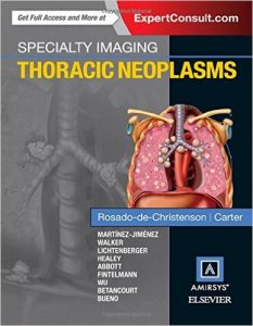
Part of the highly regarded Specialty Imaging series, this unique title by Dr. Melissa L. Rosado-de-Christenson clearly presents the imaging features of all thoracic neoplasms (including those affecting the cardiovascular system) as well as staging of malignancies and patterns of metastatic spread in a single, convenient volume. An easy-to-read bulleted format and state-of-the-art imaging examples guide you step by step through every aspect of the field, including invasive diagnostic and therapeutic procedures. This book is an ideal resource for radiologists, pulmonary medicine physicians, thoracic surgeons, thoracic oncologists, and radiation oncologists – anyone who must distinguish lung cancer and thoracic metastases from less common malignant and benign neoplasms.

Superb illustrations highlight comprehensive coverage of imaging manifestations of all benign and malignant thoracic neoplasms, including lesions in the lung, mediastinum, thymus, esophagus, cardiovascular system, pleura, and chest wall

Introductory chapters discuss the various imaging modalities used in diagnosing and evaluating thoracic neoplasms, up-to-date imaging terminology, and the imaging signs that suggest neoplasia

Thorough coverage of lung cancer offers authoritative guidance on screening, specific manifestations of various cell types, issues of staging, various tissue-sampling methods, missed lung cancer, lung cancer mimics, and imaging follow-up of treated lung cancer

High-quality images and succinct text depict patterns of thoracic metastatic spread of several important malignancies, navigational bronchoscopy and image-guided biopsy, imaging manifestations of treated patients, and other key topics

A time-saving bulleted format distills essential information for fast and easy comprehension

Download this book free here

http://upsto.re/siUrX2f

Abdominal X-rays for Medical Students 1st Edition

111860055x

[amazon template=iframe image2&asin=111860055X]

Abdominal X-rays for Medical Students is a comprehensive resource offering guidance on reading, presenting and interpreting abdominal radiographs. Suitable for medical students, junior doctors, nurses and trainee radiographers, this brand new title is clearly illustrated using a unique colour overlay system to present the main pathologies and to highlight the abnormalities in abdomen x-rays.

Abdominal X-rays for Medical Students:

  • Covers the key knowledge and skills necessary for practical use
  • Provides an effective and memorable way to analyse and present abdominal radiographs – the unique ‘ABCDE’ system as developed by the authors
  • Presents each radiograph twice, side by side: the first as seen in the clinical setting, and the second with the pathology clearly highlighted
  • Includes self-assessment to test knowledge and presentation technique

With a systematic approach covering both the analysis of radiographs and next steps mirroring the clinical setting and context, Abdominal X-rays for Medical Students is a succinct and up-to-date overview of the principles and practice of this important topic.

Download this book free here

http://upsto.re/skdbERV

1 56 57 58 59 60