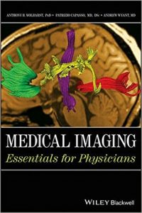
Medical Imaging: Essentials for Physicians

510d51goc4l-_sx330_bo1204203200_

[amazon template=iframe image2&asin=0470505702]

This book is a concise, one–stop resource on medical imaging written for medical professionals, addressing a field of critical importance to them, in the language that they speak. Coauthored by a medical physicist and a clinical imaging physician, the book provides a solid working knowledge of the science and technology of image formation, including coverage of imaging modalities and how they operate, how to use the resulting diagnostic information, the proper applications of imaging, and many clinical case examples.

DOWNLOAD THIS BOOK FREE HERE

http://upsto.re/EahXiCS

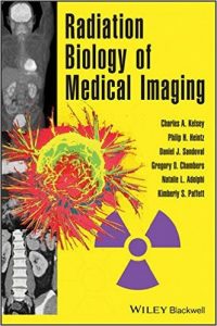
Radiation Biology of Medical Imaging

51w3qvkibol-_sx330_bo1204203200_

[amazon template=iframe image2&asin=0470551771]

This book provides a thorough yet concise introduction to quantitative radiobiology and radiation physics, particularly the practical and medical application. Beginning with a discussion of the basic science of radiobiology, the book explains the fast processes that initiate damage in irradiated tissue and the kinetic patterns in which such damage is expressed at the cellular level. The final section is presented in a highly practical handbook style and offers application–based discussions in radiation oncology, fractionated radiotherapy, and protracted radiation among others. The text is also supplemented by a Web site.

DOWNLOAD THIS BOOK FREE HERE

http://upsto.re/EahXMFe

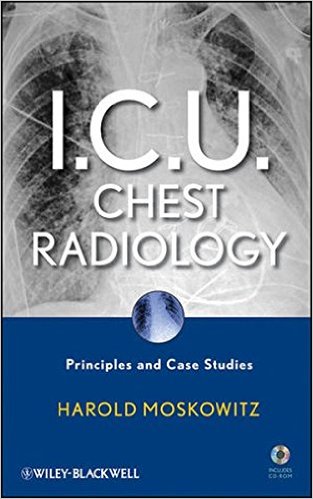
I.C.U. Chest Radiology: Principles and Case Studies Hardcover – 25 May 2010

51tvucjg7vl-_sx311_bo1204203200_

[amazon template=iframe image2&asin=0470450347]

Here is the first book in decades devoted specifically to ICU radiology, from an expert author in a major US clinical setting. It covers common, everyday problems seen in each ICU, including pneumonias, heart failure, and barotraumas, along with common technical problems that lead to interpretation errors and inappropriate interventions. This resource also features an excellent, concise presentation of the topic, with a separate section containing real–life case studies. The text is accompanied by a free CD–ROM with the case studies and radiographic images.

DOWNLOAD THIS BOOK FREE HERE

http://upsto.re/EahXfiv

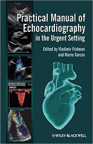
Practical Manual of Echocardiography in the Urgent Setting 1st Edition

51zlmgjtnl-_sx322_bo1204203200_

[amazon template=iframe image2&asin=0470659971]

Practical Manual of Echocardiography in the Urgent Setting covers the essentials of echocardiography in the acute setting, from ultrasound basics to descriptions of all pertinent echocardiographic views to clear stepwise advice on basic calculations and normal/abnormal ranges.

This compact new reference:

•             Provides step-by-step guidance to acquiring the correct views and making the necessary calculations to accurately diagnose cardiac conditions commonly encountered in urgent settings.

•             Presents information organized by complaint/initial presentation so that readers can work from this first knowledge of the patient through the steps required to pinpoint a diagnosis.

•             Covers echo basics, from sound wave characteristics/properties to common device settings to basic ultrasound formulas.

•             Includes diagnostic algorithms fitted to address the differential diagnosis in the most commonly-encountered clinical scenarios.

Designed and written by frontline clinicians with extensive experience treating patients, Practical Manual of Echocardiography in the Urgent Setting is the perfect pocket-sized guide for residents in cardiology, emergency medicine, and hospital medicine; trainees in echocardiography; medical students on cardiology or emergency medicine rotations; technicians, nurses, attending physicians — anyone who practices in the urgent setting and who needs reliable guidance on echocardiographic views, data and normal/abnormal ranges to aid rapid diagnosis and decision-making at the point of care.

DOWNLOAD THIS BOOK FREE HERE

http://upsto.re/EWDCyJw
Final FRCR 2B Viva: A Survival Guide (Cambridge Medicine (Paperback)) 1st Edition

31ecvhsas3l-_bo1204203200_

[amazon template=iframe image2&asin=0521183073]

Final FRCR 2B Viva: A Survival Guide presents a series of cases similar to those used in the FRCR exams and representative of everyday radiological practice. This invaluable collection of high quality images is accompanied by clear and concise explanations, enabling trainees to prepare fully for their FRCR 2B viva presentation. Covering the full range of imaging modalities and organ systems, it provides clinically important vignettes which help the reader to impress examiners and colleagues, and enhance the trainee’s ability to come up with differential diagnoses. Concise key points for each case provide additional diagnostic information which would impress an examiner. Written by a team of expert consultant radiologists and several recently successful FRCR Part 2 candidates, Final FRCR 2B Viva: A Survival Guide is an essential purchase for all radiology trainees.

DOWNLOAD THIS BOOK FREE HERE

http://upsto.re/sQycdAx

Emergency Radiology 1st Edition

51rfgvnphul-_sx331_bo1204203200_

[amazon template=iframe image2&asin=0521672473]

Emergency Radiology is a quick reference pocketbook for radiologists worldwide working in any emergency or acute care setting. It acts both as a checklist of presenting features to enable accurate interpretation of diagnostic imaging investigations and as a guide to understanding the basics of performing therapeutic or diagnostic interventional procedures. A wide range of emergencies are reviewed, giving all pertinent information in a concise presentation for on-call radiologists and trainees who encounter these emergencies out-of-hours. The text is complemented by high quality images of emergency conditions in all body systems, including head, cardiovascular, chest, abdomen, pelvis and extremities. Pediatrics, fluoroscopy, ultrasound and the use of MRI in emergencies are also discussed. Emergency Radiology is an invaluable for all radiologists, emergency physicians and radiography technicians, as well as for any healthcare practitioner involved in patient care in the emergency or acute care environment.

DOWNLOAD THIS BOOK FREE HERE

http://upsto.re/sQycJpo

Pearls and Pitfalls in Abdominal Imaging: Pseudotumors, Variants and Other Difficult Diagnoses (Cambridge Medicine (Hardcover)) 1st Edition

31-1yerhj-l-_bo1204203200_

[amazon template=iframe image2&asin=0521513774]

Research consistently suggests that 1.0 to 2.6% of radiology reports contain serious errors, many of which are avoidable, and it is clear that all radiologists can struggle with the basic questions as to whether a study is normal or abnormal. Pearls and Pitfalls in Abdominal Imaging presents over 100 conditions in the abdomen and pelvis which can commonly cause confusion and mismanagement in daily radiological practice, providing a focused textbook that can be readily used to avoid wrong diagnoses and prevent incorrect management or even malpractice litigation. It includes 700 figures and covers all the major modalities including CT, PET/CT and MRI. The pearls and pitfalls include artifacts, anatomic variants, mimics, and a miscellany of diagnoses that are under-recognized or only recently described. Conditions are presented in anatomic order from diaphragm to the symphysis pubis, with grouping by location and organ system.

DOWNLOAD THIS BOOK FREE HERE

http://upsto.re/sQycmXk

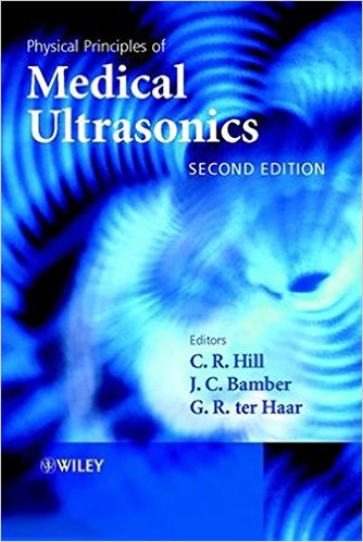
Physical Principles of Medical Ultrasonics 2nd Edition

51yweumq8cl-_sx332_bo1204203200_

[amazon template=iframe image2&asin=0471970026]

The physical properties of ultrasound, particularly its highly directional beam behaviour, and its complex interactions with human tissues, have led to its becoming a vitally important tool in both investigative and interventional medicine, and one that still has much exciting potential.

This new edition of a well-received book treats the phenomenon of ultrasound in the context of medical and biological applications, systematically discussing fundamental physical principles and concepts. Rather than focusing on earlier treatments, based largely on the simplifications of geometrical acoustics, this book examines concepts of wave acoustics, introducing them in the very first chapter.

Practical implications of these concepts are explored, first the generation and nature of acoustic fields, and then their formal descriptions and measurement. Real tissues attenuate and scatter ultrasound in ways that have interesting relationships to their physical chemistry, and the book includes coverage of these topics.

Physical Principles of Medical Ultrasonics also includes critical accounts and discussions of the wide variety of diagnostic and investigative applications of ultrasound that are now becoming available in medicine and biology. The book also encompasses the biophysics of ultrasound, its practical applications to therapeutic and surgical objectives, and its implications in questions of hazards to both patient and operator.

DOWNLOAD THIS BOOK FREE HERE

http://upsto.re/sQyccrK

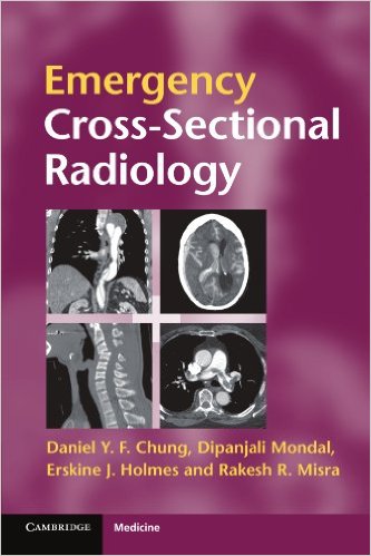
Emergency Cross-sectional Radiology (Cambridge Medicine (Paperback)) 1st Edition

51goohpch7l-_sx331_bo1204203200_

[amazon template=iframe image2&asin=0521279534]

Cross-sectional imaging plays an ever-increasing role in the management of the acutely ill patient. There is 24/7 demand for radiologists at all levels of training to interpret complex scans, and alongside this an increased expectation that the requesting physician should be able to recognise important cross-sectional anatomy and pathology in order to expedite patient management. Emergency Cross-Sectional Radiology addresses both these expectations. Part I demystifies cross-sectional imaging techniques. Part II describes a wide range of emergency conditions in an easy-to-read bullet point format. High quality images reinforce the findings, making this an invaluable rapid reference in everyday clinical practice. Emergency Cross-Sectional Radiology is a practical aide-memoire for emergency medicine physicians, surgeons, acute care physicians and radiologists in everyday reporting or emergency on-call environments.

DOWNLOAD THIS BOOK FREE HERE

http://upsto.re/sQyc8a5

Introduction to the Science of Medical Imaging 1st Edition

518hlbn3lal-_sx382_bo1204203200_

[amazon template=iframe image2&asin=0521747627]

Revolutionary advances in imaging technology that provide high resolution, 3-D, non-invasive imaging of biological subjects have made biomedical imaging an essential tool in clinical medicine and biomedical research. Key technological advances include MRI, positron emission tomography (PET) and multidetector X-ray CT scanners. Common to all contemporary imaging modalities is the creation of digital data and pictures. The evolution from analog to digital image data is driving the rapidly expanding field of digital image analysis. Scientists from numerous disciplines now require in-depth knowledge of these complex imaging modalities. Introduction to the Science of Medical Imaging presents scientific imaging principles, introduces the major biomedical imaging modalities, reviews the basics of human and computer image analysis and provides examples of major clinical and research applications. Written by one of the world’s most innovative and highly respected neuroradiologists, Introduction to the Science of Medical Imaging is a landmark text on image acquisition and interpretation.

DOWNLOAD THIS BOOK FREE HERE

http://upsto.re/sQyiBHf

1 55 56 57 58 59 62