McGraw-Hill Specialty Board Review Radiology 1st Edition
51sc72tzs4l-_sx373_bo1204203200_

[amazon template=iframe image2&asin=0071601643]

An all-in-one review for the diagnostic radiology board examination – complete with 1000+ Q&As!

McGraw-Hill Specialty Board Review: Radiology is an outstanding review for both residents-in-training and practicing radiologists. You’ll find everything you need in this one comprehensive resource . . . questions, answers, detailed explanations, and targeted coverage that emphasizes key material in a simple, straightforward manner and reinforces important concepts.

Everything you need to excel on the exam:

  • More than 1000 questions with detailed explanations for correct and incorrect answers
  • Strong focus on the fundamentals of anatomy and pathophysiology
  • An organization based on the 10 subspecialties recognized by the American Board of Radiology
  • Important overviews of imaging-based physics for ultrasound, MRI, and nuclear medicine

Content that spans the entire examination:

  • Central Nervous System
  • Pulmonary
  • Cardiac
  • Gastrointestinal Tract
  • Genitourinary Tract
  • Ultrasound
  • Musculoskeletal System
  • Breast
  • Interventional Radiology
  • Nuclear Radiology
  • Pediatric

Download this book free here

http://upsto.re/EStLvqq

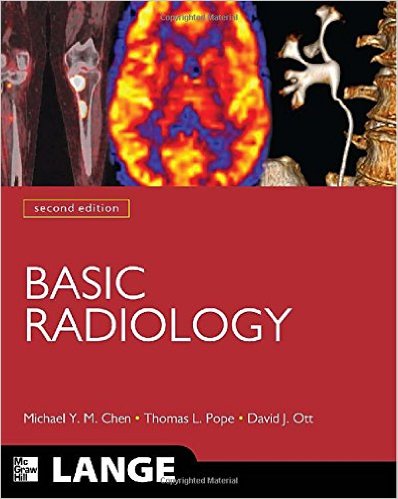
Basic Radiology, Second Edition (LANGE Clinical Medicine) 2nd Edition

51sp2w-rkl-_sx396_bo1204203200_

[amazon template=iframe image2&asin=0071627081]

A well-illustrated, systems-based primer on learning radiologic imaging

4 STAR DOODY’S REVIEW!
“Overall, this is a high quality book and a nice quick reference that is surprisingly complete for its size. The copious and well-chosen images are particularly valuable. It would be a good addition to the libraries of radiology departments, especially for medical students who are interested in radiology as a specialty and for radiology residents at the beginning of their training.”–Doody’s Review Service

Basic Radiology is the easiest and most effective way for medical students, residents, and clinicians not specializing in radiologic imaging to learn the essentials of diagnostic test selection, application, and interpretation. This trusted guide is unmatched in its ability to teach you how to select and request the most appropriate imaging modality for a patient’s presenting symptoms and familiarize yourself with the most common diseases that current radiologic imaging can best evaluate.

Features:

  • More than 800 high-quality images across all modalities
  • A logical organ-system approach
  • Consistent chapter presentation that includes: —Recap of recent developments in the radiologic imaging of the organ system discussed
    —Description of normal anatomy
    —Discussion of the most appropriate imaging technique for evaluating that organ system
    —Questions and imaging exercises designed to enhance your understanding of key principles
  • Brief list of suggested readings and general references
  • Timely chapter describing the various diagnostic imaging techniques currently available, including conventional radiography, nuclear medicine, ultrasonography, computed tomography, and magnetic resonance imaging
  • An important chapter providing an overview of the physics of radiation and its related biological effects, ultrasound, and magnetic resonance imaging

DOWNLOAD THIS BOOK FREE HERE

http://upsto.re/ESteMFG

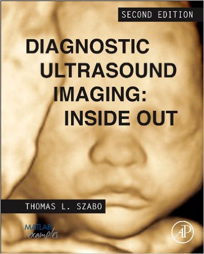
Diagnostic Ultrasound Imaging: Inside Out, Second Edition (Biomedical Engineering) 2nd Edition

51xh-unxpol-_sx400_bo1204203200_

[amazon template=iframe image2&asin=0123964873]

Diagnostic Ultrasound Imaging provides a unified description of the physical principles of ultrasound imaging, signal processing, systems and measurements. This comprehensive reference is a core resource for both graduate students and engineers in medical ultrasound research and design. With continuing rapid technological development of ultrasound in medical diagnosis, it is a critical subject for biomedical engineers, clinical and healthcare engineers and practitioners, medical physicists, and related professionals in the fields of signal and image processing.

The book contains 17 new and updated chapters covering the fundamentals and latest advances in the area, and includes four appendices, 450 figures, and almost 1,500 references. In addition to the continual influx of readers entering the field of ultrasound worldwide who need the broad grounding in the core technologies of ultrasound, this book provides those already working in these areas with clear and comprehensive expositions of these key new topics as well as introductions to state-of-the-art innovations in this field.

  • Enables practicing engineers, students and clinical professionals to understand the essential physics and signal processing techniques behind modern imaging systems as well as introducing the latest developments that will shape medical ultrasound in the future
  • Suitable for both newcomers and experienced readers, the practical, progressively organized applied approach is supported by hands-on MATLAB code and worked examples that enable readers to understand the principles underlying diagnostic and therapeutic ultrasound
  • Covers the new important developments in the use of medical ultrasound: elastography and high-intensity therapeutic ultrasound. Many new developments are comprehensively reviewed and explained, including aberration correction, acoustic measurements, acoustic radiation force imaging, alternate imaging architectures, bioeffects: diagnostic to therapeutic, Fourier transform imaging, multimode imaging, plane wave compounding, research platforms, synthetic aperture, vector Doppler, transient shear wave elastography, ultrafast imaging and Doppler, functional ultrasound and viscoelastic models
  • DOWNLOAD THIS BOOK FREE HERE
  • http://upsto.re/ELnEtCM

Genitourinary Imaging: Case Review

21n6a996tzl-_bo1204203200_

[amazon template=iframe image2&asin=0323006574]

GENITOURINARY TRACT IMAGING revisits the entire spectrum of genitourinary radiology through the presentation of radiogrpahic cases. Cases selected will help physicians and other medical personnel learn the important areas in this field. This book is a companion to the GENITOURINARY TRCT IMAGING: The Requisites textbook. Together, they cover the field, providing excellent prep for board examinations and clinical review.

DOWNLOAD THIS BOOK FREE HERE

http://upsto.re/E9WmzkV

Patient Care in Radiography: With an Introduction to Medical Imaging, 7e (Ehrlich, Patient Care in Radiography) 7th Edition


51emfiph6l-_sx403_bo1204203200_

[amazon template=iframe image2&asin=0323051782]

Confidently master vital skills that will help you provide high-quality care to your radiography patients, including safety, transfer, positioning, infection control, and assessment procedures. Comprehensive and in-depth information closely follows ASRT curriculum guidelines to guide you through key elements of patient care, as well as topics related to microbiology, emerging diseases, transcultural communication, administering medications, and bedside radiography. In addition, almost 500 illustrations and photographs visually demonstrate important procedures and assist you in acquiring both the technical and the interpersonal skills needed in the clinical environment.

  • Information from the American Society of Radiologic Technologists includes the organizational structure of ASRT and the scope of practice standards relevant to the radiographer.
  • Comprehensive overviews of department organization, job opportunities, radiation protection, clinical environment, and ethics provide a solid foundation for both students and professionals.
  • Patient care tips alongside descriptions of procedures encourage high-quality patient care in addition to technical proficiency.
  • Consistent, straightforward, engaging writing style explains and breaks down complex concepts for easier understanding.
  • Infection control content covers infectious diseases, guidelines for hand hygiene, and use of needleless devices, providing necessary information to help prevent the spread of infection.
  • Chapter on special imaging techniques and modalities discusses CT angiography, MRI, mammography, and PET imaging, keeping you up to date with a wide range of imaging modalities.
  • Procedures are described step by step, with pictures showing each step.
  • Case Studies focus on medicolegal terms, standards, and applications, preparing you to problem-solve in real life clinical situations.
  • Expanded coverage of HIPAA regulations includes relevant examples of compliance in today’s imaging departments.
  • Cultural diversity is explored in greater depth in the Communications chapter, designed to facilitate effective communication among culturally diverse healthcare professionals and patients.
  • Chapter outlines, vocabulary lists, and learning objectives help you make the most of your study and review time.
  • Check-off forms for documenting clinical objectives related to patient care are provided in an appendix.
  • More review questions are provided at the end of each chapter, and some chapters include additional critical thinking questions.
  • More Spanish phrases are included in an appendix, helping you better communicate with Spanish-speaking patients in the clinical setting.

DOWNLOAD THIS BOOK FREE HERE

http://upsto.re/E9Wmy9Z

Clinical Imaging: With Skeletal, Chest, & Abdominal Pattern Differentials, 3e 3rd Edition

519-omgqnzl-_sx388_bo1204203200_

[amazon template=iframe image2&asin=0323084958]

Clinical Imaging by Dennis Marchiori is a comprehensive text with a clear, concise writing style that allows students and practitioners to quickly develop a better understanding of diagnostic imaging. Covering soft tissue imaging and skeletal imaging, including brain and spinal cord, chest, and abdomen, Clinical Imaging seamlessly integrates plain film with MRI and CT. And with more than 3,500 illustrations all contained in one volume, this trusted text offers the most effective, realistic and comprehensive approach available today.

“In terms of value for money, the recommended price is very fair for 1,462 pages, especially when one includes the additional online content (available using a scratch card code) that includes case studies, flash cards, interactive examinations and image collections” Reviewed by RAD Magazine,Jan 2015

“For students who need to get up to speed with abnormal radiographic appearances this book is a good start.”Reviewed by RAD Magazine, Jan 2015

  • Combines the innovative pattern approach with more traditional detailed descriptions to emulate real-world patient interaction without sacrificing more in-depth content on disease states.
  • Innovative Pattern Approach uses the patterns that link similar abnormalities to help you learn to identify, and just as importantly, differentiate abnormalities.
  • Extensive cross-referencing from pattern to disease descriptions enables the reader to quickly find more detailed information.
  • Dedicated chapter on the key subject of radiology physics, including algorithms for improving film quality.
  • A glossary of nearly 500 radiological terms.
  • NEW! Over 800 new or updated images.
  • NEW! State-of-the-art MRI images deliver more comprehensive content for this growing field within imaging.
  • NEW! Updated photographs familiarize you with radiographic positioning equipment.
  • NEW! Clearer, more detailed line art visually reinforces your understanding of new concepts.
  • NEW! Additional contributors provide fresh perspectives on important topics and trends.

DOWNLOAD THIS BOOK FREE HERE

http://upsto.re/E9WieEu

Sonography Exam Review: Physics, Abdomen, Obstetrics and Gynecology, 2e 2nd Edition

51n3af2cgbl-_sx389_bo1204203200_

[amazon template=iframe image2&asin=0323100465]

Be confident that you can answer any and all questions on your registry exams correctly when you prepare with this complete review. Mosby’s Comprehensive Review for General Sonography Examinations provides study resources for all three main exams required for general ultrasound practice: physics, abdomen, and ob/gyn. Each chapter is arranged in table and outline format with 50 review questions at the end of the chapter and a mock exam at the end of each section. Access additional mock exams for each subject area on the companion CD or Evolve site. These exams give you experience with timed test taking in an electronic environment that simulates the actual registry exam experience. With this realistic preview of the exam environment and solid review of the material, you’ll be prepared to ace the exams!

“..no doubt that this is a worthwhile text which could provide a useful revision platform for sonography students in the UK.” Reviewed by Sue Halson-Brown on behalf of RAD Magazine, February 2015

  • Complete preparation for the three general ARDMS exams (physics, abdomen, and ob/gyn)
  • Content review in outline and tabular format provides a quick review of all the material you need to learn, including key terms, anatomy, functions, scanning techniques, lab values, and pathology.
  • More than 2,500 questions in Registry format cover everything you’ll be tested on in the Registry exams.
  • Rationales for answers to mock questions help you understand why an answer is correct or incorrect and increase your comprehension.
  • More than 350 ultrasound scans included in the abdominal and ob/gyn sections prepare you for exam questions that ask you to identify pathology on scans.
  • Color insert with Doppler images of the liver, biliary, and umbilical cord helps you be ready to answer questions related to Doppler imaging.
  • Companion CD provides extra timed, graded mock exams and two entertaining, interactive games: Sonography Millionaire and Tournament of Sonography.

DOWNLOAD THIS BOOK FREE HERE

http://upsto.re/E9WiYPK

The Physics of Diagnostic Imaging Second Edition 2nd Edition

51ekpfzcfnl-_sx383_bo1204203200_

[amazon template=iframe image2&asin=0340808918]

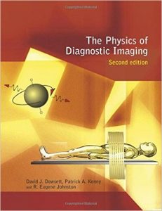
Over recent years there has been a vast expansion in the variety of imaging techniques available, and developments in machine specifications continue apace. If radiologists and radiographers are to obtain optimal image quality while minimising exposure times, a good understanding of the fundamentals of the radiological science underpinning diagnostic imaging is essential.

DOWNLOAD THIS BOOK FREE HERE

http://upsto.re/E9WP5NV

Clark’s Pocket Handbook for Radiographers 1st Edition

4168deltqjl-_sx323_bo1204203200_

[amazon template=iframe image2&asin=0340939931]

This pocket-sized guide, drawn from the twelfth edition of Clark’s Positioning in Radiography, provides clear and practical advice to help radiographers in their day-to-day work. The authors considered that it is important for radiographers and students to have access to an additional text available in a “pocket” format which is easily transportable and convenient to use during everyday radiographic practice.

Designed for rapid reference, it covers how to position the patient and the central ray, describes the essential image characteristics, and illustrates each radiographic projection with a positioning photograph and a radiograph.

The authors have included a range of additional information new to this text. This includes a protocol for evaluating images (the “10-point plan”) and a range of general advice for undertaking procedures in a professional and efficient manner. The book also includes basic information in relation to some non-imaging diagnostic tests, common medical terminology, and abbreviations. This is designed to help readers gain a better understanding of the diagnostic requirements and role of particular imaging procedures from the information presented in X-ray requests. In addition, the book discusses image evaluation, medical abbreviations, relevant normal blood values, and radiation protection.

Together with key points, this information helps the radiographer achieve the ideal image result.

DOWNLOAD THIS BOOK FREE HERE

http://upsto.re/EYQrW3L

Chest X-Ray Made Easy, 3e 3rd Edition

41hkzensmnl-_sx329_bo1204203200_

[amazon template=iframe image2&asin=0443069220]

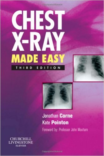
Translated into over a dozen languages, this book has been widely praised for making interpretation of the chest X-ray as simple as possible. It describes the range of conditions likely to be encountered on the wards and guides the doctor through the process of examining and interpreting the film based on the appearance of the abnormality shown. It then assists the doctor in determining the nature of the abnormality and points the clinician towards a possible differential diagnosis. It covers the common radiological problems the junior doctors are faced with starting with the appearance of the film, e.g. showing generalised shadowing or a coin lesion. It gives advice on how to examine an X-ray, how to check its technical quality and how to identify where the lesion is. All the X-rays are accompanied by a simple line diagram outlining where the abnormality is.

  • Covers the full range of common radiological problems.
  • Includes valuable advice on how to examine an X-ray.
  • Assists the doctor in determining the nature of the abnormality.
  • Points the clinician towards a possible differential diagnosis.
  • Now presented in two-colour to enhance the appearance of the text.
  • New material includes an introduction to thoracic CT scanning indicating the usefulness of these scans where appropriate.

DOWNLOAD THIS BOOK FREE HERE

http://upsto.re/EahhLom

1 54 55 56 57 58 62