Get Through First FRCR: Questions for the Anatomy Module 1st Edition

[amazon template=iframe image2&asin=1853159581]

Completely up to date with the latest exam changes, Get Through First FRCR: Questions for the Anatomy Module offers a valuable insight into the new anatomy exam. 170 high quality practice cases, each containing 5 question stems, are presented according to syllabus topics, accurately reflecting the content, style and level of difficulty of the actual examination questions.

Anatomical images are included from all modalities commonly used in current radiological practice (plain x-rays, CT, MRI, ultrasound, nuclear medicine). Each case is supported by full explanatory answers, providing appropriate anatomy knowledge and relevant radiological learning points for the candidate.

Featuring a wealth of practice questions plus one full mock examination, this book has been designed for candidates to assess their knowledge, identify topics that require further study and to build up confidence in preparation for the exam day.

Written by Specialty Trainees in Radiology, Get Through First FRCR: Questions for the Anatomy Module is the essential revision tool for all First FRCR candidates preparing for the newly revised examination.

DOWNLOAD THIS BOOK FREE HERE

http://upsto.re/v9cXkzH

Research for the Radiation Therapist: From Question to Culture 1st Edition

[amazon template=iframe image2&asin=1926895983]

This book addresses a wide range of topics, from the principles of evidence-based practice to the process and dissemination of research to unique considerations such as clinical trials, patenting, and health services research. The case for evidence-based practice and a collaborative research culture is made first, followed by a series of chapters walking the reader through the research process by way of the scientific method. One of the more unique aspects of the scope of this book is the inclusion of chapters relating to the dissemination of knowledge, manuscript publication, and how to build an academic research program. Each chapter focuses on introducing the reader to foundational principles, methodology, and terminology, and highlight case studies of radiation therapist research or experience that is relevant to provide contextual examples and inspiration to the reader.

DOWNLOAD THIS BOOK FREE HERE

http://upsto.re/v9c5uGU

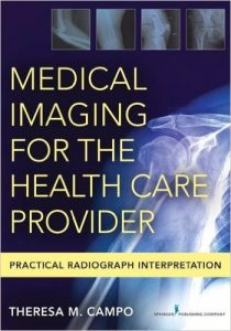
Medical Imaging for the Health Care Provider: Practical Radiograph Interpretation 1st Edition

[amazon template=iframe image2&asin=0826131263]

The only text to integrate the basics of radiology, characteristics and differences of testing modalities, and interpretation skills

This unique book fills a void in radiology interpretation texts by encompassing the foundational tools and concepts of the full range of medical imaging, including radiology, the basics of interpretation of plain radiographs, comparison with other testing modalities, the rationale for selecting the first diagnostic step, and exploration and interpretation of chest, abdomen, extremity, and spinal radiographs. A concise, easy-to-use reference, it includes written descriptions enhanced with figures, tables, and actual patient films to demonstrate concepts, and discusses―in easily accessible language―differences in testing modalities. The text also features a step-by-step guide to interpretation of radiographs.

This resource describes and compares available diagnostic modalities, including plain radiograph, CT scan, nuclear imaging, MRI, and ultrasound. It discusses pediatric considerations and includes separate chapters for the chest, abdomen, upper and lower extremities, and the cervical, thoracic, and lumbar spine. The book will be an asset to nurse practitioners and physician assistants working in all emergency, urgent, intensive, and primary care settings. It will also benefit medical students and graduate students in acute care, family, adult/gerontology, and emergency nurse practitioner programs, as well as emergency/trauma clinical nurse specialists, and hospitalists and intensivist nurse practitioners.

Key Features:

    • Integrates the basics of radiology, CT scans, nuclear imaging, MRIs, and ultrasound, their characteristics and differences among testing modalities, and basic step-by-step interpretation skills
    • Relevant to a wide range of nurse practitioners, physician assistants, and other mid-level providers in multiple settings
    • Includes a step-by-step guide to the interpretation of the radiographs


  • Delivers an easy-to-understand approach to selecting diagnostic imaging tests
  • Presents actual images and figures to demonstrate concepts

DOWNLOAD THIS BOOK FREE HERE

http://upsto.re/v9jt5DD

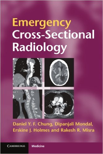
Emergency Cross-sectional Radiology (Cambridge Medicine (Paperback)) 1st Edition


[amazon template=iframe image2&asin=0521279534]

Cross-sectional imaging plays an ever-increasing role in the management of the acutely ill patient. There is 24/7 demand for radiologists at all levels of training to interpret complex scans, and alongside this an increased expectation that the requesting physician should be able to recognise important cross-sectional anatomy and pathology in order to expedite patient management. Emergency Cross-Sectional Radiology addresses both these expectations. Part I demystifies cross-sectional imaging techniques. Part II describes a wide range of emergency conditions in an easy-to-read bullet point format. High quality images reinforce the findings, making this an invaluable rapid reference in everyday clinical practice. Emergency Cross-Sectional Radiology is a practical aide-memoire for emergency medicine physicians, surgeons, acute care physicians and radiologists in everyday reporting or emergency on-call environments.

DOWNLOAD THIS BOOK FREE HERE

http://upsto.re/vuyA4SG

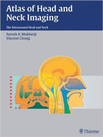
Atlas of Head and Neck Imaging: The Extracranial Head and Neck 1st Edition Edition

[amazon template=iframe image2&asin=1588901785]

Designed for easy use at the PACS station of viewbox, here is your right-hand tool and pictorial guide for locating, identifying, and accurately diagnosing lesions of the extracranial head and neck. This beautifully produced atlas employs the spaces concept of analysis, which helps radiologists directly visualize complex head and neck anatomy and pathology.

With hundreds of high quality illustrations, this book makes the identification and localization of complex neck masses relatively simple. This book provides CT and MR examples for more than 200 different diseases of the suprahyoid and infrahyoid neck, as well as clear and concise information on the epidemiology, clinical findings, pathology, and treatment guidelines for each disease.

Each space within the head and neck has its own separate section, with examples of the common pathology that arises in this area. A standard format consisting of Epidemiology, Clinical Presentation, Pathology, Treatment, and Imaging Findings, allows quick and efficient access to well-structured subjects. This uniform organization streamlines research for radiologists at any level of training.

Although well over 200 pathologies are included within this remarkable text, Atlas of Head and Neck Imaging focuses primarily on the suprahyoid and infrahyoid neck, providing exceptionally detailed information on the most challenging aspects of this field.

Radiologists and radiation oncologists will find this visual text ideal as a quick anatomic reference and diagnostic tool. Radiology residents preparing for board exams and neuroradiology fellows and staff studying for the CAQ exam will also benefit from the wealth of information.

DOWNLOAD THIS BOOK FREE HERE

http://upsto.re/vuNzTTV

Fundamentals of Diagnostic Radiology – 4 Volume Set (Brant, Fundamentals of Diagnostic Radiology) Fourth, in four volumes Edition

[amazon template=iframe image2&asin=1608319121]

This fully revised edition of Fundamentals of Diagnostic Radiology conveys the essential knowledge needed to understand the clinical application of imaging technologies. An ideal tool for all radiology residents and students, it covers all subspecialty areas and current imaging modalities as utilized in neuroradiology, chest, breast, abdominal, musculoskeletal imaging, ultrasound, pediatric imaging, interventional techniques and nuclear radiology. New and expanded topics in this edition include use of diffustion-weighted MR, new contrast agents, breast MR, and current guidelines for biopsy and intervention.  Many new images, expanded content, and full-color throughout make the fourth edition of this classic text a comprehensive review that is ideal as a first reader for beginning residents, a reference during rotations, and a vital resource when preparing for the American Board of Radiology examinations.

More than just a book, the fourth edition is a complete print and online package.  Readers will also have access to fully searchable content from the book, a downloadable image bank containing all images from the text, and study guides for each chapter that outline the key points for every image and table in an accessible format—ideal for study and review.

This is the 4 volume set.

DOWNLOAD THIS BOOK FREE HERE

PART I http://upsto.re/vuspzLy

PART II http://upsto.re/vuvvAS5

Corneal Topography in Clinical Practice Pentacam System: Basics and Clinical Interpretation 2nd Edition

[amazon template=iframe image2&asin=9350255758]

Corneal topography is a non-invasive medical imaging technique for mapping the surface curvature of the cornea, the outer structure of the eye. This procedure may be carried out with a Pentacam, which uses a rotating camera to create a 3D image of the anterior of the eye. This second edition has been fully updated to provide the latest developments in corneal topography and tomography using the Pentacam machine. Beginning with an introduction, the following sections describe the fundamentals of corneal topography and use of the Pentacam with different ophthalmic disorders. With nearly 250 high quality, colour images and illustrations, this concise guide is especially useful to graduate and postgraduate students in learning how to read and interpret corneal topography.

DOWNLOAD THIS BOOK FREE HERE

http://upsto.re/vuR8z5Y

Handbook of MRI Technique 4th Edition

[amazon template=iframe image2&asin=1118661621]

Now entering its fourth edition, the market-leading Handbook of MRI Technique has been fully revised and updated to incorporate new technologies and developments essential to good practice. Written specifically for technologists and highly illustrated, it guides the uninitiated through scanning techniques and helps more experienced technologists to improve image quality.

The first part of the book considers the main aspects of theory that relate to scanning and also includes practical tips on gating, equipment use, patient care and safety, and information on contrast media. The second half provides step-by-step instruction for examining each anatomical area, beginning with a basic anatomy section followed by sections on indications, patient positioning, equipment, artefacts and tips on optimizing image quality.

  • Written by an international team of technologists from the United States, United Kingdom and Europe
  • Suitable for users for all types of MRI systems
  • Now includes key points throughout for quick reference
  • Companion website at www.wiley.com/go/westbrook/mritechnique with self-assessment and image flashcards

Handbook of MRI Technique continues to be the ideal support both for radiographers new to MRI and for regular users looking for information on alternative techniques and suggestions on protocol modifications.

DOWNLOAD THIS BOOK FREE HERE

http://upsto.re/v4QMxeR

RF Coils for MRI 1st Edition

510ovu87vl-_sx379_bo1204203200_

[amazon template=iframe image2&asin=0470770767]

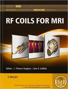
The content of this volume has been added to eMagRes (formerly Encyclopedia of Magnetic Resonance) – the ultimate online resource for NMR and MRI.

To date there is no single reference aimed at teaching the art of applications guided coil design for use in MRI. This RF Coils for MRI handbook is intended to become this reference.

Heretofore, much of the know-how of RF coil design is bottled up in various industry and academic laboratories around the world. Some of this information on coil technologies and applications techniques has been disseminated through the literature, while more of this knowledge has been withheld for competitive or proprietary advantage. Of the published works, the record of technology development is often incomplete and misleading, accurate referencing and attribution assignment being tantamount to admission of patent infringement in the commercial arena.  Accordingly, the literature on RF coil design is fragmented and confusing.  There are no texts and few courses offered to teach this material. Mastery of the art and science of RF coil design is perhaps best achieved through the learning that comes with a long career in the field at multiple places of employment…until now.

RF Coils for MRI combines the lifetime understanding and expertise of many of the senior designers in the field into a single, practical training manual. It informs the engineer on part numbers and sources of component materials, equipment, engineering services and consulting to enable anyone with electronics bench experience to build, test and interface a coil. The handbook teaches the MR system user how to safely and successfully implement the coil for its intended application. The comprehensive articles also include information required by the scientist or physician to predict respective experiment or clinical performance of a coil for a variety of common applications.  It is expected that RF Coils for MRI becomes an important resource for engineers, technicians, scientists, and physicians wanting to safely and successfully buy or build and use MR coils in the clinic or laboratory.  Similarly, this guidebook provides teaching material for students, fellows and residents wanting to better understand the theory and operation of RF coils.

Many of the articles have been written by the pioneers and developers of coils, arrays and probes, so this is all first hand information! The handbook serves as an expository guide for hands-on radiologists, radiographers, physicians, engineers, medical physicists, technologists, and for anyone with interests in building or selecting and using RF coils to achieve best clinical or experimental results.

About EMR Handbooks / eMagRes Handbooks 

The Encyclopedia of Magnetic Resonance (up to 2012) and eMagRes (from 2013 onward) publish a wide range of online articles on all aspects of magnetic resonance in physics, chemistry, biology and medicine. The existence of this large number of articles, written by experts in various fields, is enabling the publication of a series of EMR Handbooks / eMagRes Handbooks on specific areas of NMR and MRI. The chapters of each of these handbooks will comprise a carefully chosen selection of articles from eMagRes. In consultation with the eMagRes Editorial Board, the EMR Handbooks / eMagRes Handbooks  are coherently planned in advance by specially-selected Editors, and new articles are written (together with updates of some already existing articles) to give appropriate complete coverage. The handbooks are intended to be of value and interest to research students, postdoctoral fellows and other researchers learning about the scientific area in question and undertaking relevant experiments, whether in academia or industry.

DOWNLOAD THIS BOOK FREE HERE

http://upsto.re/vLFTnKn
Practical Vascular Ultrasound: An Illustrated Guide 1st Edition

41s5cuuprjl-_sx398_bo1204203200_

[amazon template=iframe image2&asin=1444181181]

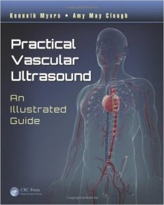
Ultrasound is used to demonstrate and classify numerous vascular diseases and provides a firm basis for deciding the most appropriate interventional treatment. Practical Vascular Ultrasound: An Illustrated Guide delivers a hands-on, practical approach to the diagnosis of vascular disease.

Providing an overview of performing vascular ultrasounds, the physiology of blood flow, and vascular pathology, the book explores the various vascular conditions most investigated through ultrasound. Conditions covered range from extracranial cerebrovascular arterial diseases to chronic venous disease in the lower limbs. For each disorder, the authors discuss:

  • Anatomy
  • Pathology
  • Clinical presentations
  • Differential diagnosis
  • What doctors need to know for each condition
  • Normal findings and criteria for disease as seen by ultrasound
  • Comprehensive protocols for scanning
  • Ultrasound images and observations that should be collected for best practice reporting

The authors also discuss interventional vascular ultrasound procedures. Using easy-to-read, point-form text, this concise text is enhanced with high-resolution ultrasound images and clear line diagrams. It provides a consistent, comprehensive, and professional approach to vascular ultrasound.

DOWNLOAD THIS BOOK FREE HERE

http://upsto.re/vSpZ2g8

1 51 52 53 54 55 62