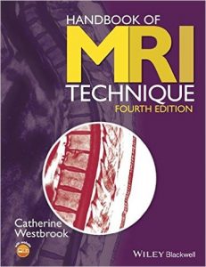
Handbook of MRI Technique 4th Edition

[amazon template=iframe image2&asin=1118661621]

Now entering its fourth edition, the market-leading Handbook of MRI Technique has been fully revised and updated to incorporate new technologies and developments essential to good practice. Written specifically for technologists and highly illustrated, it guides the uninitiated through scanning techniques and helps more experienced technologists to improve image quality.

The first part of the book considers the main aspects of theory that relate to scanning and also includes practical tips on gating, equipment use, patient care and safety, and information on contrast media. The second half provides step-by-step instruction for examining each anatomical area, beginning with a basic anatomy section followed by sections on indications, patient positioning, equipment, artefacts and tips on optimizing image quality.

  • Written by an international team of technologists from the United States, United Kingdom and Europe
  • Suitable for users for all types of MRI systems
  • Now includes key points throughout for quick reference
  • Companion website at www.wiley.com/go/westbrook/mritechnique with self-assessment and image flashcards

Handbook of MRI Technique continues to be the ideal support both for radiographers new to MRI and for regular users looking for information on alternative techniques and suggestions on protocol modifications.

DOWNLOAD THIS BOOK FREE HERE

http://upsto.re/v4QMxeR

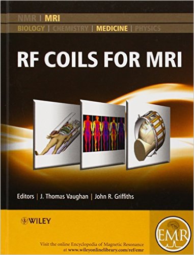
RF Coils for MRI 1st Edition

510ovu87vl-_sx379_bo1204203200_

[amazon template=iframe image2&asin=0470770767]

The content of this volume has been added to eMagRes (formerly Encyclopedia of Magnetic Resonance) – the ultimate online resource for NMR and MRI.

To date there is no single reference aimed at teaching the art of applications guided coil design for use in MRI. This RF Coils for MRI handbook is intended to become this reference.

Heretofore, much of the know-how of RF coil design is bottled up in various industry and academic laboratories around the world. Some of this information on coil technologies and applications techniques has been disseminated through the literature, while more of this knowledge has been withheld for competitive or proprietary advantage. Of the published works, the record of technology development is often incomplete and misleading, accurate referencing and attribution assignment being tantamount to admission of patent infringement in the commercial arena.  Accordingly, the literature on RF coil design is fragmented and confusing.  There are no texts and few courses offered to teach this material. Mastery of the art and science of RF coil design is perhaps best achieved through the learning that comes with a long career in the field at multiple places of employment…until now.

RF Coils for MRI combines the lifetime understanding and expertise of many of the senior designers in the field into a single, practical training manual. It informs the engineer on part numbers and sources of component materials, equipment, engineering services and consulting to enable anyone with electronics bench experience to build, test and interface a coil. The handbook teaches the MR system user how to safely and successfully implement the coil for its intended application. The comprehensive articles also include information required by the scientist or physician to predict respective experiment or clinical performance of a coil for a variety of common applications.  It is expected that RF Coils for MRI becomes an important resource for engineers, technicians, scientists, and physicians wanting to safely and successfully buy or build and use MR coils in the clinic or laboratory.  Similarly, this guidebook provides teaching material for students, fellows and residents wanting to better understand the theory and operation of RF coils.

Many of the articles have been written by the pioneers and developers of coils, arrays and probes, so this is all first hand information! The handbook serves as an expository guide for hands-on radiologists, radiographers, physicians, engineers, medical physicists, technologists, and for anyone with interests in building or selecting and using RF coils to achieve best clinical or experimental results.

About EMR Handbooks / eMagRes Handbooks 

The Encyclopedia of Magnetic Resonance (up to 2012) and eMagRes (from 2013 onward) publish a wide range of online articles on all aspects of magnetic resonance in physics, chemistry, biology and medicine. The existence of this large number of articles, written by experts in various fields, is enabling the publication of a series of EMR Handbooks / eMagRes Handbooks on specific areas of NMR and MRI. The chapters of each of these handbooks will comprise a carefully chosen selection of articles from eMagRes. In consultation with the eMagRes Editorial Board, the EMR Handbooks / eMagRes Handbooks  are coherently planned in advance by specially-selected Editors, and new articles are written (together with updates of some already existing articles) to give appropriate complete coverage. The handbooks are intended to be of value and interest to research students, postdoctoral fellows and other researchers learning about the scientific area in question and undertaking relevant experiments, whether in academia or industry.

DOWNLOAD THIS BOOK FREE HERE

http://upsto.re/vLFTnKn

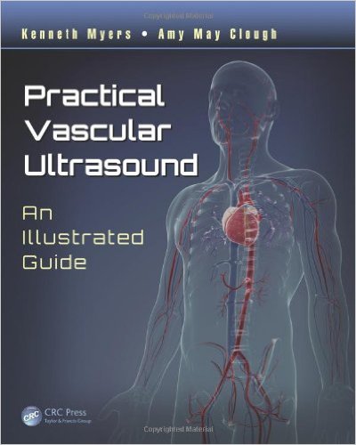
Practical Vascular Ultrasound: An Illustrated Guide 1st Edition

41s5cuuprjl-_sx398_bo1204203200_

[amazon template=iframe image2&asin=1444181181]

Ultrasound is used to demonstrate and classify numerous vascular diseases and provides a firm basis for deciding the most appropriate interventional treatment. Practical Vascular Ultrasound: An Illustrated Guide delivers a hands-on, practical approach to the diagnosis of vascular disease.

Providing an overview of performing vascular ultrasounds, the physiology of blood flow, and vascular pathology, the book explores the various vascular conditions most investigated through ultrasound. Conditions covered range from extracranial cerebrovascular arterial diseases to chronic venous disease in the lower limbs. For each disorder, the authors discuss:

  • Anatomy
  • Pathology
  • Clinical presentations
  • Differential diagnosis
  • What doctors need to know for each condition
  • Normal findings and criteria for disease as seen by ultrasound
  • Comprehensive protocols for scanning
  • Ultrasound images and observations that should be collected for best practice reporting

The authors also discuss interventional vascular ultrasound procedures. Using easy-to-read, point-form text, this concise text is enhanced with high-resolution ultrasound images and clear line diagrams. It provides a consistent, comprehensive, and professional approach to vascular ultrasound.

DOWNLOAD THIS BOOK FREE HERE

http://upsto.re/vSpZ2g8

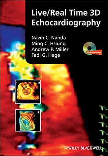
Live/Real Time 3D Echocardiography 1st Edition

51ylyxkjul-_sx346_bo1204203200_

[amazon template=iframe image2&asin=1405161418]

Now there is a source of recent information on the basic and clinical usefulness of real-time 3D echocardiography for echocardiographers, cardiologists, cardiac sonographers, and internists. This excellent reference helps clinicians improve pre- and post-surgical planning, attain better measurement of heart function, decrease exam times, and enhance patient communication. An accompanying DVD shows procedures in real time.

DOWNLOAD THIS BOOK FREE HERE

http://upsto.re/vNMceK5

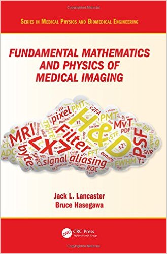
Fundamental Mathematics and Physics of Medical Imaging (Series in Medical Physics and Biomedical Engineering) 1st Edition

51cl8gn2hl-_sx327_bo1204203200_

[amazon template=iframe image2&asin=149875161X]

Authored by a leading educator, this book teaches the fundamental mathematics and physics concepts associated with medical imaging systems. Going beyond mere description of imaging modalities, this book delves into the mechanisms of image formation and image quality common to all imaging systems: contrast mechanisms, noise, and spatial and temporal resolution, making it an important reference for medical physicists and biomedical engineering students. This is an extensively revised new edition of The Physics of Medical X-Ray Imaging by Bruce Hasegawa (Medical Physics Publishing, 1991), and includes a wide range of modalities such as X-ray CT, MRI and SPECT.

DOWNLOAD THIS BOOK FREE HERE

http://upsto.re/vNMWkpj

Tutorials in Radiotherapy Physics: Advanced Topics with Problems and Solutions 1st Edition

516qemkw-gl-_sx348_bo1204203200_

[amazon template=iframe image2&asin=1482251671]

The Topics Every Medical Physicist Should Know

Tutorials in Radiotherapy Physics: Advanced Topics with Problems and Solutions covers selected advanced topics that are not thoroughly discussed in any of the standard medical physics texts. The book brings together material from a large variety of sources, avoiding the need for you to search through and digest the vast research literature. The topics are mathematically developed from first principles using consistent notation.

Clear Derivations and In-Depth Explanations

The book offers insight into the physics of electron acceleration in linear accelerators and presents an introduction to the study of proton therapy. It then describes the predominant method of clinical photon dose computation: convolution and superposition dose calculation algorithms. It also discusses the Boltzmann transport equation, a potentially fast and accurate method of dose calculation that is an alternative to the Monte Carlo method. This discussion considers Fermi–Eyges theory, which is widely used for electron dose calculations. The book concludes with a step-by-step mathematical development of tumor control and normal tissue complication probability models. Each chapter includes problems with solutions given in the back of the book.

Prepares You to Explore Cutting-Edge Research

This guide provides you with the foundation to read review articles on the topics. It can be used for self-study, in graduate medical physics and physics residency programs, or in vendor training for linacs and treatment planning systems.

DOWNLOAD THIS BOOK FREE HERE

http://upsto.re/vnojiUf

Imaging of the Cardiovascular System, Thorax, and Abdomen (Volume 2)

51rjutkgdnl-_sx346_bo1204203200_

[amazon template=iframe image2&asin=1482216264]

Magnetic resonance imaging (MRI) is a technique used in biomedical imaging and radiology to visualize internal structures of the body. Because MRI provides excellent contrast between different soft tissues, the technique is especially useful for diagnostic imaging of the brain, muscles, and heart.

In the past 20 years, MRI technology has improved significantly with the introduction of systems up to 7 Tesla (7 T) and with the development of numerous post-processing algorithms such as diffusion tensor imaging (DTI), functional MRI (fMRI), and spectroscopic imaging. From these developments, the diagnostic potentialities of MRI have improved impressively with an exceptional spatial resolution and the possibility of analyzing the morphology and function of several kinds of pathology.

Given these exciting developments, the Magnetic Resonance Imaging Handbook: Imaging of the Cardiovascular System, Thorax, and Abdomen is a timely addition to the growing body of literature in the field. Offering comprehensive coverage of cutting-edge imaging modalities, this book:

  • Discusses MRI of the heart, blood vessels, lungs, breasts, diaphragm, liver, gallbladder, spleen, pancreas, adrenal glands, and gastrointestinal tract
  • Explains how MRI can be used in vascular, posttraumatic, postsurgical, and computer-aided diagnostic (CAD) applications
  • Highlights each organ’s anatomy and pathological processes with high-quality images
  • Examines the protocols and potentialities of advanced MRI scanners such as 7 T systems
  • Includes extensive references at the end of each chapter to enhance further study

Thus, the Magnetic Resonance Imaging Handbook: Imaging of the Cardiovascular System, Thorax, and Abdomen provides radiologists and imaging specialists with a valuable, state-of-the-art reference on MRI.

DOWNLOAD THIS BOOK FREE HERE

http://upsto.re/vn5bi7b

Musculoskeletal MRI, Second Edition 2nd Edition

51ipj1uir2l-_sx362_bo1204203200_

[amazon template=iframe image2&asin=1482247801]

Musculoskeletal MRI covers the entire musculoskeletal system and related conditions, both common and rare. The text is neatly divided into sections based on the major anatomic divisions. Each section discusses anatomic subdivisions or joints, keeping sections on normal anatomy and pathologic findings close to each other, allowing radiologists to easily compare images of normal and pathologic findings.

With more than 4000 high-quality MR images, information is presented in an easy-to-read bulleted format, providing the radiologist with all the information required to make an informed diagnosis in the clinical setting. The new edition also includes a complimentary eBook as well as access to image downloads. Comprehensive and user-friendly in its approach, the book provides every radiologist, both consultant and trainee, with increased confidence in their reporting.

DOWNLOAD THIS BOOK FREE HERE

PART I http://upsto.re/vnkTYa6

PART II http://upsto.re/vnk67qi

Measurement in Ultrasound, Second Edition 2nd Edition

51koyswi6l-_sx323_bo1204203200_

[amazon template=iframe image2&asin=1482231352]

Measurement and interpretation of key ultrasound parameters are essential to differentiate normal anatomy from pathology. By using Measurement in Ultrasound, trainee radiologists and ultrasonographers can gain an appreciation of such measurements, while practitioners can use it as a valuable reference in the clinical setting.

The book follows a consistent format throughout for ease of reference and features useful information on preparation and positioning of the patient for ultrasound, the type of transducer and method to be used, the appearance of the resulting ultrasound images and the measurements to be derived from them.

Designed for frequent use in everyday practice, the book includes more than 150 high-quality ultrasound images annotated with key measurements and accompanied by concise explanatory text. Normal variants are provided, along with ranges for features that can change during development and in disease.

This new edition covers relevant developments in ultrasound. Where appropriate, updated ultrasound measurements that have arisen are also included and key references are provided as an aid to further study.

DOWNLOAD THIS BOOK FREE HERE

http://upsto.re/vnsTrD3

Monte Carlo Techniques in Radiation Therapy (Imaging in Medical Diagnosis and Therapy) 1st Edition

51zrenwu0ol-_sx369_bo1204203200_

[amazon template=iframe image2&asin=1466507926]

Modern cancer treatment relies on Monte Carlo simulations to help radiotherapists and clinical physicists better understand and compute radiation dose from imaging devices as well as exploit four-dimensional imaging data. With Monte Carlo-based treatment planning tools now available from commercial vendors, a complete transition to Monte Carlo-based dose calculation methods in radiotherapy could likely take place in the next decade. Monte Carlo Techniques in Radiation Therapy explores the use of Monte Carlo methods for modeling various features of internal and external radiation sources, including light ion beams.

The book―the first of its kind―world examples, it illustrates the use of Monte Carlo modeling and simulations in dose calculation, beam delivery, kilovoltage and megavoltage imaging, proton radiography, device design, and much more.

DOWNLOAD THIS BOOK FREE HERE

http://upsto.re/vbPtjux

1 50 51 52 53 54 60