Chest Radiology: A Resident’s Manual 1 Pap/Psc Edition
[amazon template=iframe image2&asin=3131538716]

Chest Radiology: A Resident’s Manual is a comprehensive introduction to reading and  analyzing radiologic cardiopulmonary images. Readers are guided through systemic image analysis and can further enhance their learning experience with training cases found at the end of each chapter. Cases describe and discuss frequently asked questions regarding heart failure, bronchitis, pneumonia, bronchial carcinoma, fibrosis, pleural disorders, and more. This user-friendly manual will allow the reader to confidently answer the most important and commonly encountered questions related to plain chest radiographs in daily clinical practice. The easy-to-read layout pairs explanatory text on the left page with related drawings and images on the right, allowing readers to navigate their way through each section with ease.

Features

More than 600 high-resolution images and illustrations
demonstrate a wealth of pathology
Concise descriptions explain how to examine
conventional x-ray and CT images
Numerous callout boxes in each chapter highlight key
takeaway points
A scratch-off code provides access to a searchable
online database of 250 must-know thoracic imaging cases

This practice-oriented manual is an invaluable resource and reference guide for residents and radiologists-in-training.

DOWNLOAD THIS BOOK FREE HERE

http://upsto.re/ARGfnsC

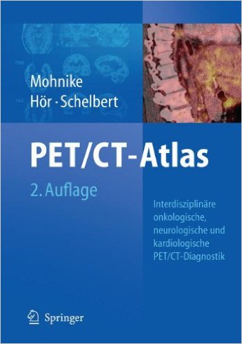
Oncologic and Cardiologic PET/CT-Diagnosis: An Interdisciplinary Atlas and Manual 2008th Edition

[amazon template=iframe image2&asin=3540740902]

“This nuclear medicine atlas provides a complete overview of the diagnostic aspect of PET-CT … . The book is well organized with abundant illustrations and teaching points. … The audience includes nuclear medicine professionals, radiologists, oncologists, cardiologists, and residents and fellows in these areas. … a valuable resource for new practitioners as well as for experienced nuclear medicine physicians who interpret PET-CT in either oncology or cardiology. … This excellent atlas will be a great addition to any nuclear medicine physician’s library … .” (Oussama Nachar, Doody’s Review Service, October, 2008)

“The portion of this book dealing with oncology is updated information based on PET/CT-Atlas, a German-language book … . Each case presented in the book includes a brief clinical history, imaging findings, and teaching points. The images are clear and well done. … I highly recommend this book as a quick reference to nuclear physicians and radiologists in practice or in training. This atlas and manual will also be useful to oncologists, cardiologists, and imaging technologists.” (E. Edmund Kim, The Journal of Nuclear Medicine, Vol. 50 (6), June, 2009)

From the Back Cover

Since 1999/2000 simultaneous imaging of anatomical features and metabolic activities using fused PET and CT images has complemented and in many cases even replaced what was originally done as separate analysis. – Is this a small revolution? – In fact, this new approach can be considered to be without a rival for imaging diagnosis. The complementary efforts of nuclear medicine physicians and radiologists have improved the diagnostic value of PET/CT scans. The successful use of PET/CT in the field of oncology has been documented world-wide, and in cardiology we can take advantage of the significant findings of PET diagnosis, particularly in the USA. New prospects are now available for paediatric oncology and molecular medicine.
Based on the experience gained by PET/CT experts with more than 10,000 patients, this manual impressively demonstrates the advantages of combined PET/CT. It also refers to publications from Europe, the USA and Asia as well as numerous studies.

DOWNLOAD THIS BOOK FREE HERE
http://upsto.re/ARrhtU4

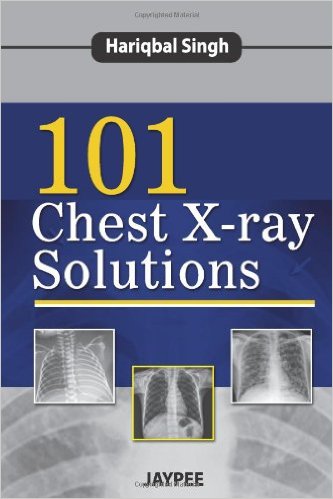
101 Chest X-ray Solutions 1st Edition

[amazon template=iframe image2&asin=9350904624]

Chest X-ray is the most commonly requested film in any medical department so it is vital for clinicians to have a good understanding of the signs in order to make an accurate diagnosis. This book presents numerous postero-anterior plain chest X-ray images with detailed descriptions, exposing trainees and clinicians to both basic and more complex diagnoses. Beginning with an introduction to chest anatomy and how to read an X-ray, the following chapters present 101 cases by chest section. The final chapter includes 20 practice X-rays or spotters for self assessment. Key points *101 cases presenting chest X-ray images with detailed descriptions * Covers simple and complex diagnoses * Cases presented in logical format by section of the chest * Includes 20 practice X-rays for self assessment

DOWNLOAD THIS BOOK FREE HERE
PART I http://upsto.re/A2qRf73

PART II http://upsto.re/A2fNxaj

PART III http://upsto.re/A2kkJxA

PART IV http://upsto.re/A2qcJwJ

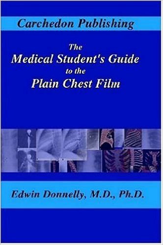
The Medical Student’s Guide to the Plain Chest Film

[amazon template=iframe image2&asin=1411695380]

This book is designed to provide the medical student a simple yet systemized approach to interpreting the plain chest radiograph. The book teaches the normal anatomy as well as a systematic approach that can (and should) be used to evaluate every chest film. Full-color volume-rendered computed tomography (CT) images are used to help the student understand the anatomy.

DOWNLOAD THIS BOOK FREE HERE
http://upsto.re/AC9hXvf

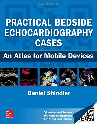
Practical Bedside Echocardiography Cases 1st Edition

[amazon template=iframe image2&asin=0071812652]

A COMPREHENSIVE REVIEW OF ECHOCARDIOGRAPHIC IMAGING ENHANCED BY HUNDREDS OF ONLINE ANIMATIONS VIEWABLE ON YOUR MOBILE DEVICE

EIGHTEEN CHAPTERS COVERS EVERY IMPORTANT CARDIAC DISEASE AND ABNORMALITY

Echocardiographic imaging has become essential to the proper diagnosis of every type of cardiac disease and disorder, and there is no more efficient or innovative way to learn the proper use of cardiac ultrasound in clinical practice than Practical Bedside Echocardiography Cases. This powerful combination text and online video collection delivers coverage of every major cardiac topic and abnormality.

There are eighteen chapters that include questions followed by detailed discussion that answers the question, as well as illustrations and text supplemented by animations that can be opened with a QR code reader using a smartphone or a tablet with Internet access. An index of all videos and direct links to them online are also included. Multiple variations on a topic are often included to illustrate the full spectrum of the abnormality being discussed. Bedside findings and the electrocardiogram, an integral part of the practice of echocardiography, are incorporated into the discussion whenever possible.

Numerous references are provided, with a strong emphasis on freely available full-text articles. These are identified by an open book icon indicating they are readable as PDFs. Other references point you to full-text cardiac physical diagnosis articles that will prove valuable at the bedside; some are annotated to further explain their value, and others were chosen for the comprehensive illustration of cardiac pathology as it relates to echocardiographic imaging. The book also includes references to classic echocardiography articles. Practical Bedside Echocardiography Cases will prove invaluable to medical students, cardiovascular practitioners at all levels of training, cardiac sonographers, and for users of hand-held, bedside, point-of-care ultrasound devices.

DOWNLOAD THIS BOOK FREE HERE
http://upsto.re/ADXrdfC

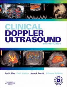
Clinical Doppler Ultrasound, 2e 2nd Edition

[amazon template=iframe image2&asin=0443101167]

The imaging possibilities of Doppler ultrasound have revolutionized radiology. This highly practical guide provides the tools you need to navigate this constantly evolving technology with confidence and skill. The major applications of Doppler ultrasound, including examination techniques and the interpretation of results, are discussed in an accessible, reader-friendly manner, making the book an ideal reference for all radiologists who wish to update and expand their knowledge and skills.

  • Discusses everything you need to know from anatomy, scanning, and technique, to normal and abnormal findings and their interpretation.
  • Explains both the advantages and the limitations of using Doppler imaging so you can make an informed decision every time.
  • Covers the basic principles behind the techniques so you can understand the why as well as the how.
  • Contains detailed protocols for acquiring optimal images and avoiding errors.
  • Uses 450 high-quality illustrations to bring techniques to life.
  • Includes a bonus CD-ROM containing real-time videos of Doppler imaging to further demonstrate key techniques and diagnoses.
  • Contains new chapters on ultrasonic echo-enhancing agents and the ultrasound of transplants.
  • Presents the latest developments in scanning technologies such as harmonic imaging and pulse inversion.

DOWNLOAD THIS BOOK FREE HERE
http://upsto.re/AF7GFZj

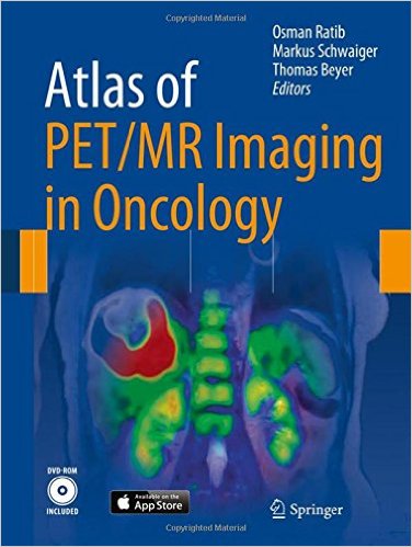
Atlas of PET/MR Imaging in Oncology 2013th Edition

[amazon template=iframe image2&asin=3642312918]

This new project on PET-MR imaging in oncology includes digital interactive software matching the cases in the book. The interactive version of the atlas is based on the latest web standard, HTML5, ensuring compatibility with any computer operating system as well as a dedicated version for Apple iPad.

The book opens with an introduction to the principles of hybrid imaging that pays particular attention to PET/MR imaging and standard PET/MR acquisition protocols. A wide range of illustrated clinical case reports are then presented. Each case study includes a short clinical history, findings, and teaching points, followed by illustrations, legends, and comments.

The multimedia version of the book includes dynamic movies that allow the reader to browse through series of rotating 3D images (MIP or volume rendered), display blending between PET and MR, and dynamic visualization of 3D image volumes. The movies can be played either continuously or sequentially for better exploration of sets of images.

The editors of this state-of-the-art publication are key opinion leaders in the field of multimodality imaging. Professor Osman Ratib (Geneva) and Professor Markus Schwaiger (Munich) were the first in Europe to initiate the clinical adoption of PET/MR imaging. Professor Thomas Beyer (Zurich) is an internationally renowned pioneering physicist in the field of hybrid imaging. Individual clinical cases presented in this book are co-authored by leading international radiologists and nuclear physicians experts in the use of PET and MRI.

DOWNLOAD THIS BOOK FREE HERE
http://upsto.re/AFuhgZf

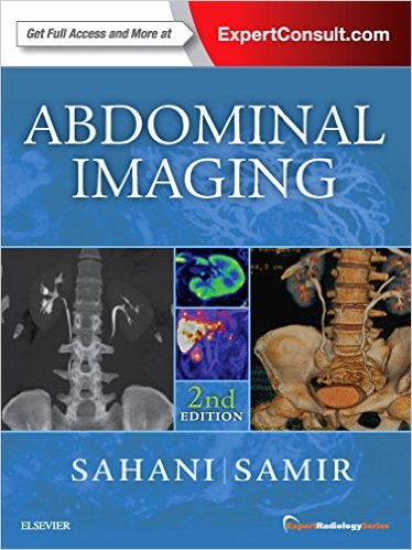
Abdominal Imaging: Expert Radiology Series, 2e 2nd Edition

[amazon template=iframe image2&asin=032337798X]

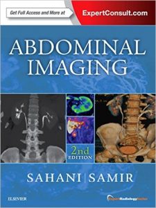
Richly illustrated and comprehensive in scope, Abdominal Imaging, 2nd Edition, by Drs. Dushyant V. Sahani and Anthony E. Samir, is your up-to-date, one-volume source for evaluating the full range of diagnostic, therapeutic, and interventional challenges in this fast-changing field. Part of the Expert Radiology series, this highly regarded reference covers all modalities and organ systems in a concise, newly streamlined format for quicker access to common and uncommon findings. Detailed, expert guidance, accompanied by thousands of high-quality digital images, helps you make the most of new technologies and advances in abdominal imaging.

    • Offers thorough coverage of all diagnostic modalities for abdominal imaging: radiographs, fluoroscopy, ultrasound, CT, MRI, PET and PET/CT.
    • Helps you select the best imaging approaches and effectively interpret your findings with a highly templated, well-organized, at-a-glance organization.
    • Covers multi-modality imaging of the esophagus, stomach, small bowel, colon, liver, pancreas, gall bladder, bile ducts, spleen, pelvic lymph nodes, kidneys, urinary tract, prostate, and peritoneum.


  • Expert Consult™ eBook version included with purchase. This enhanced eBook experience allows you to search all of the text, figures, and references from the book on a variety of devices.
  • Includes new chapters on esophageal imaging; 5RECIST, WHO, and other response criteria; and a new section on oncologic imaging.
  • Keeps you up to date with the latest developments in image-guided therapies, dual-energy CT, elastography, and much more.
  • Features more than 2,400 high-quality images, including 240 images new to this edition.

DOWNLOAD THIS BOOK FREE HERE
http://upsto.re/AvbwAiX

Diagnostic and Interventional Radiology 1st ed. 2016 Edition

[amazon template=iframe image2&asin=3662440369]

This exceptional book covers all aspects of diagnostic and interventional radiology within one volume, at a level appropriate for the specialist. From the basics through diagnosis to intervention: the reader will find a complete overview of all areas of radiology. The clear, uniform structure, with chapters organized according to organ system, facilitates the rapid retrieval of information. Features include:

  • Presentation of the normal radiological anatomy
  • Classification of the different imaging procedures according to their diagnostic relevance
  • Imaging diagnosis with many reference images
  • Precise description of the interventional options

The inclusion of many instructive aids will be of particular value to novices in decision making:

  • Important take home messages and summaries of key radiological findings smooth the path through the jungle of facts
  • Numerous tables on differential diagnosis and typical findings in the most common diseases offer a rapid overview and orientation
  • Diagnostic flow charts outline the sequence of diagnostic evaluation

All standard procedures within the field of interventional radiology are presented in a clinically relevant and readily understandable way, with an abundance of illustrations. This is a textbook, atlas, and reference in one: with more than 2500 images for comparison with the reader’s own findings. This comprehensive and totally up-to-date book provides a superb overview of everything that the radiology specialist of today needs to know.

DOWNLOAD THIS BOOK FREE HERE
http://upsto.re/AvYg3nh

Radiology Secrets Plus, 4e 4th Edition

[amazon template=iframe image2&asin=0323286380]

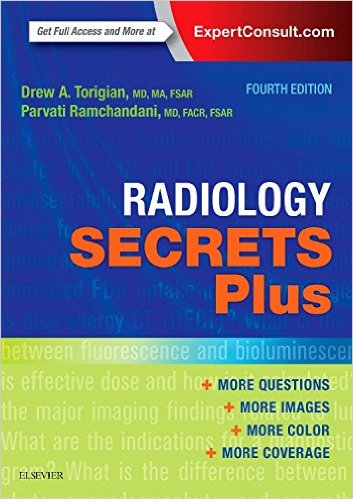
For 30 years, the highly regarded Secrets Series® has provided students and practitioners in all areas of health care with concise, focused, and engaging resources for quick reference and exam review. Radiology Secrets Plus, 4th Edition, by Drs. Drew Torigian and Parvati Ramchandani, features the Secrets’ popular question-and-answer format that also includes lists, tables, and an informal tone – making reference and review quick, easy, and enjoyable.

Top 100 Secrets and Key Points boxes provide a fast overview of the secrets you must know for success in practice and on exams.

The proven Secrets® format gives you the most return for your study time – concise, easy to read, engaging, and highly effective.

Larger page size provides the visual detail necessary for success in radiology, yet is still easy to carry with you for quick reference or review.

NEW: Expert Consult eBook features online and mobile access.

Full-color, expanded layout enhances understanding in this highly visual field.

Thorough updates throughout by a new expert author team from the highly regarded program at University of Pennsylvania and world-renowned contributors from top radiology programs.

DOWNLOAD THIS BOOK FREE HERE
http://upsto.re/AvYUMRA

1 47 48 49 50 51 62