Echocardiography: A Practical Guide for Reporting and Interpretation, Third Edition 3rd Edition

[amazon template=iframe image2&asin=1482231921]

Echocardiography: A Practical Guide for Reporting and Interpretation is a step-by-step guide to clinical echocardiography.

This new edition has been extensively revised and includes new international guidelines, grading criteria and normal data. The book presents an up-to-date discussion of echocardiography use in both acute and critical care settings and provides increased guidance for clinical interpretation, including how echocardiography can help inform surgical decisions. In addition, the content has been extended, particularly in the consideration of valve disease and cardiomyopathies.

Providing a pragmatic approach to clinical echocardiography, the book also covers examples of conditions requiring urgent clinical advice. It contains a summary of formulae as well as supporting online images and videos.

Review

“My interpretation is that the authors have clearly achieved their ambition to produce a small but comprehensive, inexpensive practical and reference aid for Echocardiographers of all grades of experience. I consider that this book should be available within all Echo departments and additionally I strongly recommend all Echocardiographers to have their own personal copy.” – Dr Gordon Williams, BSE Newsletter

“Technological advances and broadened knowledge about the pathogenesis of cardiovascular diseases have made echocardiography a very important part of the diagnosis and management of cardiovascular disorders. This book differs from the classic textbooks and instead is an easy-to-read, step-by-step guidebook. It will serve as a valuable resource when echocardiographers need quick and practical information.” –  Ahmet Afsin Oktay, MD(Ochsner Clinic Foundation), Doody Enterprises

DOWNLOAD THIS BOOK FREE HERE

http://upsto.re/AR5Gc7M

Imaging for Students Fourth Edition 4th Edition

[amazon template=iframe image2&asin=1444121820]

Imaging for Students delivers step-by-step guidance to the range of imaging techniques available, providing a clear explanation of how each imaging modality actually works, and including information on the associated risks and hazards. Throughout, the importance of patient preparation and post-procedure observation is emphasized.

Taking information from evidence-based studies and published guidelines, in line with current clinical practice, the book takes a highly logical approach to the investigation of clinical scenarios, where possible indicating the “best first test”―vital to both appropriate clinical and cost-effective decision-making.

Drawing on the extensive clinical and teaching experience of its respected author, the fourth edition of Imaging for Students gives students and junior doctors everything they need to understand the advantages, disadvantages, and possible side effects of the imaging modalities available, and how to apply them appropriately in clinical practice.

DOWNLOAD THIS BOOK FREE HERE

http://upsto.re/ARULenw

Atlas of Ultrasound-Guided Regional Anesthesia: Expert Consult – Online and Print, 2e 2nd Edition

[amazon template=iframe image2&asin=1455728187]

Safely and effectively perform regional nerve blocks with Atlas of Ultrasound-Guided Regional Anesthesia, 2nd Edition. Using a wealth of step-by-step videos and images, Dr. Andrew T. Gray shows you how to use the latest methods to improve the success rate of these techniques.

“I have read a lot of atlas type books and this is one of the best such books that I have seen. It is difficult to see how it could be improved.” Reviewed by: N. D. Edwards on behalf of The British Journal of Anaesthesia, Sept 2014


  • Master essential techniques through step-by-step videos demonstrating paravertebral block, transversus abdominis block, psoas nerve block, subgluteal nerve block, and more.
  • Test your knowledge and prepare for the ABA exam with board-style review questions.


  • Ensure correct needle placement with numerous 3-D and long-axis views that clearly depict surrounding structures.
  • Update your skills with completely rewritten chapters on Infraclavicular, Neuraxial, and Cervical Plexus Blocks as well as entirely new chapters on Fascia Iliaca, Anterior Sciatic, Transversus Abdominis Plane (TAP), and Stellate Ganglion Blocks.
  • Review a full range of nerve block techniques in an easy-to-follow, step-by-step manner using new quick-reference summary tables.
  • View author-narrated videos and access the complete contents online at www.expertconsult.com; assess your knowledge with the aid of a new “turn labels off” feature for each image

DOWNLOAD THIS BOOK FREE HERE

http://upsto.re/AR73wUD

Transesophageal Echocardiography for Congenital Heart Disease 2014th Edition

[amazon template=iframe image2&asin=1848000618]

Transesophageal Echocardiography for Congenital Heart Disease represents a unique contribution as the only contemporary reference to focus exclusively on the clinical applications of transesophageal echocardiography (TEE) in congenital heart disease (CHD). Written by numerous prominent specialists and renowned leaders in the field, it presents a comprehensive, modern, and integrated review of the subject in light of the cumulative experience and most recent advances in the technology. Topics related to CHD include: (1) physics and instrumentation of TEE, particularly as they apply to the structural evaluation; (2) specialized aspects of the examination, with emphases on technical considerations pertinent to both pediatric and adult patients with congenital cardiovascular pathology; (3) segmental approach to diagnosis and functional assessment; (4) extensive discussion of the TEE evaluation of the many anomalies encompassing the CHD spectrum; (5) use of the imaging modality in the perioperative and interventional settings; and (6) important aspects of 3D TEE evaluation.

Richly illustrated by more than 700 figures/illustrations and 400 videos, this textbook will serve as an indispensable resource for all who use TEE in the care of both children and adults with CHD, from the novice to the expert.

DOWNLOAD THIS BOOK FREE HERE

http://upsto.re/ARGqQu6

Chest Radiology: A Resident’s Manual 1 Pap/Psc Edition

[amazon template=iframe image2&asin=3131538716]

Chest Radiology: A Resident’s Manual is a comprehensive introduction to reading and  analyzing radiologic cardiopulmonary images. Readers are guided through systemic image analysis and can further enhance their learning experience with training cases found at the end of each chapter. Cases describe and discuss frequently asked questions regarding heart failure, bronchitis, pneumonia, bronchial carcinoma, fibrosis, pleural disorders, and more. This user-friendly manual will allow the reader to confidently answer the most important and commonly encountered questions related to plain chest radiographs in daily clinical practice. The easy-to-read layout pairs explanatory text on the left page with related drawings and images on the right, allowing readers to navigate their way through each section with ease.

Features

More than 600 high-resolution images and illustrations
demonstrate a wealth of pathology
Concise descriptions explain how to examine
conventional x-ray and CT images
Numerous callout boxes in each chapter highlight key
takeaway points
A scratch-off code provides access to a searchable
online database of 250 must-know thoracic imaging cases

This practice-oriented manual is an invaluable resource and reference guide for residents and radiologists-in-training.

DOWNLOAD THIS BOOK FREE HERE

http://upsto.re/ARGfnsC

Oncologic and Cardiologic PET/CT-Diagnosis: An Interdisciplinary Atlas and Manual 2008th Edition

[amazon template=iframe image2&asin=3540740902]

“This nuclear medicine atlas provides a complete overview of the diagnostic aspect of PET-CT … . The book is well organized with abundant illustrations and teaching points. … The audience includes nuclear medicine professionals, radiologists, oncologists, cardiologists, and residents and fellows in these areas. … a valuable resource for new practitioners as well as for experienced nuclear medicine physicians who interpret PET-CT in either oncology or cardiology. … This excellent atlas will be a great addition to any nuclear medicine physician’s library … .” (Oussama Nachar, Doody’s Review Service, October, 2008)

“The portion of this book dealing with oncology is updated information based on PET/CT-Atlas, a German-language book … . Each case presented in the book includes a brief clinical history, imaging findings, and teaching points. The images are clear and well done. … I highly recommend this book as a quick reference to nuclear physicians and radiologists in practice or in training. This atlas and manual will also be useful to oncologists, cardiologists, and imaging technologists.” (E. Edmund Kim, The Journal of Nuclear Medicine, Vol. 50 (6), June, 2009)

From the Back Cover

Since 1999/2000 simultaneous imaging of anatomical features and metabolic activities using fused PET and CT images has complemented and in many cases even replaced what was originally done as separate analysis. – Is this a small revolution? – In fact, this new approach can be considered to be without a rival for imaging diagnosis. The complementary efforts of nuclear medicine physicians and radiologists have improved the diagnostic value of PET/CT scans. The successful use of PET/CT in the field of oncology has been documented world-wide, and in cardiology we can take advantage of the significant findings of PET diagnosis, particularly in the USA. New prospects are now available for paediatric oncology and molecular medicine.
Based on the experience gained by PET/CT experts with more than 10,000 patients, this manual impressively demonstrates the advantages of combined PET/CT. It also refers to publications from Europe, the USA and Asia as well as numerous studies.

DOWNLOAD THIS BOOK FREE HERE

http://upsto.re/ARrhtU4

101 Chest X-ray Solutions 1st Edition

[amazon template=iframe image2&asin=9350904624]

Chest X-ray is the most commonly requested film in any medical department so it is vital for clinicians to have a good understanding of the signs in order to make an accurate diagnosis. This book presents numerous postero-anterior plain chest X-ray images with detailed descriptions, exposing trainees and clinicians to both basic and more complex diagnoses. Beginning with an introduction to chest anatomy and how to read an X-ray, the following chapters present 101 cases by chest section. The final chapter includes 20 practice X-rays or spotters for self assessment. Key points *101 cases presenting chest X-ray images with detailed descriptions * Covers simple and complex diagnoses * Cases presented in logical format by section of the chest * Includes 20 practice X-rays for self assessment

DOWNLOAD THIS BOOK FREE HERE

PART I http://upsto.re/A2qRf73

PART II http://upsto.re/A2fNxaj

PART III http://upsto.re/A2kkJxA

PART IV http://upsto.re/A2qcJwJ

The Medical Student’s Guide to the Plain Chest Film

[amazon template=iframe image2&asin=1411695380]

This book is designed to provide the medical student a simple yet systemized approach to interpreting the plain chest radiograph. The book teaches the normal anatomy as well as a systematic approach that can (and should) be used to evaluate every chest film. Full-color volume-rendered computed tomography (CT) images are used to help the student understand the anatomy.

DOWNLOAD THIS BOOK FREE HERE

http://upsto.re/AC9hXvf

Practical Bedside Echocardiography Cases 1st Edition

[amazon template=iframe image2&asin=0071812652]

A COMPREHENSIVE REVIEW OF ECHOCARDIOGRAPHIC IMAGING ENHANCED BY HUNDREDS OF ONLINE ANIMATIONS VIEWABLE ON YOUR MOBILE DEVICE

EIGHTEEN CHAPTERS COVERS EVERY IMPORTANT CARDIAC DISEASE AND ABNORMALITY

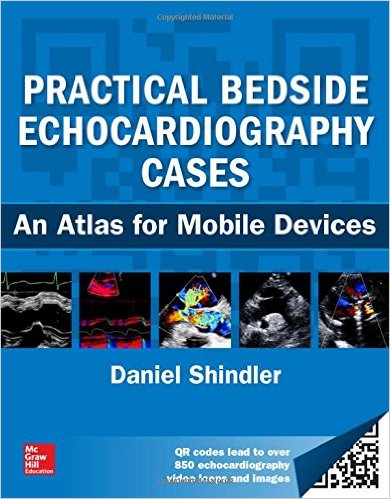
Echocardiographic imaging has become essential to the proper diagnosis of every type of cardiac disease and disorder, and there is no more efficient or innovative way to learn the proper use of cardiac ultrasound in clinical practice than Practical Bedside Echocardiography Cases. This powerful combination text and online video collection delivers coverage of every major cardiac topic and abnormality.

There are eighteen chapters that include questions followed by detailed discussion that answers the question, as well as illustrations and text supplemented by animations that can be opened with a QR code reader using a smartphone or a tablet with Internet access. An index of all videos and direct links to them online are also included. Multiple variations on a topic are often included to illustrate the full spectrum of the abnormality being discussed. Bedside findings and the electrocardiogram, an integral part of the practice of echocardiography, are incorporated into the discussion whenever possible.

Numerous references are provided, with a strong emphasis on freely available full-text articles. These are identified by an open book icon indicating they are readable as PDFs. Other references point you to full-text cardiac physical diagnosis articles that will prove valuable at the bedside; some are annotated to further explain their value, and others were chosen for the comprehensive illustration of cardiac pathology as it relates to echocardiographic imaging. The book also includes references to classic echocardiography articles. Practical Bedside Echocardiography Cases will prove invaluable to medical students, cardiovascular practitioners at all levels of training, cardiac sonographers, and for users of hand-held, bedside, point-of-care ultrasound devices.

DOWNLOAD THIS BOOK FREE HERE

http://upsto.re/ADXrdfC

Clinical Doppler Ultrasound, 2e 2nd Edition

[amazon template=iframe image2&asin=0443101167]

The imaging possibilities of Doppler ultrasound have revolutionized radiology. This highly practical guide provides the tools you need to navigate this constantly evolving technology with confidence and skill. The major applications of Doppler ultrasound, including examination techniques and the interpretation of results, are discussed in an accessible, reader-friendly manner, making the book an ideal reference for all radiologists who wish to update and expand their knowledge and skills.

  • Discusses everything you need to know from anatomy, scanning, and technique, to normal and abnormal findings and their interpretation.
  • Explains both the advantages and the limitations of using Doppler imaging so you can make an informed decision every time.
  • Covers the basic principles behind the techniques so you can understand the why as well as the how.
  • Contains detailed protocols for acquiring optimal images and avoiding errors.
  • Uses 450 high-quality illustrations to bring techniques to life.
  • Includes a bonus CD-ROM containing real-time videos of Doppler imaging to further demonstrate key techniques and diagnoses.
  • Contains new chapters on ultrasonic echo-enhancing agents and the ultrasound of transplants.
  • Presents the latest developments in scanning technologies such as harmonic imaging and pulse inversion.

DOWNLOAD THIS BOOK FREE HERE

http://upsto.re/AF7GFZj

1 45 46 47 48 49 60