Learning Abdominal Imaging (Learning Imaging) 2012th Edition
[amazon template=iframe image2&asin=354088002X]

Review

From the reviews:

“The book is very well organized, and the coverage of common disease entities is clear and succinct. It is richly illustrated and uses all of the current imaging modalities. … it will be of great benefit to the advanced medical student and resident trainee in a specialty other than radiology who is particularly interested in abdominal diseases. It also belongs in the teaching library of every medical school and institution of postgraduate training.” (Bharat Raval, Radiology, Vol. 267 (3), June, 2013)

“This book in its 256 pages is intended to give the reader a review of cases devoted to abdominal imaging, addressing in ten sections … . Ten cases have been chosen for each section, simple as well as complicated or unusual cases highlighting the topic, cases which should and could be of practical use to both physicians in training as well as expert radiologists. … a useful book to be at hand in daily practice.” (G. Beluffi, La radiologia medica, Vol. 118, 2013)

“Learning Abdominal Imaging is a concise guide to recognizing the essential elements of abdominal CT and MRI images for a fast and accurate diagnosis. … Written mainly for beginners to the field of image interpretation … . Clinicians new to interpretation of abdominal images will find this to be a concise and highly informative resource. … Gastroenterologists, emergency medicine physicians, primary care practitioners and others who require a working knowledge of abdominal imaging in their daily practice should read this book.” (Medical Science Books, May, 2012)

DOWNLOAD THIS BOOK FREE HERE

http://upsto.re/AJhRk5Z

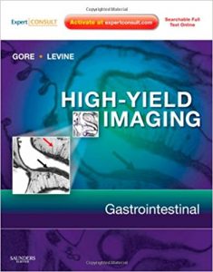
High Yield Imaging: Gastrointestinal: Expert Consult – Online and Print, 1e (HIGH YIELD in Radiology) 1 Har/Psc Edition

[amazon template=iframe image2&asin=1416055444]

This state-of-the-art Gastrointestinal title in the brand-new High-Yield Imaging series is ideally designed to let you quickly look up a diagnosis, confirm your suspicion, and move on to the next case. Renowned authorities, Drs. Gore and Levine, present 350 GI diagnoses, 1,200 optimally sized multi-modality clinical images, and at-a-glance bulleted text, to help you efficiently and accurately diagnose any GI disorder. A full-color design along with a consistent organization makes reference a snap. And, at expertconsult.com you’ll have easy access to the full text online as well as all of the book’s images for enhanced visual guidance.

  • Features online access to the full text and all the illustrations from the book, with links to Medline, for easy referencing in the viewing room.
  • Covers 350 GI diagnoses allowing you to diagnose a broader spectrum of conditions.
  • Offers all the information necessary to provide referring physicians with the complete, actionable reports they need.
  • Presents more than 1,200 optimally sized multi-modality clinical images to help you recognize the characteristic appearance of both common and uncommon GI disorders.
  • Structures every chapter consistently to include Findings, Imaging Modalities, Clinical Presentation, Pathology, Incidence/Prevalence and Epidemiology, What the Referring Physician Needs to Know, Suggested Readings, and Selected Images to make reference fast and easy.

DOWNLOAD THIS BOOK FREE HERE

http://upsto.re/Aw5DRRE

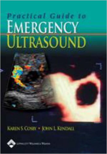
Practical Guide to Emergency Ultrasound 1st Edition

[amazon template=iframe image2&asin=0781778581]

Featuring over 700 illustrations, this book is a practical, visual guide to performing and interpreting ultrasound and using ultrasound findings for making clinical decisions in the emergency department. Consistently formatted chapters cover both common and less common uses of ultrasound in the emergency department. Each chapter includes clinical applications, anatomy and landmarks, image acquisition, pathology, clinical decision making, incidental findings, and clinical examples.

High-quality images include patient photographs demonstrating the correct probe placement and large ultrasound images allowing findings to be easily seen. Labels on ultrasound scans and side-by-side anatomic drawings help readers locate the key parts of all images.

I find this textbook is consistently well written with well-organized chapters. Among the references for emergency ultrasound, it stands out by having tons of high quality real images and excellent schematic diagrams to illustrate important anatomy. I also find particularly helpful the pitfalls section of each chapter. Overall, I highly recommend this reference as a standard for every ED and residency program.

DOWNLOAD THIS BOOK FREE HERE

http://upsto.re/Ah3MRsB

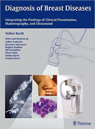
Diagnosis of Breast Diseases: Integrating the Findings of Clinical Presentation, Mammography, and Ultrasound 1st Edition

[amazon template=iframe image2&asin=3131438312]

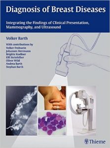
This atlas provides radiologists with essential
information for the differential diagnosis of breast diseases on the basis of
clinical presentation, mammography, and ultrasound. It begins with
chapters on tumor biology, prognostic factors, and histology. The authors then
provide a thorough evaluation of various methods for early detection and
accurate diagnosis, including analog and digital mammography, ultrasound, MR
imaging, PET/CT, and interventional procedures. They discuss in detail the
strengths and limitations of each imaging modality, aspects of quality control,
test intervals, peri- and postoperative management principles, and follow-up
care.

Highlights:


  • Presentation of difficult cases that effectively
    demonstrate the diagnostic hurdles and forensic pitfalls in breast diagnosis
  • Special sections on breast cancer in men and young
    women, with discussion of women who are pregnant or lactating
  • Color-coded practical tips and clinical notes for
    optimal comprehension of the material
  • Extensive Q&A sections for self-testing in two
    major chapters
  • More than 1,700 high-quality illustrations, including
    clinical color photographs, ultrasound images, and mammograms
  • From the Back Cover

    This atlas provides radiologists with essential information for the differential diagnosis of breast diseases on the basis of clinical presentation, mammography, and ultrasound. It begins with chapters on tumor biology, prognostic factors, and histology. The authors then provide a thorough evaluation of various methods for early detection and accurate diagnosis, including analog and digital mammography, ultrasound, MR imaging, PET/CT, and interventional procedures. They discuss in detail the strengths and limitations of each imaging modality, aspects of quality control, test intervals, peri- and postoperative management principles, and follow-up care.

    Highlights: Presentation of difficult cases that effectively demonstrate the diagnostic hurdles and forensic pitfalls in breast diagnosis Special sections on breast cancer in men and young women, with discussion of women who are pregnant or lactating Color-coded practical tips and clinical notes for optimal comprehension of the material Extensive Q&A sections for self-testing in two major chapters More than 1,700 high-quality illustrations, including clinical color photographs, ultrasound images, and mammograms

    “I believe that this outstanding modern book will be highly valued by radiologists, oncologists, surgeons and many other specialists.British Journal of Radiology”

    “Every page contains an opportunity to increase the user’s knowledge, .Radiologic Technology”

    DOWNLOAD THIS BOOK FREE HERE

    http://upsto.re/A6MiUbi

RadCases Head and Neck Imaging 1 Pap/Psc Edition

[amazon template=iframe image2&asin=1604061936]

Head and Neck Imaging presents the challenging cases that are most likely to be encountered by residents and radiologists focusing on imaging of the head and neck. It helps radiologists correctly interpret images and thus quickly make initial diagnoses on both common and less common disorders of the head and neck. This book can also be used as an effective review guide for those studying for the radiology board exams.

    Key Features:

  • Content covers the imaging of the temporal bone, skull base, orbits, paranasal sinuses, and all the structures of the head and neck
  • Examples of critical cases that must be accurately diagnosed in daily practice and on exams
  • Clearly labeled, high-quality images help you quickly absorb key findings
  • A scratch-off code provides free access to a searchable online database of must-know cases, the 100 cases that are in the book, plus additional cases available only online
  • From the Back Cover

    Head and Neck Imaging presents the challenging cases that are most likely to be encountered by residents and radiologists focusing on imaging of the head and neck. It helps radiologists correctly interpret images and thus quickly make initial diagnoses on both common and less common disorders of the head and neck. This book can also be used as an effective review guide for those studying for the radiology board exams.
    Key Features:
    Content covers the imaging of the temporal bone, skull base, orbits, paranasal sinuses, and all the structures of the head and neckExamples of critical cases that must be accurately diagnosed in daily practice and on examsClearly labeled, high-quality images help you quickly absorb key findings A scratch-off code provides free access to a searchable online database of must-know cases, the 100 cases that are in the book, plus additional cases available only online”

    DOWNLOAD THIS BOOK FREE HERE

    http://upsto.re/A66fFYq

100 Challenges in Echocardiography, 1e 1st Edition

[amazon template=iframe image2&asin=0443069271]

This book offers both experienced cardiologists and trainees alike the opportunity to identify and address the most common pitfalls encountered with echocardiology in routine clinical practice. The book covers a series of challenges ranging from technical difficulties to problems with echocardiographic interpretation with each section presenting a series of simulations to test the reader’s understanding of the problem. The text is written in a highly practical and didactic style which will help the busy physician by presenting the information in a succinct, ‘essentials only’ manner. The heavy use of high-quality illustrations serves to ensure that the concepts explained are supported by real-life examples and thus readily applicable to the clinical setting.

  • Edited by an international expert.
  • Abundant use of full-colour allows accurate representation of images to afford detailed discussion of the techniques involved.
  • Didactic approach ensures delivery of key information in manageable portions thus saving time for the busy cardiologist.
  • Includes the most frequent pitfalls associated with a range of techniques thus ensuring applicability to a variety of clinical settings and equipment availability.
  • About the Author

    est cardiologue, praticien hospitalier des hôpitaux de l’AP de Paris (Charles-Foix à Ivry-sur-Seine, Émile-Roux à Limeil-Brévannes) – Groupements hospitaliers universitaires.

    Dr Petros Nihoyannopoulos, FRCP, is a senior Consultant Cardiologist at Hammersmith Hospital and Reader at the Imperial College London and National Heart and Lung Institute. He qualified in Medicine in 1979 from Strasbourg University in France (Universite Luis Pasteur) and completed his formal cardiac training in London, at the Hammersmith Hospital.

    Dr Nihoyannopoulos, as Clinical Firm Leader, is heavily involved with everyday clinical practice involving patients with a variety of cardiac conditions. He is also Fellow of many national and international societies and associations including the American Heart Asociation, American College of Cardiology and European Society of Cardiology. He is founding member and past president of the British Society of Echocardiography and is the President-Elect of the European Association of Echocardiography, a Registered Branch of the European Society of Cardiology.

    Dr Nihoyannopoulos’ main research interest is in the study of heart muscle disorders, valvular heart disease as well as acute and chronic coronary syndromes using state-of-the-art echocardiography. He has published more than 160 peered reviewed papers in major international journals as well as more than 40 review articles and book chapters. As part of his academic position, Dr Nihoyannopoulos is committed to teaching under- and post-graduate students on both clinical cardiology and echocardiology courses, where he has made his name as one of the international leading experts in the field.

    DOWNLOAD THIS BOOK FREE HERE

    http://upsto.re/AR57M7a

Echocardiography: A Practical Guide for Reporting and Interpretation, Third Edition 3rd Edition

[amazon template=iframe image2&asin=1482231921]

Echocardiography: A Practical Guide for Reporting and Interpretation is a step-by-step guide to clinical echocardiography.

This new edition has been extensively revised and includes new international guidelines, grading criteria and normal data. The book presents an up-to-date discussion of echocardiography use in both acute and critical care settings and provides increased guidance for clinical interpretation, including how echocardiography can help inform surgical decisions. In addition, the content has been extended, particularly in the consideration of valve disease and cardiomyopathies.

Providing a pragmatic approach to clinical echocardiography, the book also covers examples of conditions requiring urgent clinical advice. It contains a summary of formulae as well as supporting online images and videos.

Review

“My interpretation is that the authors have clearly achieved their ambition to produce a small but comprehensive, inexpensive practical and reference aid for Echocardiographers of all grades of experience. I consider that this book should be available within all Echo departments and additionally I strongly recommend all Echocardiographers to have their own personal copy.” – Dr Gordon Williams, BSE Newsletter

“Technological advances and broadened knowledge about the pathogenesis of cardiovascular diseases have made echocardiography a very important part of the diagnosis and management of cardiovascular disorders. This book differs from the classic textbooks and instead is an easy-to-read, step-by-step guidebook. It will serve as a valuable resource when echocardiographers need quick and practical information.” –  Ahmet Afsin Oktay, MD(Ochsner Clinic Foundation), Doody Enterprises

DOWNLOAD THIS BOOK FREE HERE

http://upsto.re/AR5Gc7M

Imaging for Students Fourth Edition 4th Edition

[amazon template=iframe image2&asin=1444121820]

Imaging for Students delivers step-by-step guidance to the range of imaging techniques available, providing a clear explanation of how each imaging modality actually works, and including information on the associated risks and hazards. Throughout, the importance of patient preparation and post-procedure observation is emphasized.

Taking information from evidence-based studies and published guidelines, in line with current clinical practice, the book takes a highly logical approach to the investigation of clinical scenarios, where possible indicating the “best first test”―vital to both appropriate clinical and cost-effective decision-making.

Drawing on the extensive clinical and teaching experience of its respected author, the fourth edition of Imaging for Students gives students and junior doctors everything they need to understand the advantages, disadvantages, and possible side effects of the imaging modalities available, and how to apply them appropriately in clinical practice.

DOWNLOAD THIS BOOK FREE HERE

http://upsto.re/ARULenw

Atlas of Ultrasound-Guided Regional Anesthesia: Expert Consult – Online and Print, 2e 2nd Edition

[amazon template=iframe image2&asin=1455728187]

Safely and effectively perform regional nerve blocks with Atlas of Ultrasound-Guided Regional Anesthesia, 2nd Edition. Using a wealth of step-by-step videos and images, Dr. Andrew T. Gray shows you how to use the latest methods to improve the success rate of these techniques.

“I have read a lot of atlas type books and this is one of the best such books that I have seen. It is difficult to see how it could be improved.” Reviewed by: N. D. Edwards on behalf of The British Journal of Anaesthesia, Sept 2014


  • Master essential techniques through step-by-step videos demonstrating paravertebral block, transversus abdominis block, psoas nerve block, subgluteal nerve block, and more.
  • Test your knowledge and prepare for the ABA exam with board-style review questions.


  • Ensure correct needle placement with numerous 3-D and long-axis views that clearly depict surrounding structures.
  • Update your skills with completely rewritten chapters on Infraclavicular, Neuraxial, and Cervical Plexus Blocks as well as entirely new chapters on Fascia Iliaca, Anterior Sciatic, Transversus Abdominis Plane (TAP), and Stellate Ganglion Blocks.
  • Review a full range of nerve block techniques in an easy-to-follow, step-by-step manner using new quick-reference summary tables.
  • View author-narrated videos and access the complete contents online at www.expertconsult.com; assess your knowledge with the aid of a new “turn labels off” feature for each image

DOWNLOAD THIS BOOK FREE HERE

http://upsto.re/AR73wUD

Transesophageal Echocardiography for Congenital Heart Disease 2014th Edition

[amazon template=iframe image2&asin=1848000618]

Transesophageal Echocardiography for Congenital Heart Disease represents a unique contribution as the only contemporary reference to focus exclusively on the clinical applications of transesophageal echocardiography (TEE) in congenital heart disease (CHD). Written by numerous prominent specialists and renowned leaders in the field, it presents a comprehensive, modern, and integrated review of the subject in light of the cumulative experience and most recent advances in the technology. Topics related to CHD include: (1) physics and instrumentation of TEE, particularly as they apply to the structural evaluation; (2) specialized aspects of the examination, with emphases on technical considerations pertinent to both pediatric and adult patients with congenital cardiovascular pathology; (3) segmental approach to diagnosis and functional assessment; (4) extensive discussion of the TEE evaluation of the many anomalies encompassing the CHD spectrum; (5) use of the imaging modality in the perioperative and interventional settings; and (6) important aspects of 3D TEE evaluation.

Richly illustrated by more than 700 figures/illustrations and 400 videos, this textbook will serve as an indispensable resource for all who use TEE in the care of both children and adults with CHD, from the novice to the expert.

DOWNLOAD THIS BOOK FREE HERE

http://upsto.re/ARGqQu6

1 46 47 48 49 50 62