Contrast-Enhanced Ultrasound in Clinical Practice: Liver, Prostate, Pancreas, Kidney and Lymph Nodes

[amazon template=image&asin=8847003040]

The value of ultrasound contrast agents (USCA) in everyday clinical practice depends on the pharmacokinetics, the signal processing, and the contrast-specific imaging modalities.

Second-generation USCA, are blood pool agents that do not leak into the organ tissue to be examined but remain in the intravascular compartment increasing the Doppler signal amplitude during their dynamic vascular phase. Taking advantage of the stability of their microbubbles, they can withstand the acoustic pressure of insonation much better than first-generation contrast media, which results in an increased half-life of the agent and, consequently, in a prolonged diagnostic window.

Concomitant with the improvement of contrast agents, different contrast-specific imaging modalities have been developed which, used in combination with USCA and a low mechanical index, allow continuous real-time grey-scale imaging. These recent technical improvements have opened new possibilities in the use of USCA in a variety of indications. Written by internationally renowned experts, the contributions gathered in this book give an overview of current and possible future new applications of USCA in routine and clinical practice.


DOWNLOAD THIS BOOK FREE HERE

https://upsto.re/FUWAqHR

PET/CT in Cancer of Unknown Primary (Clinicians’ Guides to Radionuclide Hybrid Imaging) 1st ed

[amazon template=image&asin=3319564234]

About the Author

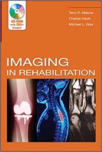
Imaging In Rehabilitation 1st Edition

[amazon template=image&asin=0071447784]

“This is a need to know reference to orient therapists to the role of imaging procedures….it is a useful resource and offers nice clinical pearls to apply the information presented.”–Doody’s Review Service

“…a very useful reference text for both the practicing physical therapist as well as the physical therapy student….The illustrations, and especially the CD-ROM, do an excellent job of depicting points brought up in the text and greatly enhance the clarity of the information. I have found the CD-ROM images to be valuable tools in patient education and in clarifying pathology for physical therapy students.”–Journal of Orthopaedic and Sports Physical Therapy

This unique book enables therapists to understand the various imaging technologies and weigh the benefits and drawbacks of each in the context of their own screening and evaluation process. It also serves as an indispensable resource for meaningfully discussing the results of these tests as part of the patient education process.

DOWNLOAD THIS BOOK FREE HERE

https://upsto.re/FUYUaeX

Functional Neuroimaging in Child Psychiatry 1st Edition

[amazon template=image&asin=0521126584]

Book Description

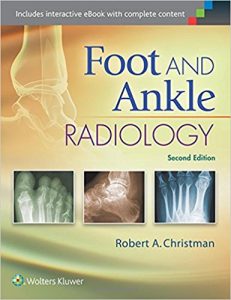
Foot and Ankle Radiology Second Edition

[amazon template=image&asin=1451192835]

Clearly written and abundantly illustrated, Foot and Ankle Radiology, 2nd Edition provides a solid understanding of diagnostic radiology as it applies to podiatric medicine, including both normal and pathological presentations of the foot and ankle.  This highly regarded text has been completely updated with all-new chapters on MR and CT imaging, digital radiography, musculoskeletal ultrasound, and more; full-color illustrations; and significant revisions throughout.  With coverage of both adult and pediatric imaging, it’s your one-stop resource for the radiographic presentations of pathologic conditions of the foot and ankle.

Features:

  • Get comprehensive coverage of everything you need to know about the principles of radiographic interpretation, normal and variant radiographic anatomy and development of the foot and ankle, and systematic evaluation of bone and joint disorders, as well as bone and joint abnormalities.
  • Make the most of all imaging procedures with new content on MR, CT, PET, SPECT, and musculoskeletal ultrasound.
  • Address challenging issues with confidence, thanks to new chapters on The Diabetic Foot and Postoperative Evaluation and Complications.
  • Visualize key concepts, disorders, and procedures easily with more than 1,000 high-quality radiographs and full-color illustrations.
  • Quickly see how to systematically analyze a radiograph and identify conditions that are intrinsic to the foot or that represent manifestations of extrinsic disease.
  • Gain a better understanding of both normal and variant radiographic anatomy, as well as both adult and pediatric imaging of the foot and ankle.

Now with the print edition, enjoy the bundled interactive eBook edition, offering tablet, smartphone, or online access to:

  • Complete content with enhanced navigation.
  • Powerful search tools and smart navigation cross-links that pull results from content in the book, your notes, and even the web.
  • Cross-linked pages, references, and more for easy navigation.
  • A highlighting tool for easier reference of key content throughout the text.
  • The ability to take and share notes with friends and colleagues.
  • Quick reference tabbing to save your favorite content for future use.

DOWNLOAD THIS BOOK FREE HERE

https://upsto.re/FMLkJ9f

Living without an Amygdala 1st Edition

[amazon template=image&asin=1462525946]

Bringing together leading researchers, this book comprehensively covers what is known about the amygdala, with a unique focus on what happens when this key brain region is damaged or missing. Offering a truly comparative approach, the volume presents research on rats, monkeys, and humans. It reports on compelling cases of people living without an amygdala, whether due to genetic conditions, disease, or other causes. The consequences for an individual’s ability to detect danger and regulate emotions–and for broader cognitive and social functions–are explored, as are lessons learned about brain pathways and plasticity. The volume delves into the role of the amygdala in psychiatric disorders and identifies important directions for future research. Illustrations include six color plates.

About the Author

Cone Beam CT and 3D imaging: A Practical Guide

[amazon template=image&asin=8847053188]

Cone beam computed tomography (CBCT) has become the standard of reference in dental imaging. The distribution of CBCT devices is increasingly wide, and the number of required examinations is constantly growing. In this setting, it is now essential that medical and technical staff receive specific training in the use of CBCT and that technical guidelines for CBCT examinations are established.

This clearly structured book on CBCT will be an ideal aid in daily clinical practice. It clearly explains basic CBCT anatomy, examination technique, and the use of 3D reformatting software. A wide range of cases are presented, covering the most frequent and relevant conditions and pathologies, including dental anomalies, inflammatory and degenerative disease, tumors, and implants.

DOWNLOAD THIS BOOK FREE HERE

https://upsto.re/FUgUBFj

Quantitative Ultrasound in Soft Tissues

[amazon template=image&asin=9400769512]

From the Back Cover

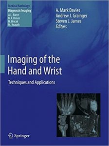
Imaging of the Hand and Wrist: Techniques and Applications (Medical Radiology)

[amazon template=image&asin=3642111432]

From the Back Cover

Choosing the Correct Radiologic Test: Case-Based Teaching Files

[amazon template=image&asin=3642157718]

From the Back Cover

1 32 33 34 35 36 62