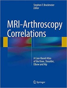
MRI-Arthroscopy Correlations: A Case-Based Atlas of the Knee, Shoulder, Elbow and Hip 1st ed

[amazon template=image&asin=1493926446]

Integrating MRI findings associated with the spectrum of problems seen in the most commonly treated joints in sports medicine with the diagnostic findings seen during arthroscopy of the same joint in the same patient, this unique text correlates this pathology and applies these findings to the clinic, the radiology reading room and the operating suite. Representing a microcosm of daily patient care, this type of interactive correlation is an exceedingly effective tool for education and continued learning, an impetus for interdisciplinary research collaboration and a critical part of an approach to optimum patient care. Furthermore, this case-based correlation between MRI imaging and arthroscopic findings and treatment is a well-received and effective method for teaching and discussion at meetings and instructional courses.

MRI-Arthroscopy Correlations is organized into four sections highlighting the four major joints in which MRI and arthroscopy are most commonly used in sports medicine: knee, shoulder, elbow and hip. Chapters are formatted to present an overview of the specific disease entity first, followed by selected cases chosen by the chapter authors that best illustrate common or noteworthy disease entities or pathology with an emphasis on the parallel MRI imaging and arthroscopic findings. Each of the section editors, as well as the volume editor, are nationally recognized experts, teachers and pioneers in their respective areas of sports medicine and have covered the gamut of topics in each of their sections. Taken together, this will be an invaluable resource for sports medicine specialists, orthopedic surgeons and musculoskeletal radiologists alike, promoting increasingly accurate diagnoses of pathology and advanced treatment options to aid in the optimization of patient care and recovery.

DOWNLOAD THIS BOOK FREE HERE

https://upsto.re/Fpb5igs

A Practical Approach to Medical Image Processing (Series in Medical Physics and Biomedical Engineering)

[amazon template=image&asin=1584888245]

The ability to manipulate and analyze pictorial information to improve medical diagnosis, monitoring, and therapy via imaging is a valuable tool that every professional working in radiography, medical imaging, and medical physics should utilize. However, previous texts on the subject have only approached the subject from a programming or computer science viewpoint at a mathematically inaccessible level. Unlike these previous publications, A Practical Approach to Medical Imaging Processing provides hands-on instruction, using the freely available software program ImageJ, on all of the skills needed to perform filtering and image enhancement techniques used in structured image discrimination.

In this unique text, the author focuses exclusively on image processing and treats medical images in a generic way to highlight the features that all digital images have in common. The book first introduces the main topics in image processing and as it progresses, you will discover relevant points of good practice. The author validates each technique with a corresponding case study, which originates from a published journal article. The case studies demonstrate how the concepts of image processing are applied to real-life situations, such as how to uncover information suffering from distortion and pixel-size limitations. The accompanying CD-ROM contains the Windows version of the ImageJ software, digital images, and documents to be used during the practical activities included in each chapter.

With its highly functional workbook approach, A Practical Approach to Medical Image Processing allows you to build your skills in image manipulation and to enjoy the benefits of this valuable field without having to code or develop your own program.

DOWNLOAD THIS BOOK FREE HERE

https://upsto.re/FpN5kay

Image-Guided Surgery: Fundamentals and Clinical Applications in Otolaryngology 1st Edition

[amazon template=image&asin=1597567191]

Image-guided surgery (IGS) is the term used for surgical procedures in which the surgeon employs tracked surgical instruments with preoperative or intraoperative images in order to navigate during the procedure. It is part of the wider field of computer-assisted surgery. In the field of otolaryngology, IGS is primarily used by rhinologists during sinus surgery and by skull-base surgeons often in conjunction with neurosurgeons. While widely utilized, surgeons’ understanding of the technology is often inadequate to appreciate its strengths and weaknesses.
Image-Guided Surgery: Fundamentals and Clinical Applications in Otolaryngology was written to overcome this knowledge gap to educate practicing surgeons as well as provide an introduction to the field for engineers. Coauthored by a practicing otolaryngologist with a doctorate in bioengineering and a computer scientist with a doctorate in physics whose career has focused on error analysis in IGS, the book includes chapters on history, CT and MRI, tracking, registration, error analysis, evidence-based practices, FDA-cleared IGS systems, and future applications, as well as more than 90 full-color figures.
The book also comes with a complimentary access to a PluralPlus companion website with videos and animations.
Contents
Introduction
Acknowledgments
Chapter 1. Brief History of Image-Guided Surgery
Chapter 2. CT and MRI
Chapter 3. Tracking Systems
Chapter 4. Registration
Chapter 5. Error Analysis
Chapter 6. Best Practices for Use of IGS
Chapter 7. Surgical Systems
Chapter 8. What Does the Future Hold?
Index

NOTE:This book comes with supplementary content on a PluralPlus companion website. If you purchase or rent a used copy of the printed book, the code to access the website printed inside the book may have been previously redeemed/used or be incorrect and you will not be able to use it. To guarantee access to the website, it is recommended you purchase a new copy of this book directly from Amazon or the publisher, Plural Publishing.

DOWNLOAD THIS BOOK FREE HERE

https://upsto.re/FpNNYHo

Imaging of Cerebrovascular Disease: A Practical Guide 1st Edition

[amazon template=image&asin=1626232482]

…a good starting point for those early in their neuroradiology training. For others, a study of the images and the accompanying legends is a good review of the commonly encountered aspects of CV disease. — American Journal of Neuroradiology

…an image-rich, information-rich compendium that serves well both a resident starting to cope with complex central nervous system vascular anatomy and pathology and the practicing radiologist in day-to-day clinical work….I enthusiastically recommend this textbook to all residents, neuroradiology fellows, and general diagnostic radiologists. — Investigative Radiology

Imaging plays an integral role in the diagnosis and intervention of debilitating, often fatal vascular diseases of the brain, such as ischemic and hemorrhagic stroke, aneurysms, and arteriovenous malformations (AVMs). Written by a world renowned neuroradiologist and pioneer in the early adoption of magnetic resonance (MR) technology, Imaging of Cerebrovascular Disease is a concise yet remarkably thorough textbook that advances the reader’s expertise on this subject.

The text draws on the author’s vast personal experience, case studies, and traditional educational sources, offering didactic dialogue with accompanying images. A practical clinical resource organized into six chapters, this book offers unparalleled breadth in delineating the diagnostic and treatment usages of modern imaging techniques. Chapter one sets a foundation with extensive coverage of modalities and their applications, including MR, computed tomography (CT) and digital subtraction angiography (DSA). Subsequent chapters cover utilization of imaging techniques specific to underlying pathologies.

Key Features:

  • In-depth discussion of medical and neuroradiologic/neurosurgical interventions, focusing on the use of imaging prior to, during, and following treatment
  • Comprehensive text enhanced with more than 700 high-quality images
  • Detailed evaluation of normal brain anatomy, as well as gyral anatomy in brain ischemia, an important subtopic
  • Advantages, disadvantages, mortality, and morbidity of surgery (clipping) versus endovascular techniques (coiling and flow diversion) for aneurysms

Presented in a style that facilitates cover-to-cover reading, this is an essential tool for residents and fellows, and provides a robust study guide prior to sitting for relevant certification exams. It is also a quick, invaluable reference for radiologists, neurosurgeons, and neurologists in the midst of a busy clinical day.

DOWNLOAD THIS BOOK FREE HERE

https://upsto.re/FpNuhrc

Contrast-Enhanced Ultrasound in Clinical Practice: Liver, Prostate, Pancreas, Kidney and Lymph Nodes

[amazon template=image&asin=8847003040]

The value of ultrasound contrast agents (USCA) in everyday clinical practice depends on the pharmacokinetics, the signal processing, and the contrast-specific imaging modalities.

Second-generation USCA, are blood pool agents that do not leak into the organ tissue to be examined but remain in the intravascular compartment increasing the Doppler signal amplitude during their dynamic vascular phase. Taking advantage of the stability of their microbubbles, they can withstand the acoustic pressure of insonation much better than first-generation contrast media, which results in an increased half-life of the agent and, consequently, in a prolonged diagnostic window.

Concomitant with the improvement of contrast agents, different contrast-specific imaging modalities have been developed which, used in combination with USCA and a low mechanical index, allow continuous real-time grey-scale imaging. These recent technical improvements have opened new possibilities in the use of USCA in a variety of indications. Written by internationally renowned experts, the contributions gathered in this book give an overview of current and possible future new applications of USCA in routine and clinical practice.

DOWNLOAD THIS BOOK FREE HERE

https://upsto.re/FUWAqHR

PET/CT in Cancer of Unknown Primary (Clinicians’ Guides to Radionuclide Hybrid Imaging) 1st ed

[amazon template=image&asin=3319564234]

About the Author

Imaging In Rehabilitation 1st Edition

[amazon template=image&asin=0071447784]

“This is a need to know reference to orient therapists to the role of imaging procedures….it is a useful resource and offers nice clinical pearls to apply the information presented.”–Doody’s Review Service

“…a very useful reference text for both the practicing physical therapist as well as the physical therapy student….The illustrations, and especially the CD-ROM, do an excellent job of depicting points brought up in the text and greatly enhance the clarity of the information. I have found the CD-ROM images to be valuable tools in patient education and in clarifying pathology for physical therapy students.”–Journal of Orthopaedic and Sports Physical Therapy

This unique book enables therapists to understand the various imaging technologies and weigh the benefits and drawbacks of each in the context of their own screening and evaluation process. It also serves as an indispensable resource for meaningfully discussing the results of these tests as part of the patient education process.

DOWNLOAD THIS BOOK FREE HERE

https://upsto.re/FUYUaeX

Functional Neuroimaging in Child Psychiatry 1st Edition

[amazon template=image&asin=0521126584]

Book Description

Foot and Ankle Radiology Second Edition

[amazon template=image&asin=1451192835]

Clearly written and abundantly illustrated, Foot and Ankle Radiology, 2nd Edition provides a solid understanding of diagnostic radiology as it applies to podiatric medicine, including both normal and pathological presentations of the foot and ankle.  This highly regarded text has been completely updated with all-new chapters on MR and CT imaging, digital radiography, musculoskeletal ultrasound, and more; full-color illustrations; and significant revisions throughout.  With coverage of both adult and pediatric imaging, it’s your one-stop resource for the radiographic presentations of pathologic conditions of the foot and ankle.

Features:

  • Get comprehensive coverage of everything you need to know about the principles of radiographic interpretation, normal and variant radiographic anatomy and development of the foot and ankle, and systematic evaluation of bone and joint disorders, as well as bone and joint abnormalities.
  • Make the most of all imaging procedures with new content on MR, CT, PET, SPECT, and musculoskeletal ultrasound.
  • Address challenging issues with confidence, thanks to new chapters on The Diabetic Foot and Postoperative Evaluation and Complications.
  • Visualize key concepts, disorders, and procedures easily with more than 1,000 high-quality radiographs and full-color illustrations.
  • Quickly see how to systematically analyze a radiograph and identify conditions that are intrinsic to the foot or that represent manifestations of extrinsic disease.
  • Gain a better understanding of both normal and variant radiographic anatomy, as well as both adult and pediatric imaging of the foot and ankle.

Now with the print edition, enjoy the bundled interactive eBook edition, offering tablet, smartphone, or online access to:

  • Complete content with enhanced navigation.
  • Powerful search tools and smart navigation cross-links that pull results from content in the book, your notes, and even the web.
  • Cross-linked pages, references, and more for easy navigation.
  • A highlighting tool for easier reference of key content throughout the text.
  • The ability to take and share notes with friends and colleagues.
  • Quick reference tabbing to save your favorite content for future use.

DOWNLOAD THIS BOOK FREE HERE

https://upsto.re/FMLkJ9f

Living without an Amygdala 1st Edition

[amazon template=image&asin=1462525946]

Bringing together leading researchers, this book comprehensively covers what is known about the amygdala, with a unique focus on what happens when this key brain region is damaged or missing. Offering a truly comparative approach, the volume presents research on rats, monkeys, and humans. It reports on compelling cases of people living without an amygdala, whether due to genetic conditions, disease, or other causes. The consequences for an individual’s ability to detect danger and regulate emotions–and for broader cognitive and social functions–are explored, as are lessons learned about brain pathways and plasticity. The volume delves into the role of the amygdala in psychiatric disorders and identifies important directions for future research. Illustrations include six color plates.

About the Author

1 30 31 32 33 34 60