Ultrasound Imaging – Medical Applications

This book provides an overview of ultrafast ultrasound imaging, 3D high-quality ultrasonic imaging, correction of phase aberrations in medical ultrasound images, etc. Several interesting medical and clinical applications areas are also discussed in the book, like the use of three dimensional ultrasound imaging in evaluation of Asherman’s syndrome, the role of 3D ultrasound in assessment of endometrial receptivity and follicular vascularity to predict the quality oocyte, ultrasound imaging in vascular diseases and the fetal palate, clinical application of ultrasound molecular imaging, Doppler abdominal ultrasound in small animals and so on.

DOWNLOAD THIS BOOK FREE HERE

https://upsto.re/FyDsmYQ

Multimodal Imaging in Uveitis 1st ed. 2018 Edition

[amazon template=iframe image2&asin=331923689X]

About the Author

Mnemonics for Radiologists and FRCR 2B Viva Preparation: A Systematic Approach (Masterpass) 1st Edition

[amazon template=image&asin=1908911956]

Revising for the final FRCR examination is a challenging task. The candidate has to balance their revision time between image interpretation practice, learning facts and refining presentation skills. Whilst it is better to gain an understanding of the underlying mechanisms that result in a radiological abnormality, it is an unavoidable truth that some things just have to be rote learned. This revision guide demonstrates a proven mechanism for organising thoughts, directing revision towards the more important diagnoses and providing clarity when confronted with the mountain of information expected to retained and recalled during the FRCR viva exam. Mnemonics typically uses word-based cues to enhance memory, reduce cognitive load and aid learning. This guide relies on acronyms and acrostics – proven to be highly effective for rote learning and focused revision. FRCR examination candidates will appreciate the unified structure throughout, with extensive use of sample images followed by sets of differential diagnoses and mnemonic listings. For each stem of the mnemonic, the main radiological findings and useful distinguishing features are listed and a model answer is given. A short discussion of the main diagnoses follows, increasing comprehension and aiding examination confidence.

DOWNLOAD THIS BOOK FREE HERE

https://upsto.re/FGuvAaK

Dynamic Radiology of the Abdomen: Normal and Pathologic Anatomy 5th Edition

[amazon template=image&asin=0387988459]

Meyers’ Dynamic Radiology of the Abdomen, extensively revised and updated, is the classic text covering radiology of the abdomen as it relates to the progression of disease within an organ and from one organ to another. The book provides a systematic application of anatomic and dynamic principles to the practical understanding and diagnosis of intraabdominal disease. The full range of imaging modalities is addressed, from plain films and conventional contrast studies to CT, US, MRI and endoscopic ultrasonography. Highly selected, ample images including CT and MRI support the thoroughly descriptive text. Expanded references, citing both the classic and recent contributions, and a detailed cross-referenced index are presented. For radiologists, general surgeons, gastroenterologists, and others seeking insight into the clinical practice of radiology, this text continues to be the gold standard in the field.

DOWNLOAD THIS BOOK FREE HERE

https://upsto.re/F75EcWV

Musculoskeletal Imaging: A Core Review First Edition

[amazon template=image&asin=1451192673]

Preparing for the new Core ExamMainenance of Certification Exam, or Certifying Exam?  Excel on your boards with this review book written for the new exam format. Featuring high-quality radiographs and CT, MRI, and ultrasound images, this practical, easy-to-use resource includes key musculoskeletal radiology questions formatted to model the new core exam.

Key Features:

  • 300 questions are formatted to model the new Core Exam
  • Questions are divided into topic sections for quick and easy review
  • Each question includes an explanation of why the correct answer is correct and the other options incorrect.

Now with the print edition, enjoy the bundled interactive eBook edition, offering tablet, smartphone, or online access to:

  • Complete content with enhanced navigation
  • A powerful search that pulls results from content in the book, your notes, and even the web
  • Cross-linked pages, references, and more for easy navigation
  • Highlighting tool for easier reference of key content throughout the text
  • Ability to take and share notes with friends and colleagues
  • Quick reference tabbing to save your favorite content for future use
  • 300 interactive exam questions

DOWNLOAD THIS BOOK FREE HERE

https://upsto.re/F75Ff25

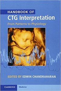
Handbook of CTG Interpretation: From Patterns to Physiology 1st Edition

[amazon template=image&asin=1107485509]

This book promotes an evidence-based paradigm of fetal heart rate monitoring during labour, with a move away from the traditional ‘pattern-based’ interpretation to physiology-based interpretation. Chapters are presented in a systematic and accessible format, covering topics such as non-hypoxic causes of fetal brain injury, pre-existing hypoxia, types of intrapartum hypoxia, erroneous monitoring of maternal heart rate, fetal scalp blood sampling and fetal ECG (STAN), intermittent auscultation and medico-legal issues. Sections on ‘pearls’ and ‘pitfalls’ highlight good practice and common errors to promote best practice. End-of-chapter exercises allow the reader to engage with the theory and test their knowledge of key areas. This is a highly practical manual aimed at obstetricians and midwives, particularly those undergoing CTG training. The book will also be very useful to midwifery and medical students and to all those involved in multiprofessional intrapartum care.

DOWNLOAD THIS BOOK FREE HERE

https://upsto.re/FpMVo7j

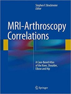
MRI-Arthroscopy Correlations: A Case-Based Atlas of the Knee, Shoulder, Elbow and Hip 1st ed

[amazon template=image&asin=1493926446]

Integrating MRI findings associated with the spectrum of problems seen in the most commonly treated joints in sports medicine with the diagnostic findings seen during arthroscopy of the same joint in the same patient, this unique text correlates this pathology and applies these findings to the clinic, the radiology reading room and the operating suite. Representing a microcosm of daily patient care, this type of interactive correlation is an exceedingly effective tool for education and continued learning, an impetus for interdisciplinary research collaboration and a critical part of an approach to optimum patient care. Furthermore, this case-based correlation between MRI imaging and arthroscopic findings and treatment is a well-received and effective method for teaching and discussion at meetings and instructional courses.

MRI-Arthroscopy Correlations is organized into four sections highlighting the four major joints in which MRI and arthroscopy are most commonly used in sports medicine: knee, shoulder, elbow and hip. Chapters are formatted to present an overview of the specific disease entity first, followed by selected cases chosen by the chapter authors that best illustrate common or noteworthy disease entities or pathology with an emphasis on the parallel MRI imaging and arthroscopic findings. Each of the section editors, as well as the volume editor, are nationally recognized experts, teachers and pioneers in their respective areas of sports medicine and have covered the gamut of topics in each of their sections. Taken together, this will be an invaluable resource for sports medicine specialists, orthopedic surgeons and musculoskeletal radiologists alike, promoting increasingly accurate diagnoses of pathology and advanced treatment options to aid in the optimization of patient care and recovery.

DOWNLOAD THIS BOOK FREE HERE

https://upsto.re/Fpb5igs

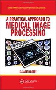
A Practical Approach to Medical Image Processing (Series in Medical Physics and Biomedical Engineering)

[amazon template=image&asin=1584888245]

The ability to manipulate and analyze pictorial information to improve medical diagnosis, monitoring, and therapy via imaging is a valuable tool that every professional working in radiography, medical imaging, and medical physics should utilize. However, previous texts on the subject have only approached the subject from a programming or computer science viewpoint at a mathematically inaccessible level. Unlike these previous publications, A Practical Approach to Medical Imaging Processing provides hands-on instruction, using the freely available software program ImageJ, on all of the skills needed to perform filtering and image enhancement techniques used in structured image discrimination.

In this unique text, the author focuses exclusively on image processing and treats medical images in a generic way to highlight the features that all digital images have in common. The book first introduces the main topics in image processing and as it progresses, you will discover relevant points of good practice. The author validates each technique with a corresponding case study, which originates from a published journal article. The case studies demonstrate how the concepts of image processing are applied to real-life situations, such as how to uncover information suffering from distortion and pixel-size limitations. The accompanying CD-ROM contains the Windows version of the ImageJ software, digital images, and documents to be used during the practical activities included in each chapter.

With its highly functional workbook approach, A Practical Approach to Medical Image Processing allows you to build your skills in image manipulation and to enjoy the benefits of this valuable field without having to code or develop your own program.

DOWNLOAD THIS BOOK FREE HERE

https://upsto.re/FpN5kay

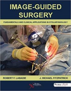
Image-Guided Surgery: Fundamentals and Clinical Applications in Otolaryngology 1st Edition

[amazon template=image&asin=1597567191]

Image-guided surgery (IGS) is the term used for surgical procedures in which the surgeon employs tracked surgical instruments with preoperative or intraoperative images in order to navigate during the procedure. It is part of the wider field of computer-assisted surgery. In the field of otolaryngology, IGS is primarily used by rhinologists during sinus surgery and by skull-base surgeons often in conjunction with neurosurgeons. While widely utilized, surgeons’ understanding of the technology is often inadequate to appreciate its strengths and weaknesses.
Image-Guided Surgery: Fundamentals and Clinical Applications in Otolaryngology was written to overcome this knowledge gap to educate practicing surgeons as well as provide an introduction to the field for engineers. Coauthored by a practicing otolaryngologist with a doctorate in bioengineering and a computer scientist with a doctorate in physics whose career has focused on error analysis in IGS, the book includes chapters on history, CT and MRI, tracking, registration, error analysis, evidence-based practices, FDA-cleared IGS systems, and future applications, as well as more than 90 full-color figures.
The book also comes with a complimentary access to a PluralPlus companion website with videos and animations.
Contents
Introduction
Acknowledgments
Chapter 1. Brief History of Image-Guided Surgery
Chapter 2. CT and MRI
Chapter 3. Tracking Systems
Chapter 4. Registration
Chapter 5. Error Analysis
Chapter 6. Best Practices for Use of IGS
Chapter 7. Surgical Systems
Chapter 8. What Does the Future Hold?
Index

NOTE:This book comes with supplementary content on a PluralPlus companion website. If you purchase or rent a used copy of the printed book, the code to access the website printed inside the book may have been previously redeemed/used or be incorrect and you will not be able to use it. To guarantee access to the website, it is recommended you purchase a new copy of this book directly from Amazon or the publisher, Plural Publishing.

DOWNLOAD THIS BOOK FREE HERE

https://upsto.re/FpNNYHo

Imaging of Cerebrovascular Disease: A Practical Guide 1st Edition

[amazon template=image&asin=1626232482]

…a good starting point for those early in their neuroradiology training. For others, a study of the images and the accompanying legends is a good review of the commonly encountered aspects of CV disease. — American Journal of Neuroradiology

…an image-rich, information-rich compendium that serves well both a resident starting to cope with complex central nervous system vascular anatomy and pathology and the practicing radiologist in day-to-day clinical work….I enthusiastically recommend this textbook to all residents, neuroradiology fellows, and general diagnostic radiologists. — Investigative Radiology

Imaging plays an integral role in the diagnosis and intervention of debilitating, often fatal vascular diseases of the brain, such as ischemic and hemorrhagic stroke, aneurysms, and arteriovenous malformations (AVMs). Written by a world renowned neuroradiologist and pioneer in the early adoption of magnetic resonance (MR) technology, Imaging of Cerebrovascular Disease is a concise yet remarkably thorough textbook that advances the reader’s expertise on this subject.

The text draws on the author’s vast personal experience, case studies, and traditional educational sources, offering didactic dialogue with accompanying images. A practical clinical resource organized into six chapters, this book offers unparalleled breadth in delineating the diagnostic and treatment usages of modern imaging techniques. Chapter one sets a foundation with extensive coverage of modalities and their applications, including MR, computed tomography (CT) and digital subtraction angiography (DSA). Subsequent chapters cover utilization of imaging techniques specific to underlying pathologies.

Key Features:

  • In-depth discussion of medical and neuroradiologic/neurosurgical interventions, focusing on the use of imaging prior to, during, and following treatment
  • Comprehensive text enhanced with more than 700 high-quality images
  • Detailed evaluation of normal brain anatomy, as well as gyral anatomy in brain ischemia, an important subtopic
  • Advantages, disadvantages, mortality, and morbidity of surgery (clipping) versus endovascular techniques (coiling and flow diversion) for aneurysms

Presented in a style that facilitates cover-to-cover reading, this is an essential tool for residents and fellows, and provides a robust study guide prior to sitting for relevant certification exams. It is also a quick, invaluable reference for radiologists, neurosurgeons, and neurologists in the midst of a busy clinical day.

DOWNLOAD THIS BOOK FREE HERE
https://upsto.re/FpNuhrc

1 31 32 33 34 35 62