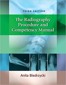
The Radiography Procedure and Competency Manual 3rd Edition
[amazon template=iframe image2&asin=0803660952]

Be prepared to meet the ARRT competency requirements! These procedure checklists make it easy. To qualify for your certification exam, you must demonstrate your competency in all 36 mandatory procedures and in at least 15 of the 30 elective procedures—and your instructors must verify your proficiencies.


First, you can use the checklists to review the procedures in preparation for the exam and to develop decision-making skills that will produce the highest quality radiographs while considering the needs and limitations of the patient. Then, your instructors can use them to record their evaluation of your competency for each procedure. And, finally, program directors can use them to verify to the ARRT that the you have demonstrated the required competencies and proficiencies.

DOWNLOAD THIS BOOK FREE HERE

https://upsto.re/FnCwpbH

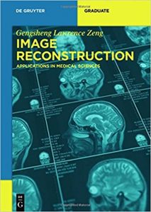
Image Reconstruction: Applications in Medical Sciences (De Gruyter Textbook) 1st Edition

[amazon template=iframe image2&asin=3110500485]

This book introduces the classical and modern image reconstruction technologies. It covers topics in two-dimensional (2D) parallel-beam and fan-beam imaging, three-dimensional (3D) parallel ray, parallel plane, and cone-beam imaging. Both analytical and iterative methods are presented. The applications in X-ray CT, SPECT (single photon emission computed tomography), PET (positron emission tomography), and MRI (magnetic resonance imaging) are discussed. Contemporary research results in exact region-of-interest (ROI) reconstruction with truncated projections, Katsevich’s cone-beam filtered backprojection algorithm, and reconstruction with highly under-sampled data are included.

The last chapter of the book is devoted to the techniques of using a fast analytical algorithm to reconstruct an image that is equivalent to an iterative reconstruction. These techniques are the author’s most recent research results.

This book is intended for students, engineers, and researchers who are interested in medical image reconstruction. Written in a non-mathematical way, this book provides an easy access to modern mathematical methods in medical imaging.

DOWNLOAD THIS BOOK FREE HERE

https://upsto.re/FNsTSRY

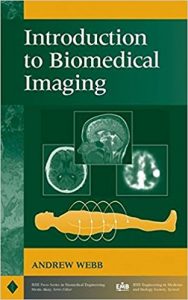
Introduction to Biomedical Imaging 1st Edition

[amazon template=iframe image2&asin=0471237663]

An integrated, comprehensive survey of biomedical imaging modalities

An important component of the recent expansion in bioengineering is the area of biomedical imaging. This book provides in-depth coverage of the field of biomedical imaging, with particular attention to an engineering viewpoint.

Suitable as both a professional reference and as a text for a one-semester course for biomedical engineers or medical technology students, Introduction to Biomedical Imaging covers the fundamentals and applications of four primary medical imaging techniques: magnetic resonance imaging, ultrasound, nuclear medicine, and X-ray/computed tomography.

Taking an accessible approach that includes any necessary mathematics and transform methods, this book provides rigorous discussions of:

  • The physical principles, instrumental design, data acquisition strategies, image reconstruction techniques, and clinical applications of each modality
  • Recent developments such as multi-slice spiral computed tomography, harmonic and sub-harmonic ultrasonic imaging, multi-slice PET scanning, and functional magnetic resonance imaging
  • General image characteristics such as spatial resolution and signal-to-noise, common to all of the imaging modalities

DOWNLOAD THIS BOOK FREE HERE

https://upsto.re/FHz6FHA

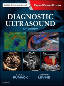
Diagnostic Ultrasound, 2-Volume Set, 5e 5th Edition

[amazon template=iframe image2&asin=0323401716]

Now fully updated with more than 2,000 new images, 200 new videos, and new content throughout, Diagnostic Ultrasound, 5th Edition, by Drs. Carol M. Rumack and Deborah Levine, remains the most comprehensive and authoritative ultrasound resource available. Spanning a wide range of medical specialties and practice settings, it provides complete, detailed information on the latest techniques for ultrasound imaging of the whole body; image-guided procedures; fetal, obstetric, and pediatric imaging; and much more. Up-to-date guidance from experts in the field keep you abreast of expanding applications of this versatile imaging modality and help you understand the “how” and “why” of ultrasound use and interpretation.

  • Covers all aspects of diagnostic ultrasound with sections for Physics; Abdominal, Pelvic, Small Parts, Vascular, Obstetric, and Pediatric Sonography.
  • Uses a straightforward writing style and extensive image panels with correlative findings.
    • Features 5,000 images – more than 2,000 brand-new – including new 2D and 3D imaging as well as the use of contrast agents and elastography.
    • Includes a new virtual chapter on artifacts with individually labelled images from throughout the book, displaying artifacts with descriptive legends by category and how they can be used in diagnosis or corrected for better quality imaging.
    • Features more images and new uses for contrast agents in the liver, breast, and in pediatric applications. Includes current information on imaging more diagnostic dilemmas, such as Zika virus in the fetus and newborn.
    • Includes 400 video clips showing real-time scanning of anatomy and pathology.


  • Expert Consult™ eBook version included with purchase. This enhanced eBook experience allows you to search all of the text, figures, Q&As, and references from the book on a variety of devices.

DOWNLOAD THIS BOOK FREE HERE

https://upsto.re/FkKxb2c

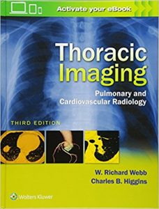
Thoracic Imaging: Pulmonary and Cardiovascular Radiology Third Edition

[amazon template=iframe image2&asin=1496321049]

Written by two of the world’s most respected specialists in the field, Thoracic Imaging: Pulmonary and Cardiovascular Radiology, Third Edition brings you completely up to date with all you need to know for optimal imaging of the heart and lungs. This comprehensive title provides practical, authoritative guidance for the radiologic assessment and diagnosis of both congenital and acquired cardiovascular and pulmonary diseases. A must-have reference for more than ten years, Thoracic Imaging is your one-stop source for current, accurate coverage of pulmonary infections, diffuse lung diseases, mediastinal masses, coronary artery CT, myocardial disease, pericardial disease, CT of ischemic heart disease,  and much more.

Key Features:

  • Today’s most comprehensive text-reference in the field, written by pulmonary imaging expert W. Richard Webb, MD and cardiac imaging expert Charles B. Higgins, MD.
  • More than 2700 high-quality images, many in full color, help you visualize key aspects of chest imaging.
  • A new chapter on MR and CT in in Atrial and Ventricular Arrhythmias brings you up to date on the latest imaging techniques.
  • Updated images and content throughout, including expanded coverage of PET/CT.

Now with the print edition, enjoy the bundled interactive eBook edition, which can be downloaded to your tablet and smartphone or accessed online and includes features like:

  • Complete content with enhanced navigation
  • Powerful search tools and smart navigation cross-links that pull results from content in the book, your notes, and even the web
  • Cross-linked pages, references, and more for easy navigation
  • Highlighting tool for easier reference of key content throughout the text
  • Ability to take and share notes with friends and colleagues
  • Quick reference tabbing to save your favorite content for future use

DOWNLOAD THIS BOOK FREE HERE

https://upsto.re/FyFo7WL

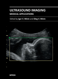
Ultrasound Imaging – Medical Applications

This book provides an overview of ultrafast ultrasound imaging, 3D high-quality ultrasonic imaging, correction of phase aberrations in medical ultrasound images, etc. Several interesting medical and clinical applications areas are also discussed in the book, like the use of three dimensional ultrasound imaging in evaluation of Asherman’s syndrome, the role of 3D ultrasound in assessment of endometrial receptivity and follicular vascularity to predict the quality oocyte, ultrasound imaging in vascular diseases and the fetal palate, clinical application of ultrasound molecular imaging, Doppler abdominal ultrasound in small animals and so on.

DOWNLOAD THIS BOOK FREE HERE

https://upsto.re/FyDsmYQ

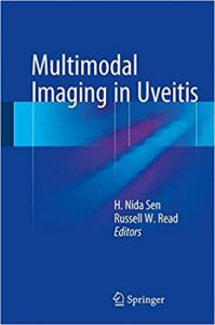
Multimodal Imaging in Uveitis 1st ed. 2018 Edition

[amazon template=iframe image2&asin=331923689X]

About the Author

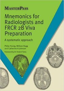
Mnemonics for Radiologists and FRCR 2B Viva Preparation: A Systematic Approach (Masterpass) 1st Edition

[amazon template=image&asin=1908911956]

Revising for the final FRCR examination is a challenging task. The candidate has to balance their revision time between image interpretation practice, learning facts and refining presentation skills. Whilst it is better to gain an understanding of the underlying mechanisms that result in a radiological abnormality, it is an unavoidable truth that some things just have to be rote learned. This revision guide demonstrates a proven mechanism for organising thoughts, directing revision towards the more important diagnoses and providing clarity when confronted with the mountain of information expected to retained and recalled during the FRCR viva exam. Mnemonics typically uses word-based cues to enhance memory, reduce cognitive load and aid learning. This guide relies on acronyms and acrostics – proven to be highly effective for rote learning and focused revision. FRCR examination candidates will appreciate the unified structure throughout, with extensive use of sample images followed by sets of differential diagnoses and mnemonic listings. For each stem of the mnemonic, the main radiological findings and useful distinguishing features are listed and a model answer is given. A short discussion of the main diagnoses follows, increasing comprehension and aiding examination confidence.

DOWNLOAD THIS BOOK FREE HERE

https://upsto.re/FGuvAaK

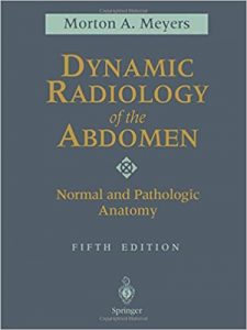
Dynamic Radiology of the Abdomen: Normal and Pathologic Anatomy 5th Edition

[amazon template=image&asin=0387988459]

Meyers’ Dynamic Radiology of the Abdomen, extensively revised and updated, is the classic text covering radiology of the abdomen as it relates to the progression of disease within an organ and from one organ to another. The book provides a systematic application of anatomic and dynamic principles to the practical understanding and diagnosis of intraabdominal disease. The full range of imaging modalities is addressed, from plain films and conventional contrast studies to CT, US, MRI and endoscopic ultrasonography. Highly selected, ample images including CT and MRI support the thoroughly descriptive text. Expanded references, citing both the classic and recent contributions, and a detailed cross-referenced index are presented. For radiologists, general surgeons, gastroenterologists, and others seeking insight into the clinical practice of radiology, this text continues to be the gold standard in the field.

DOWNLOAD THIS BOOK FREE HERE

https://upsto.re/F75EcWV

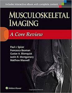
Musculoskeletal Imaging: A Core Review First Edition

[amazon template=image&asin=1451192673]

Preparing for the new Core ExamMainenance of Certification Exam, or Certifying Exam?  Excel on your boards with this review book written for the new exam format. Featuring high-quality radiographs and CT, MRI, and ultrasound images, this practical, easy-to-use resource includes key musculoskeletal radiology questions formatted to model the new core exam.

Key Features:

  • 300 questions are formatted to model the new Core Exam
  • Questions are divided into topic sections for quick and easy review
  • Each question includes an explanation of why the correct answer is correct and the other options incorrect.

Now with the print edition, enjoy the bundled interactive eBook edition, offering tablet, smartphone, or online access to:

  • Complete content with enhanced navigation
  • A powerful search that pulls results from content in the book, your notes, and even the web
  • Cross-linked pages, references, and more for easy navigation
  • Highlighting tool for easier reference of key content throughout the text
  • Ability to take and share notes with friends and colleagues
  • Quick reference tabbing to save your favorite content for future use
  • 300 interactive exam questions

DOWNLOAD THIS BOOK FREE HERE

https://upsto.re/F75Ff25

1 29 30 31 32 33 60