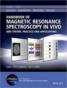
Handbook of Magnetic Resonance Spectroscopy In Vivo: MRS Theory, Practice and Applications (eMagRes Books) 1st Edition

This handbook covers the entire field of magnetic resonance spectroscopy (MRS), a unique method that allows the non-invasive identification, quantification and spatial mapping of metabolites in living organisms–including animal models and patients.

Comprised of three parts:

  • Methodology covers basic MRS theory, methodology for acquiring, quantifying spectra, and spatially localizing spectra, and equipment essentials, as well as vital ancillary issues such as motion suppression and physiological monitoring.
  • Applications focuses on MRS applications, both in animal models of disease and in human studies of normal physiology and disease, including cancer, neurological disease, cardiac and muscle metabolism, and obesity.
  • Reference includes useful appendices and look up tables of relative MRS signal-to-noise ratios, typical tissue concentrations, structures of common metabolites, and useful formulae.

About eMagRes Handbooks

eMagRes (formerly the Encyclopedia of Magnetic Resonance) publishes a wide range of online articles on all aspects of magnetic resonance in physics, chemistry, biology and medicine. The existence of this large number of articles, written by experts in various fields, is enabling the publication of a series of eMagRes Handbooks on specific areas of NMR and MRI. The chapters of each of these handbooks will comprise a carefully chosen selection of eMagRes articles. In consultation with the eMagRes Editorial Board, the eMagRes Handbooks are coherently planned in advance by specially-selected Editors, and new articles are written to give appropriate complete coverage. The handbooks are intended to be of value and interest to research students, postdoctoral fellows and other researchers learning about the scientific area in question and undertaking relevant experiments, whether in academia or industry.

Have the content of this handbook and the complete content of eMagRes at your fingertips!
Visit the eMagRes Homepage

DOWNLOAD THIS BOOK FREE HERE

https://upsto.re/3vUk43t

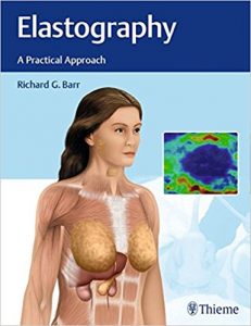
Elastography: A Practical Approach 1st Edition

…explained in simple and reader-friendly terms…accurate and concise…images are appropriately labeled, helpful, and easily interpretable.— AJNR

I would highly recommend this book… — Acta Radiologica

This is an excellent book on the relatively newer diagnostic technique of elastography. This work is authoritative as well as comprehensive… — BIZINDIA

This practical guide is a compilation of firsthand expertise from leading authorities around the world on the use of ultrasound elastography. The stiffness or softness of the imaged tissue derived from elastography provides accurate radiologic diagnosis for disease processes including cancer, inflammation, and fibrosis. It is an efficacious and accurate diagnostic imaging modality that helps avoid invasive biopsies.

The first two chapters cover basic fundamental principles of elastography, with subsequent chapters exploring pathology-specific utilization. The authors cover the extensively validated and implemented use of elastography for diffuse liver disease, and diseases of the breast andthyroid gland. They also discuss the potential benefits and limitations for the prostate, spleen, pancreas, kidneys, musculoskeletal system, salivary glands, lymph nodes, and testes. The book concludes with a chapter on potential future applications of this ever-evolving technology.

Key Highlights

  • Discussion of key differences between strain elastography and shear wave elastography by individual organ systems
  • Clinical pearls on how to accurately perform elastography and tips for avoiding false-positive or false-negative results
  • Case studies elucidate the targeted use of elastographic findings by specific pathology
  • Illustrations in the breast and liver chapters demonstrate precise transducer techniques
  • MRI elastography as an emerging and safe assessment tool, primarily for the diagnosis of liver disease, with emergent potential for additional organs

This book provides key knowledge on visualizing quantifiable differences in tissue elasticity and applying this data to improved treatment strategies for diverse pathologies. It is essential reading for radiologists, sonographers, and imaging technicians.

DOWNLOAD THIS BOOK FREE HERE

https://upsto.re/3vG7WwP

Imaging of Bones and Joints: A Concise, Multimodality Approach 1st Edition

…easy to read, interesting, and informative… — Radiologic Technology

…a useful layout… — Doody’s Reviews (starred review)

One of the outstanding values of this book is the large number of images used….These greatly help in viewing and analyzing the conditions and diseases within the bones, in the bone marrow, in the joints, in regular and soft tissue, and in other places. — BIZ INDIA

This book is unique. It will guide you through the essentials of musculoskeletal imaging using a multimodality approach. Organized by categories of musculoskeletal disorders, it uses a “findings within-the-image” method to help you identify the typical imaging features of each condition.

As a comprehensive reference compiled by well-known specialists in the field, it is useful for both practicing radiologists and those in training.

Focus on the essentials

Provides a solid foundation of what the radiologist needs to know when interpreting musculoskeletal imaging studies, including the indications for when to use various imaging modalities.

“Findings within the image”

An excellent presentation method for learning to interpret bone and joint images.

Find it quickly

In addition to a detailed text and high-quality images, important points are summarized in boxes, tables, and illustrative figures for quick reference.

Extra features are included on the Thieme MediaCenter

An additional 338 images along with supplemental text and references are provided online on the Thieme MediaCenter.

Special Features

  • All chapters are written by leading international authors.
  • A comprehensive, multimodality approach is used.
  • Over 2100 brilliant, state-of-the-art images are provided, including a multitude of MR images.

DOWNLOAD THIS BOOK FREE HERE

https://upsto.re/3s3FmHc

Advanced Thyroid and Parathyroid Ultrasound 1st ed

This text provides a comprehensive review of ultrasound in thyroid and parathyroid diseases. These topics are presented from a vantage point of complex decision-making encountered in real clinical scenarios. The sections are organized according to a logical structure covering benign and malignant thyroid conditions, parathyroid disease, and ultrasound technology, ultrasound-guided interventions, and innovations. The style of the chapters provide practical, actionable information that is richly illustrated with figures and links to video cine-clips. The chapter topics aim to show how different specialists uniquely apply ultrasound in given clinical scenarios. The text illustrates the optimal incorporation of current practice guidelines, as this remains varied and inconsistent among clinicians. The content is written by invited experts who perform ultrasound in their daily clinical practices and participate in teaching ultrasound nationally and internationally. It conveys the most up-to-date scientific and clinical information in an interactive and visual format.

Advanced Thyroid and Parathyroid Ultrasound fills a gap in currently available resources by serving as a single resource unifying information relevant to multiple specialists interested in advanced thyroid and parathyroid ultrasound. It provides a practical, concise yet comprehensive summary of the current status of the field that will help guide patient management.

DOWNLOAD THIS BOOK FREE HERE

https://upsto.re/3sDgAF4

Quantification of Biophysical Parameters in Medical Imaging 1st ed. 2018 Edition

About the Author

Musculoskeletal Ultrasound in Rheumatology Review 1st ed

This book provides a comprehensive clinical review of diagnostic and interventional applications of musculoskeletal ultrasound at the point-of-care. As more rheumatologists and other musculoskeletal providers in training and in practice learn the skill of musculoskeletal ultrasound, an increasing number of them will seek study materials for exam preparation and practical knowledge that apply to their clinical practice. Each chapter covers a standardized protocol of joint images with probe placement, and includes numerous examples of common ultrasound pathologies, clearly addressing what kind of pathology to look for with specific ultrasound image views. Review topics are emphasized, and study tools such as key-concept overviews, lists of important studies in the field, and extensive questions for self-assessment are included throughout. Because ultrasound training is moving toward becoming a mandatory part of rheumatology fellowship and has become mandatory in physical medicine and rehabilitation residencies, this book is a valuable educational resource for rheumatologists, physiatrists, and musculoskeletal providers seeking a practical review guide for preparation of certification exams and use in clinical practice.

DOWNLOAD THIS BOOK FREE HERE

https://upsto.re/FYqRSuV

Neuroimaging: Anatomy Meets Function 1st ed. 2018 Edition

About the Author

PET/MRI in Oncology: Current Clinical Applications 1st ed. 2018 Edition

About the Author

Combined Scintigraphic and Radiographic Diagnosis of Bone and Joint Diseases

The third edition of this book has been comprehensively rewritten and rearranged. In addition to the bone and joint diseases described in the two earlier editions, it now encompasses hitherto unpublished novel applications of pinhole scanning to the diagnosis of a broader spectrum of skeletal disorders. The book has been considerably expanded to discuss five new themes and is complemented by the addition of some 90 recently acquired cases.

DOWNLOAD THIS BOOK FREE HERE

https://upsto.re/FYgpXXi

Clinical Emergency Radiology 2nd Edition

[amazon template=iframe image2&asin=9781107065796]

This book is a highly visual guide to the radiographic and advanced imaging modalities – such as computed tomography and ultrasonography – that are frequently used by physicians during the treatment of emergency patients. Covering practices ranging from ultrasound at the point of care to the interpretation of CT scan results, this book contains over 2,200 images, each with detailed captions and line-art that highlight key findings. Within each section, particular attention is devoted to practical tricks of the trade and tips for avoiding common pitfalls. Overall, this book is a useful source for experienced clinicians, residents, mid-level providers, or medical students who want to maximize the diagnostic accuracy of each modality without losing valuable time.

DOWNLOAD THIS BOOK FREE HERE

https://upstore.net/F4TuLAX

1 28 29 30 31 32 61