Lever’s Histopathology of the Skin Eleventh Edition
[amazon template=image&asin=1451190379]

Rely on Lever’s for more accurate, more efficient diagnoses!  Continuously in publication for more than 65 years, Lever’s Histopathology of the Skin remains your authoritative source for comprehensive coverage of those skin diseases in which histopathology plays an important role in diagnosis.  This edition maintains the proven, clinicopathologic classification of cutaneous disease while incorporating a “primer” on pattern-algorithm diagnosis. More than 1800 full-color illustrations, including photomicrographs and clinical photographs, help you visualize and make the most of the clinical diagnostic process.

Key Features

  • New Clinical Summaries for most disease entities provide a concise clinical review before presenting histologic features.
  • New Principles of Management section summarizes today’s complex treatment modalities in one convenient place.
  • Ultrastructural, immunohistochemical, and molecular techniques are discussed where they have value in identifying particular diseases.
  • Updated chapter dedicated to algorithmic classification of skin diseases according to histologic pattern features helps you develop a differential diagnosis for unknown cases.
Now with the print edition, enjoy the bundled interactive eBook edition, offering tablet, smartphone, or online access to:
  • Complete content with enhanced navigation
  • Powerful search tools and smart navigation cross-links that pull results from content in the book, your notes, and even the web
  • Cross-linked pages, references, and more for easy navigation
  • Highlighting tool for easier reference of key content throughout the text
  • Ability to take and share notes with friends and colleagues
  • Quick reference tabbing to save your favorite content for future use

DOWNLOAD THIS BOOK FREE HERE

http://upsto.re/FjvPGiE

Compendium of Histology: A Theoretical and Practical Guide 1st ed. 2017 Edition

[amazon template=iframe image2&asin=3319418718]

About the Author

Fundamentals of Oral Histology and Physiology 1st Edition

[amazon template=iframe image2&asin=1118342917]

Fundamentals of Oral Histology and Physiology is a landmark new text streamlining the essentials of histology and physiology into one clinically accessible textbook. Written for predoctoral dental students, the book brings together structure, function, and clinical correlations for maximum retention and ease of use.

Assuming a background in basic biologic sciences, this text focuses on the histology and physiology that students need to know to practice dentistry and to understand and evaluate the current literature, without repeating basic information learned in other courses. Fundamentals of Oral Histology and Physiology concentrates on Oral Structures and Features, including Development, Teeth, Tooth and Jaw Support, Mucosal Structure and Function, and Effectors.

DOWNLOAD THIS BOOK FREE HERE

http://upsto.re/FCuVBTX

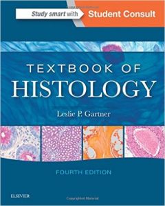
Textbook of Histology, 4e 4th Edition

[amazon template=iframe image2&asin=0323355633]

Thoroughly updated to reflect all of the latest concepts and advances in the field, this concise, extensively illustrated text presents the basic science and clinical application of cellular and molecular biology. Functioning as a combined text and atlas, it is essentially two books for the price of one providing a comprehensive, visually engaging histology education from the ground up. Illustrations, tables, PowerPoint™ presentations, chapter summaries and multiple choice questions facilitate comprehension of concepts, and clinical correlations underscore the practical relevance of the material.

    • Superb illustrations―including photomicrographs, electron micrographs, schematic diagrams and drawings―provide a visual grasp and easier retention of difficult concepts.
    • Online PowerPoint™ slides, prepared by the author, offer a convenient way to review the most essential information from the text.
    • Clinical correlations throughout the text demonstrate clinical applications and reinforce the idea that histology is pertinent not only to pathology and physiology, but in fact comprises one of the essential bases of clinical practice.
    • New! “Pathological Considerations” section at the end of each chapter.
    • Helpful tables provide an at-a-glance summary statement of key points.
    • Bolded key terms, bulleted lists and chapter summaries emphasize the “need to know” information in every chapter.
    • A combination of USMLE-style questions and image based questions in each chapter.
  • Student Consult eBook version included with purchase. This enhanced eBook experience allows you to search all of the text, figures, and references from the book on a variety of devices.

DOWNLOAD THIS BOOK FREE HERE

http://upsto.re/Ao774hh

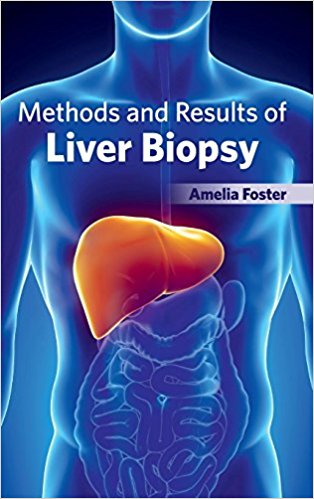
Methods and Results of Liver Biopsy

[amazon template=iframe image2&asin=1632422778]

This book primarily focuses on the methods as well as results of the process of liver biopsy. Liver biopsy is the process of obtaining a small piece of liver tissue and analyzing it in the laboratory. Liver biopsy may be used to examine a mass seen on images, diagnose lesser known liver diseases or abnormal liver function tests and to assess the complexity of several liver diseases including hepatitis, fibrosis, and steatosis. Liver biopsy is an invasive procedure and its complications are rare but potentially fatal. Therefore, it is essential to perform liver biopsy safely from clinical aspect and to take specimen steadily from a pathological one. This book deals with various prospects of liver biopsy including role, indication, technique and assessment. It also discusses novel non-invasive alternatives encompassing elastography and computer analysis of liver fibrosis.

DOWNLOAD THIS BOOK FREE HERE

http://upsto.re/A8qYVrT


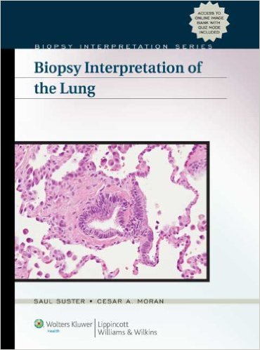
Biopsy Interpretation of the Lung (Biopsy Interpretation Series) 1 Har/Psc Edition

[amazon template=iframe image2&asin=0781784670]

Biopsy Interpretation of the Lung aids pathologists in accurately evaluating disease processes. It includes detailed chapters on interstitial lung disease, lung cancer, and neuroendocrine tumors. Organized by traditional disease groups, Biopsy Interpretation of the Lung provides a solid approach to formulating surgical and medical decisions.

This text is geared toward surgical pathologists, pathology residents, pathology fellows, pulmonologists, pulmonology fellows, and thoracic surgeons, and includes the latest information on the best use of immunohistochemical markers, special stains, and molecular assays to aid in more accurate and cost-efficient diagnoses.

DOWNLOAD THIS BOOK FREE HERE

http://upsto.re/v4VYRzu

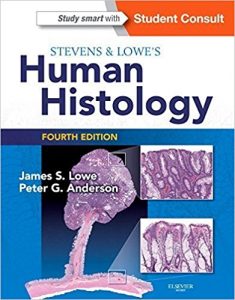
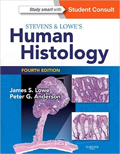
Stevens & Lowe’s Human Histology, 4e (HUMAN HISTOLOGY (STEVENS)) 4th Edition

[amazon template=iframe image2&asin=9780723435020]

“A well produced book that our trainees and students like.” By PathLab.org, March 2015


  • Learn from an easy-to-read writing style and well-designed, full-color layout to present of all histology’s need-to-know content.
  • Conveniently access important information through a design that sets off the key laboratory, clinical, and high-level scientific material in boxes.


  • Take advantage of an increased amount of clinical content and photos.
  • Master the basics of the field with an enhanced focus on cell biology.
  • Quickly review important information with reviews available at the end of each chapter and online, Key Facts boxes throughout the chapters, MCQs in the text, plus 180 additional review questions online.
  • Easily visualize complex procedures and concepts with nearly 900 illustrations, photos, and graphics.
  • Student Consult eBook version included with purchase. This enhanced eBook experience allows you to search all of the text, figures, references, and videos from the book on a variety of devices.

DOWNLOAD THIS BOOK FREE HERE

http://upsto.re/v4RMTWs

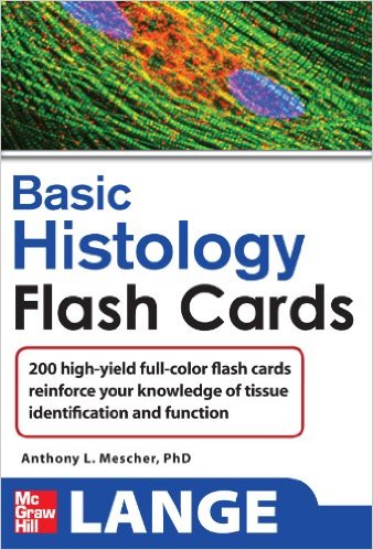
Lange Basic Histology Flash Cards (LANGE FlashCards) 1st Edition

51eeqi7ytvl-_sx336_bo1204203200_

[amazon template=iframe image2&asin=0071637982]

Flashcards derived from one of the best histology texts ever published

Based on Junquira’s Basic Histology

5 STAR DOODY’S REVIEW!
“This elegant set of glossy, full-color histology flashcards is a joy to use….This is a wonderful resource for students of medicine and the life sciences. The author brings together high-resolution color images and essential information from the book in a practical format that will help students learn. Students become active learners. Learning histology becomes fun. These flash cards summarize the body of information that students need to know to understand the foundations of pathology and succeed on course and licensing examinations. This is one of the best flashcard products currently available for medical education.”–Doody’s Review Service

Junquira’s High-Yield Histology Flash Cards are a quick, fun, and fast way for you to review the clinical relevance between histological structure and function. Derived from Junquira’s Basic Histology, the field’s leading text and atlas, each of the cards features an image and a question on one side and the answer and commentary on the flip side.

Features

  • 200 cards per pack
  • One side of the card includes an image with arrows asking you to identify specific areas; the flip side provides the answer and includes the medical relevance to the image on the front

DOWNLOAD THIS BOOK FREE HERE

http://upsto.re/EStWDVS

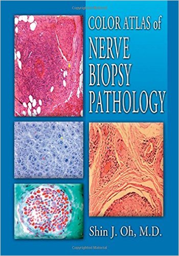
Color Atlas of Nerve Biopsy Pathology 1st Edition

0849316766

[amazon template=iframe image2&asin=0849316766]

As a professional working in the frontlines of tissue diagnosis and in everyday practice, you need a reference that gives you practical information in an easy-to-use format. Containing over 300 photographs, micrographs, and line drawings, including over 60 color illustrations, Color Atlas of Nerve Biopsy Pathology supplies a clear picture of commonly used techniques for nerve biopsies. You get new diagnostic techniques and clinicopathological correlations all in a framework designed for quick reference and easy learning.

Providing all the practical information you need from A to Z, Color Atlas of Nerve Biopsy Pathology describes how to:

  • perform a nerve biopsy
  • handle specimens
  • process the nerve by frozen sections, by paraffin sections, and by semi-thin sections

    The book delineates the general pathological features of peripheral nerve disorders and the diagnostic capability and limitations of the nerve biopsy. It details the typical pathological features of each disease by presenting color photographs and example cases that provide a brief clinical history and pertinent electrophysiological and other laboratory data. The exhaustively illustrated color format provides you with immediate information in reaching a definitive diagnosis.

    Unlike other books on this subject, this one contains frozen section materials, case histories, teased nerve illustrations, and newly described neuropathies. The author brings twenty-five years of experience in the lab and teaching to the development of this atlas. In the lab or in the classroom, on the front lines of tissue diagnosis, Color Atlas of Nerve Biopsy Pathology provides you with the information you need clearly, concisely, and conveniently.

DOWNLOAD THIS BOOK FREE HERE

http://upsto.re/sFsqm4j


1 2 3 4