Atlas of Hybrid Imaging Sectional Anatomy for PET/CT, PET/MRI and SPECT/CT Vol. 1: Brain and Neck

Atlas of Hybrid Imaging Sectional Anatomy for PET/CT, PET/MRI and SPECT/CT Vol. 1: Brain and Neck

For the first time, in this atlas nuclear physicians and radiologists cover the entire hybrid nuclear medicine (PET/CT, SPECT/CT and PET/MRI), based on their own case studies.

The structure in three volumes represents an user friendly guide for interpreting PET and SPECT in relation to co-registered CT and/or MRI.

Three companion volumes with a practical structure in two-page unit offer to the reader a navigational tool, based on anatomical districts, with labeled and explained low-dose multiplanar CT or MRI views merged with PET fusion imaging on the right hand and contrast enhanced CT or MRI on the other side. This new format enables rapid identification of hybrid nuclear medicine findings which are now routine at leading medical centers.

Volume 1 is focused on brain and neck PET imaging, with emphasis on PET/MRI; Volume 2 concerns thorax, abdomen and pelvis, with particular attention on lung and liver segmental anatomy and evaluation of peritoneum. Special chapters on heart, lymph nodes and musculoskeletal system, are collected in the Volume 3.

Each chapter begins with three-dimensional CT and/or MRI views of the evaluated anatomical region, bringing forward sectional tables.

Clinical cases, tricks and pitfalls linked to several PET or SPECT radiopharmaceuticals help to introduce the reader to peculiar molecular pathways and to improve confidence in cross-sectional imaging, that is vital for the accurate diagnosis and treatment of diseases.

  • Compact, comprehensive, easy to read guide to sectional imaging and multiplanar evaluation of hybrid PET and SPECT
  • Includes 856 fully colored, labeled, high quality original images of axial, coronal and sagittal CT, contrast enhanced CT, PET/CT and/or PET/MRI
  • Displays 200 clinical cases, showcasing both common and unusual findings that nuclear physicians and radiologists could encounter in their clinical practice
  • Costant orthogonal references on 3D images allow orientation on axial, coronal and sagittal views
  • Peculiar aspects of 18 PET and SPECT radiopharmaceuticals, defined by distinctive color scales
  • Terms and descriptions mostly based on Terminologia Anatomica
  • Specific text boxes explain anatomical variants, radiological advices and physiological findings linked to tracer bio-distribution
  • Supports interpretation and report of hybrid nuclear medicine scans
  • For professionals and vocationals
DOWNLOAD THIS MEDICAL BOOK

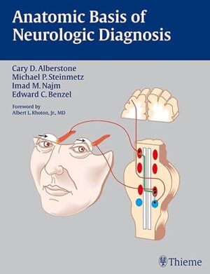
Anatomic Basis of Neurologic Diagnosis 1st Edition

Anatomic Basis of Neurologic Diagnosis 1st Edition

A visual guide to diagnosing neurologic disorders

2010 Benjamin Franklin Silver Award Winner!

Anatomic Basis of Neurologic Diagnosis is a lavishly illustrated book that places special emphasis on the paramount importance of signs and symptoms for the accurate diagnosis of neurologic disorders. It opens with a comprehensive review of neuroembryology, enabling readers to gain knowledge of normal nervous system development and related developmental disorders. The second section of the book comprises an easily accessible presentation of the anatomy of regional parts and to-the-point information on the cardinal manifestations of disease. Separate chapters in the third section of the book present the anatomy of different functional systems and provide practical approaches to diagnosing patients with system disorders. A final chapter covers the anatomy of the vascular system and cerebrospinal fluid.

Highlights:

  • Practical organization of chapters, according to regions and functional systems, reflects the clinician’s approach to patient care
  • Full-color illustrations provide an indispensable visual aid to learning and reviewing clinically relevant neurologic anatomy and pathways
  • Numerous tables summarize key points

Ideal for reading cover-to-cover, this book is essential for residents and students seeking to fully understand the complexity of clinical neuroanatomy. Seasoned clinicians will find the book a valuable refresher.

DOWNLOAD THIS MEDICAL BOOK

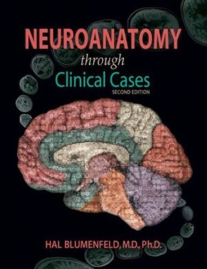
Neuroanatomy Through Clinical Cases, Second Edition 2nd Edition

Neuroanatomy Through Clinical Cases, Second Edition 2nd Edition

Neuroanatomy through Clinical Cases brings a pioneering interactive approach to the teaching of neuroanatomy, using over 100 actual clinical cases and high-quality radiologic images to bring the subject to life. The Second Edition is fully updated with the latest advances in the field, and includes several exciting new cases. This approach allows students to appreciate the clinical relevance of structural details as they are being learned, and to integrate knowledge of disparate functional systems, since a single lesion may affect several different neural structures and pathways. Most of the book comprises chapters that explain the major neuroanatomical systems. Each chapter first presents background material including an overview of relevant neuroanatomical structures and pathways, and a brief discussion of related clinical disorders. The second half of each chapter is devoted to clinical cases. The cases begin with a narrative of how the patient developed symptoms, and what deficits were found upon neurological examination. Boldface type highlights important symptoms and signs. A series of questions challenges the reader to deduce the neuroanatomical location of the patient s lesion, and the diagnosis. Discussion and answers follow, and an epilogue reveals the actual outcome. One of the book s most innovative features is the inclusion of CT and MRI scans that depict each patient s lesion. These radiographs help the reader develop skills in interpreting the same kinds of diagnostic images employed in clinical practice.

DOWNLOAD THIS MEDICAL BOOK

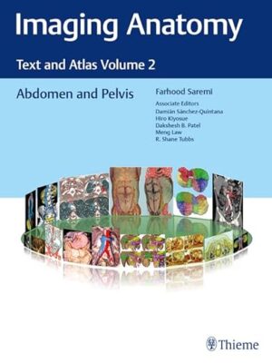
Imaging Anatomy: Text and Atlas Volume 2: Abdomen and Pelvis 1st Edition

Imaging Anatomy: Text and Atlas Volume 2: Abdomen and Pelvis 1st Edition

Unique anatomic atlas provides an indispensable virtual desk dissection experience

Normal imaging anatomy and variants, including diagnostic and surgical anatomy, are the cornerstones of radiologic knowledge. Imaging Anatomy: Text and Atlas Volume 2, Abdomen and Pelvis is the second in a series of four richly illustrated radiologic references edited by distinguished radiologist Farhood Saremi. The atlas is coedited by esteemed colleagues Damián Sánchez-Quintana, Hiro Kiyosue, Dakshesh B. Patel, Meng Law, and R. Shane Tubbs with contributions from an impressive cadre of international authors. Succinctly written text and superb images provide readers with a virtual, user-friendly dissection experience.

This exquisitely crafted atlas combines fundamental core anatomy principles with modern imaging and postprocessing methods to increase understanding of intricate anatomical features. Twenty-two concise chapters cover the abdominal wall, alimentary tract, liver, biliary system, pancreas, spleen, peritoneum, genitourinary system, pelvic floor, neurovasculature, and surface anatomy. Relevant anatomical components of the abdomen and pelvis are discussed, including musculature, arteries, veins, lymphatics, ducts, and innervation.

Key Highlights

  • High-quality cross-sectional multiplanar and volumetric color-coded CT, MRI, and angiography imaging techniques provide detailed insights on specific anatomy
  • Cross-sectional and topographic cadaveric views by internationally known anatomists coupled with more than 1,600 illustrations clearly elucidate difficult anatomical concepts
  • Consistently formatted chapters include an introduction, embryology, review of anatomy, discussion of anatomical variants, postsurgical anatomy, and congenital and acquired pathologies

This unique resource provides an excellent desk reference for differentiating normal versus pathologic anatomy. It is essential reading for medical students, radiology residents and veteran radiologists, internists, and general surgeons, as well as vascular and transplant surgeons.

DOWNLOAD THIS MEDICAL BOOK

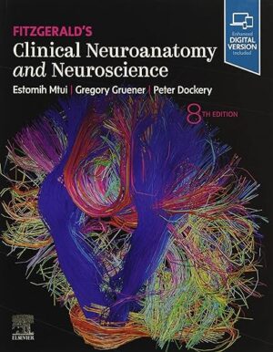
Fitzgerald’s Clinical Neuroanatomy and Neuroscience 8th Edition

Fitzgerald’s Clinical Neuroanatomy and Neuroscience 8th Edition

Ideal for both medical students and those in non-medical courses, Fitzgerald’s Clinical Neuroanatomy and Neuroscience, 8th Edition, uses clear, understandable text and outstanding artwork to make a complex subject easily accessible. This award-winning title is known for superb illustrations and high readability, expertly integrating clinical neuroanatomy with the clinical application of neuroscience.

  • Organizes chapters by anatomical area, with integrated analyses of sensory, motor, and cognitive systems.
  • Breaks complex concepts and subjects into easily digestible content with clear images and concise, straightforward explanations.
  • Features explanatory illustrations drawn by the same meticulous artists who illustrated Gray’s Anatomy.
  • Includes new Basic Science Panels that highlight an emerging or relevant basic science concept to expand your learning in specific content areas.
  • Provides access to the Student Consult enhanced eBook, which contains tutorials for each chapter, hundreds of multiple-choice questions and answers, MRI images with explanatory text, and case studies.
  • Contains learning helps in every chapter, including bulleted points, clinical boxes, opening summaries, and concluding core information boxes.
  • Enhanced eBook version included with purchase. Your enhanced eBook allows you to access all of the text, figures, and references from the book on a variety of devices.

DOWNLOAD THIS MEDICAL BOOK

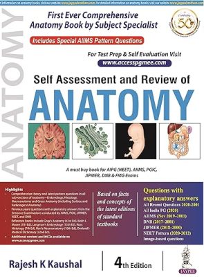
Self Assessment and Review of Anatomy 4th Edition

Self Assessment and Review of Anatomy 4th Edition

Self-assessment and review of anatomy is an adaption to the present challenge of the recent-pattern questions, which tends to explore vast extremes of the subject ‘anatomy’. it includes theory and questions in all subsections of Anatomy-Embryology, histology, neuroanatomy and gross anatomy (including surface and radiological anatomy). the book will maximize confidence and scoring in basic and advanced concepts in anatomy and related subjects in PG entrance examinations conducted by AIIMS, pgic, JIPMER, NEET and DNB.

DOWNLOAD THIS MEDICAL BOOK

Human Anatomy & Physiology Laboratory Manual, Fetal Pig Version 13th Edition

Human Anatomy & Physiology Laboratory Manual, Fetal Pig Version 13th Edition

Human Anatomy & Physiology Laboratory Manual features a wide variety of experiments for students concentrating in healthcare programs. Each exercise leads you toward an understanding of the structure and function of the human body. The lab manual uses artwork from Marieb/Hoehn’s Human Anatomy & Physiology text, but can accompany any A&P textbook. Available in 3 versions (Main, Cat, Fetal Pig), the Cat and Pig versions respectively include cat and fetal pig dissection exercises.

The 13th Edition includes many new, full-color figures and photos and revamped clinical application and critical thinking questions.

DOWNLOAD THIS MEDICAL BOOK

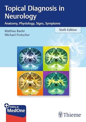
Topical Diagnosis in Neurology: Anatomy, Physiology, Signs, Symptoms 6th Edition

Topical Diagnosis in Neurology: Anatomy, Physiology, Signs, Symptoms 6th Edition


The perennial neurology classic, updated and expanded

The sixth edition of Duus’ classic Topical Diagnosis in Neurology builds on the clear, integrated presentation of anatomy, function, and disorders of the central nervous system that made it a success for almost over five decades, serving as a quick reference for practitioners and trainees alike. It elucidates the neuroanatomical pathways that lead to specific clinical syndromes, and demonstrates how comprehensive anatomical knowledge combined with a thorough neurological examination can help localize and categorize a lesion and arrive at a differential diagnosis.

The time-tested logical, thematic structure with useful summaries, at the beginning of each chapter, and color-coded section headings enable readers to distinguish neuroanatomical from clinical material at a glance and enable efficient and time saving study.

Features of the sixth edition:

  • New tables and clinical case examples
  • Improved descriptions of neuroanatomical/neurophysiological basics in the context of clinical symptoms
  • Twenty-three new illustrations, to a total of more than 400 illustrations
  • A wide range of study aids and clinical correlations that support the emphasis on integrative medicine in the current medical school curricula

Topical Diagnosis in Neurology is an ideal reference for neurologists and neuroscientists who correlate neurological diseases with anatomical location to arrive at a diagnosis or understand a clinical syndrome. It is also an essential tool for trainees and advanced students who need a solid grounding in key neurofunctional relationships.

This book includes complimentary access to a digital copy on https://medone.thieme.com.

DOWNLOAD THIS MEDICAL BOOK

Laboratory Manual for Anatomy and Physiology 7th Edition

Laboratory Manual for Anatomy and Physiology 7th Edition

Laboratory Manual for Anatomy & Physiology, 7th Edition, contains dynamic and applied activities and experiments that help students both visualize anatomical structures and understand complex physiological topics. Lab exercises are designed in a way that requires students to first apply information they learned and then critically evaluate it. With many different format options available, and powerful digital resources, it’s easy to customize this laboratory manual to best fit your course. While the Laboratory Manual for Anatomy and Physiology is designed to complement the latest 16th edition of Principles of Anatomy & Physiology, it can be used with any two-semester A&P text.

DOWNLOAD THIS MEDICAL BOOK

1 2 3 4 5 22