Anatomy and Physiology of Speech and Hearing Illustrated Edition
Anatomy and Physiology of Speech and Hearing Illustrated Edition
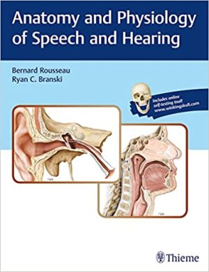
Anatomy and Physiology of Speech and Hearing
Anatomy and Physiology of Speech and Hearing by Bernard Rousseau and Ryan C. Branski fulfills a growing need for a contemporary resource for students in speech and hearing science training programs. Extending well beyond traditional speech science and human anatomy, this publication encompasses the latest advances in the understanding of human physiology, basic cell functions, biological control systems, and coordinated body functions.
Anatomy and Physiology of Speech and Hearing includes award-winning anatomic artwork from Thieme’s Atlas of Anatomy, adding a rich visual basis to the clinical facets of speech, language, swallowing, hearing, and balance. The book begins with fundamentals of human anatomy and physiology such as embryology and development of speech and hearing mechanisms. The second section details nervous system functions including central and peripheral motor control. The physiology of respiration, phonation, articulation and resonance, hearing, swallowing, and balance are covered in the last six chapters.
Key Features
- Highlighted key terms, review questions, learning objectives, and summaries enable instructors and students to consolidate information
- Textboxes offer meaningful examples of clinical disorders in a context conducive to applying newly learned concepts
- Over 400 high-quality, detailed anatomical illustrations maximize comprehension of anatomical and physiological aspects of speech, language, swallowing, hearing, balance and related functions
- Online access to Q&A content and anatomy figures provides labels on/off functionality for interactive study and review
This core textbook is essential reading for undergraduate and graduate students in communication sciences and disorders. The connection between basic and clinical science enables students to maximize learning and apply this new knowledge during clinical placements and externships.

 Abrahams’ and McMinn’s Clinical Atlas of Human Anatomy, 8th Edition delivers the