Ryan’s Retina 7th Edition
Ryan’s Retina 7th Edition
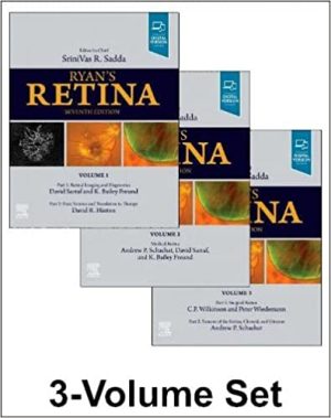
 Through six outstanding and award-winning editions, Ryan’s Retina has offered unsurpassed coverage of this complex subspecialty―everything from basic science through the latest research, therapeutics, technology, and surgical techniques. The fully revised 7th Edition, edited by Drs. SriniVas R. Sadda, Andrew P. Schachat, Charles P. Wilkinson, David R. Hinton, Peter Wiedemann, K. Bailey Freund, and David Sarraf, continues the tradition of excellence, balancing the latest scientific research and clinical correlations and covering everything you need to know on retinal diagnosis, treatment, development, structure, function, and pathophysiology. More than 300 global contributors share their knowledge and expertise to create the most comprehensive reference available on retina today.
Features sweeping content updates, including new insights into the fundamental pathogenic mechanisms of age-related macular degeneration, advances in imaging including OCT angiography and intraoperative OCT, new therapeutics for retinal vascular disease and AMD, novel immune-based therapies for uveitis, and the latest in instrumentation and techniques for vitreo-retinal surgery.
Includes five new chapters covering Artificial Intelligence and Advanced Imaging Analysis, Pachychoroid Disease and Its Association with Polypoidal Choroidal Vasculopathy, Retinal Manifestations of Neurodegeneration, Microbiome and Retinal Disease, and OCT-Angiography.
Includes more than 50 video clips (35 new to this edition) highlighting the latest surgical techniques, imaging guidance, and coverage of complications of vitreoretinal surgery. New videos cover Scleral Inlay for Recurrent Optic Nerve Pit Masculopathy, Trauma with Contact Lens, Recurrent Retinal Detachment due to PVR, Asteroid Hyalosis, and many more.
Contains more than 2,000 high-quality images (700 new to this edition) including anatomical illustrations, clinical and surgical photographs, diagnostic imaging, decision trees, and graphs.
Enhanced eBook version included with purchase. Your enhanced eBook allows you to access all of the text, figures, and references from the book on a variety of devices.


