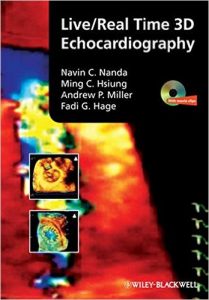
Live/Real Time 3D Echocardiography 1st Edition

51ylyxkjul-_sx346_bo1204203200_

[amazon template=iframe image2&asin=1405161418]

Now there is a source of recent information on the basic and clinical usefulness of real-time 3D echocardiography for echocardiographers, cardiologists, cardiac sonographers, and internists. This excellent reference helps clinicians improve pre- and post-surgical planning, attain better measurement of heart function, decrease exam times, and enhance patient communication. An accompanying DVD shows procedures in real time.

DOWNLOAD THIS BOOK FREE HERE

http://upsto.re/vNMceK5

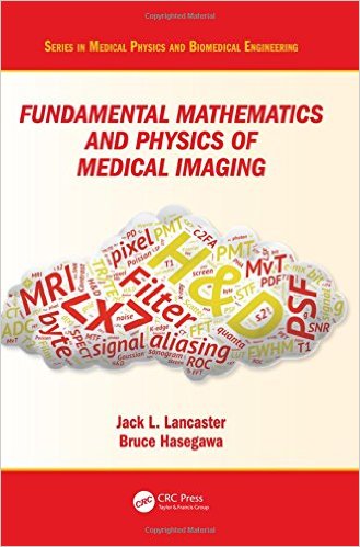
Fundamental Mathematics and Physics of Medical Imaging (Series in Medical Physics and Biomedical Engineering) 1st Edition

51cl8gn2hl-_sx327_bo1204203200_

[amazon template=iframe image2&asin=149875161X]

Authored by a leading educator, this book teaches the fundamental mathematics and physics concepts associated with medical imaging systems. Going beyond mere description of imaging modalities, this book delves into the mechanisms of image formation and image quality common to all imaging systems: contrast mechanisms, noise, and spatial and temporal resolution, making it an important reference for medical physicists and biomedical engineering students. This is an extensively revised new edition of The Physics of Medical X-Ray Imaging by Bruce Hasegawa (Medical Physics Publishing, 1991), and includes a wide range of modalities such as X-ray CT, MRI and SPECT.

DOWNLOAD THIS BOOK FREE HERE

http://upsto.re/vNMWkpj

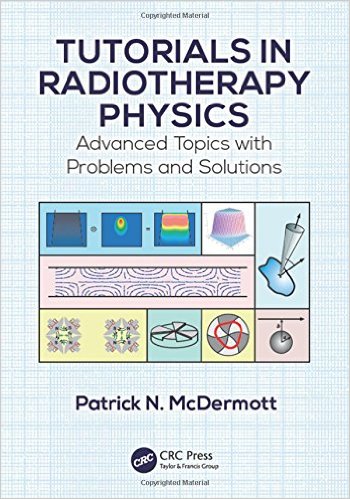
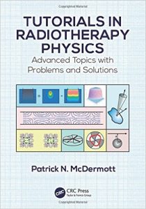
Tutorials in Radiotherapy Physics: Advanced Topics with Problems and Solutions 1st Edition

516qemkw-gl-_sx348_bo1204203200_

[amazon template=iframe image2&asin=1482251671]

The Topics Every Medical Physicist Should Know

Tutorials in Radiotherapy Physics: Advanced Topics with Problems and Solutions covers selected advanced topics that are not thoroughly discussed in any of the standard medical physics texts. The book brings together material from a large variety of sources, avoiding the need for you to search through and digest the vast research literature. The topics are mathematically developed from first principles using consistent notation.

Clear Derivations and In-Depth Explanations

The book offers insight into the physics of electron acceleration in linear accelerators and presents an introduction to the study of proton therapy. It then describes the predominant method of clinical photon dose computation: convolution and superposition dose calculation algorithms. It also discusses the Boltzmann transport equation, a potentially fast and accurate method of dose calculation that is an alternative to the Monte Carlo method. This discussion considers Fermi–Eyges theory, which is widely used for electron dose calculations. The book concludes with a step-by-step mathematical development of tumor control and normal tissue complication probability models. Each chapter includes problems with solutions given in the back of the book.

Prepares You to Explore Cutting-Edge Research

This guide provides you with the foundation to read review articles on the topics. It can be used for self-study, in graduate medical physics and physics residency programs, or in vendor training for linacs and treatment planning systems.

DOWNLOAD THIS BOOK FREE HERE

http://upsto.re/vnojiUf

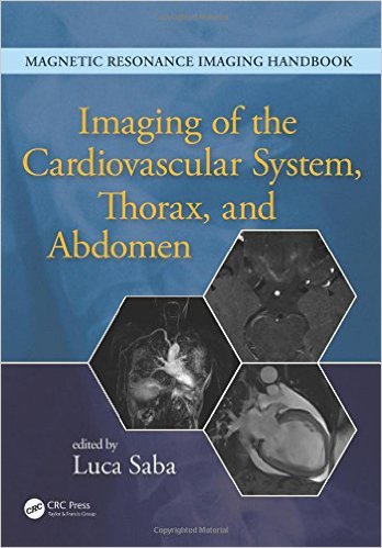
Imaging of the Cardiovascular System, Thorax, and Abdomen (Volume 2)

51rjutkgdnl-_sx346_bo1204203200_

[amazon template=iframe image2&asin=1482216264]

Magnetic resonance imaging (MRI) is a technique used in biomedical imaging and radiology to visualize internal structures of the body. Because MRI provides excellent contrast between different soft tissues, the technique is especially useful for diagnostic imaging of the brain, muscles, and heart.

In the past 20 years, MRI technology has improved significantly with the introduction of systems up to 7 Tesla (7 T) and with the development of numerous post-processing algorithms such as diffusion tensor imaging (DTI), functional MRI (fMRI), and spectroscopic imaging. From these developments, the diagnostic potentialities of MRI have improved impressively with an exceptional spatial resolution and the possibility of analyzing the morphology and function of several kinds of pathology.

Given these exciting developments, the Magnetic Resonance Imaging Handbook: Imaging of the Cardiovascular System, Thorax, and Abdomen is a timely addition to the growing body of literature in the field. Offering comprehensive coverage of cutting-edge imaging modalities, this book:

  • Discusses MRI of the heart, blood vessels, lungs, breasts, diaphragm, liver, gallbladder, spleen, pancreas, adrenal glands, and gastrointestinal tract
  • Explains how MRI can be used in vascular, posttraumatic, postsurgical, and computer-aided diagnostic (CAD) applications
  • Highlights each organ’s anatomy and pathological processes with high-quality images
  • Examines the protocols and potentialities of advanced MRI scanners such as 7 T systems
  • Includes extensive references at the end of each chapter to enhance further study

Thus, the Magnetic Resonance Imaging Handbook: Imaging of the Cardiovascular System, Thorax, and Abdomen provides radiologists and imaging specialists with a valuable, state-of-the-art reference on MRI.

DOWNLOAD THIS BOOK FREE HERE

http://upsto.re/vn5bi7b
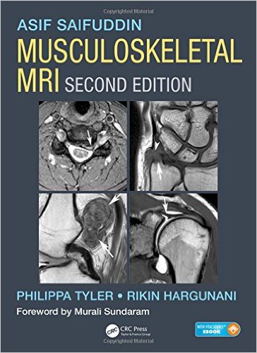
Musculoskeletal MRI, Second Edition 2nd Edition

51ipj1uir2l-_sx362_bo1204203200_

[amazon template=iframe image2&asin=1482247801]

Musculoskeletal MRI covers the entire musculoskeletal system and related conditions, both common and rare. The text is neatly divided into sections based on the major anatomic divisions. Each section discusses anatomic subdivisions or joints, keeping sections on normal anatomy and pathologic findings close to each other, allowing radiologists to easily compare images of normal and pathologic findings.

With more than 4000 high-quality MR images, information is presented in an easy-to-read bulleted format, providing the radiologist with all the information required to make an informed diagnosis in the clinical setting. The new edition also includes a complimentary eBook as well as access to image downloads. Comprehensive and user-friendly in its approach, the book provides every radiologist, both consultant and trainee, with increased confidence in their reporting.

DOWNLOAD THIS BOOK FREE HERE

PART I http://upsto.re/vnkTYa6

PART II http://upsto.re/vnk67qi

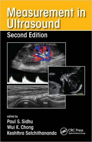
Measurement in Ultrasound, Second Edition 2nd Edition

51koyswi6l-_sx323_bo1204203200_

[amazon template=iframe image2&asin=1482231352]

Measurement and interpretation of key ultrasound parameters are essential to differentiate normal anatomy from pathology. By using Measurement in Ultrasound, trainee radiologists and ultrasonographers can gain an appreciation of such measurements, while practitioners can use it as a valuable reference in the clinical setting.

The book follows a consistent format throughout for ease of reference and features useful information on preparation and positioning of the patient for ultrasound, the type of transducer and method to be used, the appearance of the resulting ultrasound images and the measurements to be derived from them.

Designed for frequent use in everyday practice, the book includes more than 150 high-quality ultrasound images annotated with key measurements and accompanied by concise explanatory text. Normal variants are provided, along with ranges for features that can change during development and in disease.

This new edition covers relevant developments in ultrasound. Where appropriate, updated ultrasound measurements that have arisen are also included and key references are provided as an aid to further study.

DOWNLOAD THIS BOOK FREE HERE

http://upsto.re/vnsTrD3

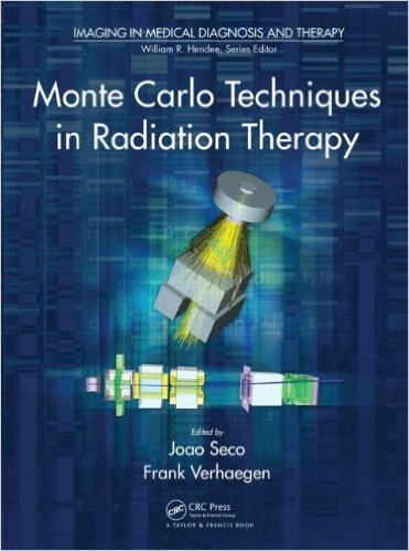
Monte Carlo Techniques in Radiation Therapy (Imaging in Medical Diagnosis and Therapy) 1st Edition

51zrenwu0ol-_sx369_bo1204203200_

[amazon template=iframe image2&asin=1466507926]

Modern cancer treatment relies on Monte Carlo simulations to help radiotherapists and clinical physicists better understand and compute radiation dose from imaging devices as well as exploit four-dimensional imaging data. With Monte Carlo-based treatment planning tools now available from commercial vendors, a complete transition to Monte Carlo-based dose calculation methods in radiotherapy could likely take place in the next decade. Monte Carlo Techniques in Radiation Therapy explores the use of Monte Carlo methods for modeling various features of internal and external radiation sources, including light ion beams.

The book―the first of its kind―world examples, it illustrates the use of Monte Carlo modeling and simulations in dose calculation, beam delivery, kilovoltage and megavoltage imaging, proton radiography, device design, and much more.

DOWNLOAD THIS BOOK FREE HERE

http://upsto.re/vbPtjux

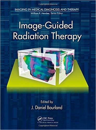
Image-Guided Radiation Therapy (Imaging in Medical Diagnosis and Therapy) 1st Edition

51axt17tsyl-_sx366_bo1204203200_

[amazon template=iframe image2&asin=1439802734]

Image-Guided Radiation Therapy presents key image-guided radiation treatment (IGRT) technologies for external beam radiotherapy. The book explores the decades-long technological developments that have occurred in the realm of image-guided conformal, customized radiation treatment.

Expert authors, all of whom have actively participated in the development or implementation of IGRT, imaging, and enabling technologies, share their first-hand experiences on the science, clinical uses, and impact of these technologies. They describe kilovoltage and megavoltage imaging as well as radiological, ultrasound, and optical technologies for determining and validating target and patient positioning. The book examines how anatomical and biological imaging using CT and PET has contributed to the understanding of target volume boundaries and biological behavior. It also explores such innovations as 4D PET/CT and digital tomosynthesis.

Advancing patient care, this book focuses on a wealth of hybrid IGRT technologies and devices for coupled imaging and treatment inside the radiation treatment room. It thoroughly covers the modalities, software tools, and imaging treatment geometries that constitute IGRT.

DOWNLOAD THIS BOOK FREE HERE

http://upsto.re/vbCzDDc

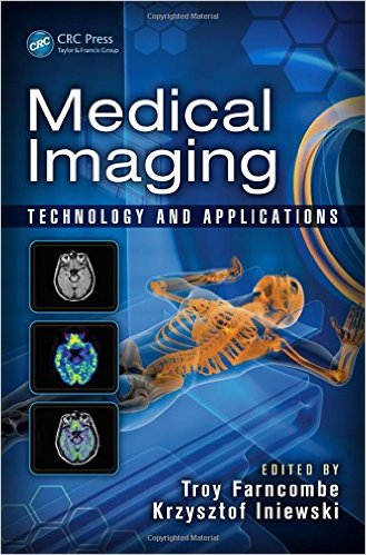
Medical Imaging: Technology and Applications (Devices, Circuits, and Systems) 1st Edition

51o3t8fkyl-_sx327_bo1204203200_

[amazon template=iframe image2&asin=1466582626]

The book has two intentions. First, it assembles the latest research in the field of medical imaging technology in one place. Detailed descriptions of current state-of-the-art medical imaging systems (comprised of x-ray CT, MRI, ultrasound, and nuclear medicine) and data processing techniques are discussed. Information is provided that will give interested engineers and scientists a solid foundation from which to build with additional resources. Secondly, it exposes the reader to myriad applications that medical imaging technology has enabled.

DOWNLOAD THIS BOOK FREE HERE

http://upsto.re/vbCS8SY

Breast Imaging

31yv0iipe3l-_sx328_bo1204203200_

[amazon template=iframe image2&asin=331941545X]

This book constitutes the refereed proceedings of the 13th International Workshop on Breast Imaging, IWDM 2016, held in Malmö, Sweden, in June 2016.

The 35 revised full papers and 50 revised poster papers presented together with 6 invited talks were carefully reviewed and selected from 89 submissions. The papers are organized in topical sections on screening; CAD; mammography, tomosynthesis, and breast CT; novel technology; density assessment and tissue analysis; dose and classification; image processing, CAD, breast density, and new technology; contrast-enhanced imaging; phase contrast breast imaging; simulations and virtual clinical trials.

DOWNLOAD THIS BOOK FREE HERE

http://upsto.re/vbdVhLC

1 52 53 54 55 56 62