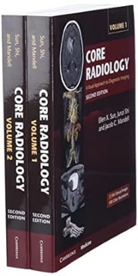
Core Radiology: A Visual Approach to Diagnostic Imaging 2nd Edition

Core Radiology: A Visual Approach to Diagnostic Imaging 2nd Edition

Embodying the principle of ‘everything you need but still easy to read’, this fully updated edition of Core Radiology is an indispensable aid for learning the fundamentals of radiology and preparing for the American Board of Radiology Core exam. Containing over 2,100 clinical radiological images with full explanatory captions and color coded annotations, streamlined formatting ensures readers can follow discussion points effortlessly. Bullet pointed text concentrates on essential concepts, with text boxes, tables and over 400 color illustrations supporting readers’ understanding of complex anatomic topics. Real world examples are presented for the readers, encompassing the vast majority of entitles likely encountered in board exams and clinical practice. Divided into two volumes, this edition is more manageable whilst remaining comprehensive in its coverage of topics, including expanded pediatric cardiac surgery descriptions, updated brain tumor classifications. non invasive vascular imaging. Highly accessible and informative, this is the go to introductory textbook for radiology residents worldwide.

Diagnostic Imaging: Interventional Radiology 3rd Edition

Diagnostic Imaging: Interventional Radiology 3rd Edition

Covering the entire spectrum of this rapidly evolving field, the third edition of Diagnostic Imaging: Interventional Radiology is an invaluable resource for interventional and diagnostic radiologists, trainees, and all proceduralists who desire an easily accessible, highly visual reference for this complex specialty. Dr. Brandt C. Wible and his team of highly regarded experts provide up-to-date information on more than 100 interventional radiologic procedures to help you make informed decisions at the point of care. Chapters are well organized, referenced, and lavishly illustrated, comprising a useful learning tool for readers at all levels of experience as well as a handy reference for daily practice.

• Provides a comprehensive, expert reference for review and preparation of common and infrequently performed procedures, with detailed “step-by-step” instructions for conducting image-guided interventions in various clinical scenarios

• Covers vascular venous, arterial, and lymphatic procedures, with specific attention to thromboembolic, posttransplant, and oncologic therapies

• Addresses emerging nonvascular image-guided treatments in pain management, neurologic and musculoskeletal procedures, and others

• Contains new procedures chapters on endovascular treatments for pulmonary embolisms and deep vein thrombosis, prostate artery embolization, pelvic venous disorders, and percutaneous/endovascular arteriovenous fistula (AVF) creation

• Features sweeping updates throughout, including updated guidelines and recommendations from the Society of Interventional Radiology

• Offers more than 3,200 images (in print and online), including radiologic

images, full-color medical illustrations, instructional photo essays, and clinical

and histologic photographs

• Clearly demonstrates procedural steps, complications, treatment alternatives, variant anatomy, and more?all fully annotated to highlight the most important diagnostic information

• Organized by procedure type, allowing for quick comparison of different procedural techniques that may have complementary or alternative rolesin managing specific disease states

• Builds on the award-winning second edition, which won first prize in the British Medical Association’s Medical Book Awards, Radiology category

• Includes the enhanced eBook version, which allows you to search all text, figures, and references on a variety of devices

DOWNLOAD THIS MEDICAL BOOK

Bone and Joint Disorders (Differential Diagnosis in Conventional Radiology) 2nd Edition

Bone and Joint Disorders (Differential Diagnosis in Conventional Radiology) 2nd Edition

The second edition of Bone and Joint Disorders, derived from the critically acclaimed series Differential Diagnosis in Conventional Radiology, provides the essential information you need to make the conventional x-ray an effective tool in diagnosing disorders affecting the bones and joints.
The book is organized according to classes of radiologic findings, with separate chapters addressing the differential diagnosis of osteopenia, osteosclerosis, trauma and fractures, arthritis, and tumors, as well as the differential diagnosis of lesions specific to anatomic sites. Concise but comprehensive tables outline key information about findings and etiology, and provide additional commentary on pertinent clinical data, such as common locations, frequency, and prevalence of the described disease or disorder.

DOWNLOAD THIS MEDICAL BOOK

Felson’s Principles of Chest Roentgenology, A Programmed Text (Goodman, Felson’s Principles of Chest Roentgenology) 4th Edition

Felson’s Principles of Chest Roentgenology, A Programmed Text (Goodman, Felson’s Principles of Chest Roentgenology) 4th Edition

Popular for its easy-to-use format, Felson?s Principles of Chest Roentgenology remains the must-have primer of chest radiology. With the inclusion of the latest imaging approaches and terminology, its unique programmed learning approach ?presented in a highly interactive style?demystifies reading and interpreting radiologic images. High-quality images and diagrams are accompanied by multiple-choice review questions to reinforce key concepts. Additional online images plus self-assessment tests help you sharpen your skills and build confidence!

“A must for anyone who fancies him/herself as a competent general radiologist.” Reviewed byJohn Reid, Borders General Hospital on behalf of RAD Magazine, December 2015

  • Quickly grasp the radiology fundamentals you need to know―including basic science, image interpretation, and terminology―with the popular “programmed learning” approach, which promotes fast learning and reference.

  • Discern the nuances between modalities by comparing CT and MR images as well as traditional radiographs.
  • View detailed clinical images covering all the image types you’ll see on the boards including digital quality radiographs and an introduction of PET imaging, plus more advanced imaging such as CT and MRI than ever before.

  • Test your skills and simulate the exam experience with updated content aligned with the new MCQ-format Board exam for easy preparation and review.

  • Benefit the from more robust interactive offerings in an e-book format―with easy-to-access quizzes and board questions provided online.

  • Expert Consult eBook version included with purchase. This enhanced eBook experience allows you to search all of the text, figures, references, and videos from the book on a variety of devices.

DOWNLOAD THIS MEDICAL BOOK

Endodontic Radiology 2nd Edition

Endodontic Radiology 2nd Edition

Endodontic Radiology, 2nd edition, is a unique reference that examines all aspects of radiographic imaging related to endodontics. Dr. Bettina Basrani and a team of prestigious international contributors build upon traditional radiographic techniques and include the latest information available on digital radiographs and cone beam computed tomography. More than an overview of equipment, the book delves into radiographic interpretation, differential diagnosis, technical difficulties and special circumstances when taking radiographs during the endodontic treatment, and how to choose the correct radiographic technique to obtain the desired images. Chapters explain general radiographic techniques; intraoral techniques; standard radiographs and interpretation; digital radiographs and their manipulation, storage, and interpretation; and CBCT principles, techniques, and clinical considerations.

Human Osteology and Skeletal Radiology: An Atlas and Guide 1st Edition

Human Osteology and Skeletal Radiology: An Atlas and Guide 1st Edition

Human Osteology and Skeletal Radiology: An Atlas and Guide features nearly 700 photographs, line drawings, and radiographs demonstrating individual bones, or collections of bones, from both a distant perspective and more detailed angles. This atlas of skeletal anatomy covers general and specific anatomic terms, includes comparative images of bones in photographic and radiographic form to aid in recognition, and notes important comparisons among adult, juvenile, and fetal bones. It discusses each bone on an individual basis and describes how to “side” bones and identify fragments.

Intended as a field guide for investigations and a lab guide in gross anatomy and skeletal specimen studies, this atlas provides easy and rapid identification of bone material. It takes you far beyond the bare bones of anatomy to aid in skeletal recognition in any situation.

DOWNLOAD THIS MEDICAL BOOK

Paediatric Radiography 1st Edition

Paediatric Radiography 1st Edition

Radiography is an integral part of paediatric health care. It is frequently requested to assist in the diagnosis, management and treatment of childhood disease and illness. Accurate interpretation of paediatric radiographs can depend entirely on the quality of images produced by the radiographer, yet there are few books available on this crucial aspect of radiographic practice.
Paediatric Radiography fills a gap. It explores radiographic practice within the context of the modern health service and focuses on how our knowledge and understanding of paediatric growth, development and illness can inform and influence radiographic procedures. It includes detailed coverage of specific paediatric techniques and good practice models, including the role of multi-modality imaging, and looks specifically at radiation protection, the chest and upper airways, the abdomen, neonatal radiography, trauma, orthopaedics, and non-accidental injury.

DOWNLOAD THIS MEDICAL BOOK

Chest Radiology: PreTest Self- Assessment and Review 1st Edition

Chest Radiology: PreTest Self- Assessment and Review 1st Edition

Diagnostic Imaging of the Foot and Ankle 1st Edition

Diagnostic Imaging of the Foot and Ankle 1st Edition

The foot has a special place in musculoskeletal diagnosis due to its complex anatomy and because many similar symptoms can have different causes, each requiring a different approach to treatment. The evaluation of foot disorders and diseases requires close clinical–radiological correlation and communication with foot experts. Foot disorders and injuries increase with age, due in part to the rising popularity of recreational sports in all age groups. Diagnostic Imaging of the Foot and Ankle will help you train your eye to recognize disorders and diseases of the foot and ankle, including those that are often misdiagnosed or overlooked.

Key Features:

  • By practitioners for practitioners: First-hand knowledge from leading surgical and orthopedic foot experts and radiologists
  • Clear and concise: A textbook and reference in a user-friendly layout focused on the foot and ankle
  • Uniform format: Entities are described by definition, clinical presentation, imaging modalities, typical imaging features, differential diagnosis, treatment options, course, and pitfalls
  • Clinical aspects and treatment: Clinical–radiological correlation plus a concise review of treatment options
  • The new standard: This information on the foot and ankle is available nowhere else in such a condensed form
  • Highest quality images: More than 500 superb illustrations including high-resolution images acquired with high-field MRI and multi-channel coils

DOWNLOAD THIS MEDICAL BOOK

Fundamentals of Body MRI (Fundamentals of Radiology) 2nd Edition

Fundamentals of Body MRI (Fundamentals of Radiology) 2nd Edition

Effectively perform and interpret MR body imaging with this concise, highly illustrated resource! Fundamentals of Body MRI, 2nd Edition, by Drs. Christopher Roth and Sandeep Deshmukh, covers the essential concepts residents, fellows, and practitioners need to know, laying a solid foundation for understanding the basics and making accurate diagnoses. This easy-to-use title in the Fundamentals of Radiology series covers all common body MR imaging indications and conditions, while providing new content on physics and noninterpretive skills with an emphasis on quality and safety.

  • More than 1,400 detailed MRI images and 100 algorithms and diagrams highlight key findings and help you grasp visual nuances of images you’re likely to encounter.

  • All common body MR imaging content is covered, along with discussion of how physics, techniques, hardware, and artifacts affect results.

  • Expert Consult™ eBook version included with purchase. This enhanced eBook experience allows you to search all of the text, figures, and references from the book on a variety of devices.
  • Newly streamlined format helps you retrieve important information more quickly.

  • Extensively revised content on the liver, including new MRI contrast agents; new coverage of the spleen; and new safety tips and guidelines keep you up to date.

  • New chapters on GI imaging, the prostate, and the male genitourinary system make this a one-stop reference to address the full range of body MRI.


DOWNLOAD THIS MEDICAL BOOK


1 2 3 4 5 6 61