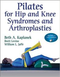
Pilates for Hip and Knee Syndromes and Arthroplasties With Web Resource Pap/Psc Edition
[amazon template=iframe image2&asin=0736092242]

As hip and knee conditions continue to become more prevalent, so does the demand for a rapid and complete return to function in these lower-extremity joints. Pilates for Hip and Knee Syndromes and Arthroplasties provides foundational guidelines and protocols—with specific modifications—for the use of Pilates in increasing core strength, balance, and flexibility and restoring function and range of motion with pre- and postoperative knee and hip syndromes and arthroplasties. Written for Pilates instructors, manual therapists, personal trainers, and physicians, this text introduces Pilates as a safe fitness and rehabilitation tool for individuals with knee or hip conditions.

Developed over 90 years ago by Joseph H. Pilates, the Pilates method is a unique system of stretching and strengthening exercises that have been shown to tone muscles and improve posture, flexibility, range of motion, and balance. Low impact and completely adaptable according to specific syndromes or fitness level, Pilates exercises are well suited for use in pre- and postoperative exercise regimens, and Pilates mat exercises can be easily incorporated into home programs.

Pilates for Hip and Knee Syndromes and Arthroplasties begins with a review of the anatomy of the hip and knee, a discussion of the most common conditions, and an overview of nonoperative and operative treatments. Building this background information will help readers gain a better understanding of why certain exercises are applied at various points in the rehabilitation time line.

The next portion of the text is dedicated to specific Pilates techniques and mat exercises and includes baseline recommendations for range of motion and both pre- and postoperative modifications for the knee and hip. Reference tables outline classical Pilates mat exercises and place them in specific rehabilitation time lines from six weeks to three months, three months to six months, and beyond six months postoperative. More than 600 photos clearly demonstrate the exercises and feature detailed instructions for correct execution of the techniques. To assist with clients who have never performed Pilates exercises or are in the very early stages after surgery, pre-Pilates exercises are also presented to help build core strength and range of motion. Case scenarios and sample Pilates mat programs provide additional guidelines on the correct application of the exercises, while an exercise finder located in the front of the text quickly directs readers to the appropriate exercises for each postop time line.

As a bonus, a Web resource included with the text provides fully trained Pilates instructors with guidelines on using the Pilates equipment to develop programs for clients with hip or knee conditions. Instructors will learn what equipment is appropriate to incorporate at the optimal time for rehabilitation. In addition, a resource finder is included to assist readers in finding a qualified Pilates training program and a qualified Pilates instructor.

DOWNLOAD THIS BOOK FREE HERE

http://upsto.re/FdzvidU

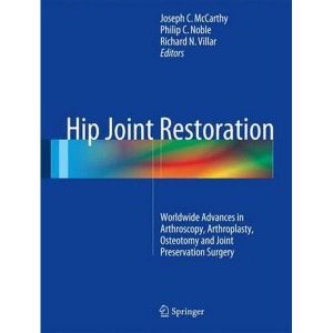
Hip Joint Restoration: Worldwide Advances in Arthroscopy, Arthroplasty, Osteotomy and Joint Preservation Surgery 1st ed. 2017 Edition

[amazon template=iframe image2&asin=1461406935]

Hip Joint Restoration is a comprehensive yet practical guide to the basic science and clinical applications of arthroscopy, arthroplasty, osteotomy and preservation surgery for the treatment of diseases and conditions of the hip. This generously illustrated text offers a comprehensive introduction to essential features of hip evaluation, the medical management of hip procedures, and treatment of specific conditions, and covers practical topics such as surgical anatomy of the hip, surgical approaches, instrumentation, and indications for arthroscopy and other surgical procedures aimed at restoration of the hip joint. Additional chapters cover clinical outcomes and equality of life following hip surgery, the current state of research and education of arthroscopic hip procedures throughout the world, other topics such as complications and rehabilitation in different patient populations. This book will be a useful resource for Orthopedic Surgeons and Osteopaths who perform open and arthroscopic hip preservation and total joint replacement, as well as for orthopedic residents and researchers.

DOWNLOAD THIS BOOK FREE HERE

http://upsto.re/Fd5gjur

Polymyalgia Rheumatica and Giant Cell Arteritis 3rd ed. 2017 Edition

[amazon template=iframe image2&asin=3319522213]

This book, now in an extensively updated and expanded third edition, offers important insights into polymyalgia rheumatica (PMR) and giant cell arteritis (GCA), providing an overview of their pathophysiology, genetics, immunology, and endocrinological mechanisms but above all presenting the state of the art in diagnosis and treatment.  Both PMR and GCA are diseases of advanced age. Early diagnosis and treatment can save one of the most precious senses – vision. In this book, the role of imaging techniques is thoroughly examined, with particular attention to ultrasonography and FDG PET. All aspects of management of both diseases are discussed, including the current use of glucocorticoids, the efficacy of modern biological treatment, such as infliximab, tocilizumab, and rituximab, and the available monitoring tools. Diagnostic criteria for PMR and GCA are outlined, with discussion of differential diagnosis. Among the other topics addressed are disease classification, the histopathology of GCA, the ACR criteria for temporal arteritis, the value of temporal artery biopsy in GCA, serum cytokine profiles, chronobiology, and sine syndromes. The reader will find that the book aids in achieving early diagnosis and providing faster and more effective disease management. General readers with an interest in rheumatology, internal medicine, ophthalmology and gerontology will find this book particularly valuable.

DOWNLOAD THIS BOOK FREE HERE

http://upsto.re/FdFTxnG

Broken Bones: The Radiologic Atlas of Fractures and Dislocations 2nd Edition

[amazon template=iframe image2&asin=1107499232]

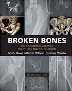
Broken Bones contains 434 individual cases and 1,101 radiologic images illustrating the typical and less typical appearances of fractures and dislocations throughout the body. The first chapter describes fractures and dislocations of the fingers, starting with fractures of the phalangeal tufts and progressing through the distal, middle, and proximal phalanges and the DIP and PIP joints. Subsequent chapters cover the metacarpals, the carpal bones, the radius and ulna, the elbow and upper arm, and the shoulder and thoracic cage. The cervical spine and the thoracic and lumbosacral spine are covered in separate chapters, followed by the pelvis, the femur, the knee and lower leg, the ankle, the tarsal bones, and the metatarsals and toes. The final three chapters cover the face, fractures and dislocations in children, and fractures and dislocations caused by bullets and nonmilitary blasts.

DOWNLOAD THIS BOOK FREE HERE

http://upsto.re/FCubYNB

Osteoarthritic Joint Pain

[amazon template=iframe image2&asin=0470867612]

Osteoarthritis is a chronic degenerative disease associated with joint pain and loss of joint function. It has an estimated incidence of 4 out of every 100 people and significantly reduces the quality of life in affected individuals. The major symptoms are chronic pain, swelling and stiffness; severe, chronic joint pain is often the central factor that causes patients to seek medical attention. Within the affected joint, there is focal degradation and remodelling of articular cartilage, new bone formation (osteophytes) and mild synovitis.

Several mechanisms are thought to contribute to osteoarthritic joint pain. These include mild synovial inflammation, bone oedema, ligament stretching, osteophyte formation and cartilage-derived mediators. Changes in joint biomechanics and muscle strength also influence the severity and duration of joint pain in osteoarthritis. Within the nervous system, the relative contributions of peripheral afferent nociceptive fibres and central mechanisms remain to be defined, and there is limited information on the phenotype of sensory neurons in the OA joint. Importantly, there is no relation between clinical severity, as measured by radiographic changes, and the presence and severity of joint pain. Patients with severe joint pain may have normal joint architecture as determined by X-ray, whereas patients with considerable evidence of joint remodelling may not have significant joint pain. Treatments for osteoarthritic joint pain include non-steroidal anti-inflammatory compounds, exercise, corrective shoes and surgical intervention. There remains a critical need for improved control of joint pain in osteoarthritis.

This book brings together contributions from key investigators in the area of osteoarthritic joint pain.  It covers the clinical presentation of joint pain, the pathways involved in joint pain, osteoarthritis disease processes and pain, experimental models and pain control. The discussions provide insights into the nature of osteoarthritic joint pain, identify key studies needed to advance understanding of the problem, highlight possible intervention points and indicate future pathways towards a better treatment of osteoarthritic joint pain.

DOWNLOAD THIS BOOK FREE HERE

http://upsto.re/FCMLjD9

Master Techniques in Orthopaedic Surgery: Orthopaedic Oncology and Complex Reconstruction First Edition

[amazon template=iframe image2&asin=1608310434]

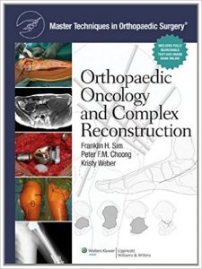
Master Techniques in Orthopaedic Surgery: Orthopaedic Oncology and Complex Reconstruction focuses on bone and soft tissue tumors, which are among the most challenging problems for orthopaedic surgeons to manage. The book is broad in scope and includes coverage of massive reconstruction following trauma. The international authorship includes outstanding surgeons from Germany, Austria, Italy, Australia, and Great Britain in addition to the United States.

The contributors describe their preferred techniques in step-by-step detail, point out pertinent anatomy, and offer pearls and tips for improving results. The book is thoroughly illustrated with full-color, sequential, surgeon’s-eye view intraoperative photographs, as well as drawings by noted medical illustrators.

DOWNLOAD THIS BOOK FREE HERE

http://upsto.re/AV4JWod

Master Techniques in Orthopaedic Surgery: Reconstructive Knee Surgery Fourth Edition

[amazon template=iframe image2&asin=1496318277]

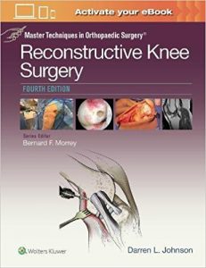
Take your mastery to the next level! Master Techniques in Orthopaedic Surgery©: Reconstructive Knee Surgeryis your ideal source for perfecting today’s most advanced and effective surgical techniques for knee reconstruction. Each chapter presents a world-leading orthopaedic surgeon’s preferred approach to a specific knee problem, replete with expert technical pearls to help you achieve optimal patient outcomes.

Key Features

  • Overcome a full range of challenges in reconstructive knee surgery with in-depth sections on extensor mechanism/patellofemoral problems, meniscus surgery, ligament injuries/instability, and reconstruction of the articular cartilage and synovium.
  • Update your skills with comprehensive updates throughout reflecting all of the latest advances in the field, including many new chapters, new topics, and new surgical innovations.
  • See exactly how to proceed thanks to thousands of high-quality, step-by-step photographs and drawings.
  • Produce the best outcomes through comprehensive discussions of indications and contraindications, decision making, operation room methodology, and pitfalls.

Your book purchase includes a complimentary download of the enhanced eBook for iOS, Android, PC & Mac. Take advantage of these practical features that will improve your eBook experience:

  • The ability to download the eBook on multiple devices at one time — providing a seamless reading experience online or offline
  • Powerful search tools and smart navigation cross-links that allow you to search within this book, or across your entire library of VitalSource eBooks
  • Multiple viewing options that enable you to scale images and text to any size without losing page clarity as well as responsive design
  • The ability to highlight text and add notes with one click

DOWNLOAD THIS BOOK FREE HERE

http://upsto.re/AVQJmjG

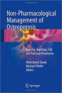
Non-Pharmacological Management of Osteoporosis: Exercise, Nutrition, Fall and Fracture Prevention 1st ed. 2017 Edition

[amazon template=iframe image2&asin=3319540149]

This practical guide presents the most up-to-date information on the application of non-pharmacological and physical therapeutic measures, either used independently or in combination with pharmacotherapy, for the management of osteoporosis. Pharmacotherapy remains the primary treatment for osteoporosis, but to improve the biomechanical competence of bone and improve quality of life, there needs to be more comprehensive management approach involving non-pharmacological methods. The book opens with a discussion of the diagnosis, pathophysiology, complications and consequences of osteoporosis. Exercise, nutrition, orthotics, and other rehabilitation measures such as whole body vibration and electrical muscle stimulation, each described in details in chapters of their own, have had a beneficial impact on fall and fracture prevention as well as recovery post-fracture. In addition, the application of acupuncture for pain management and movement-based mind-body therapies like tai chi and the Feldenkrais method are explored.

Providing a description of independent and adjuvant techniques and practices for treatment and improving quality of life, Non-Pharmacological Management of Osteoporosis is an excellent resource for endocrinologists, bone specialists, physical therapists, occupational therapists and all clinical practitioners and staff working with osteoporosis patients.

DOWNLOAD THIS BOOK FREE HERE

http://upsto.re/Aohh8Uo

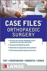
Case Files Orthopaedic Surgery (LANGE Case Files) 1st Edition

[amazon template=iframe image2&asin=0071790306]

SHARPEN YOUR CRITICAL THINKING SKILLS AND PREPARE FOR REAL-WORLD PRACTICE WITH ORTHOPAEDIC SURGERY CASES

Experience with clinical cases is key to excelling in your rotations and post-graduate training. Case Files: Orthopaedic Surgery gives you 45 true-to-life cases that illustrate concepts critical to managing common musculoskeletal injuries and conditions. Each case includes a concise and accurate patient presentation, key exam findings, and clear radiologic images where applicable. Additionally, cases include in-depth discussions of the injury or condition represented, replete with evidence-based practice recommendations, basic procedural tips and techniques, and discussion of potential complications, pitfalls, and ultimate patient outcomes. Review questions and clinical pearls reinforce learning.

  • Learn from 45 high-yield cases, each with review questions
  • Master key concepts with clinical pearls
  • Polish your approach to clinical problems and think like a surgeon
  • Perfect for students, orthopaedic interns, and residents who encounter orthopaedic conditions in daily practice

DOWNLOAD THIS BOOK FREE HERE

http://upsto.re/A9RRTXC

1 18 19 20 21 22 27