Atlas of Deep Endometriosis: MRI and Laparoscopic Correlations 1st ed. 2018 Edition
[amazon_link asins=’3319716964′ template=’ProductAd’ store=’aishabano-20′ marketplace=’US’ link_id=’653932cf-4987-11e8-9dea-53aa537d19fe’]

About the Author

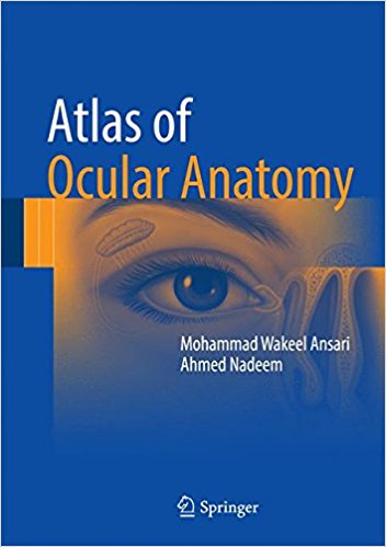
Atlas of Ocular Anatomy 1st ed

[amazon_link asins=’3319427806′ template=’ProductAd’ store=’aishabano-20′ marketplace=’US’ link_id=’a361f92a-4948-11e8-aedc-550e7417d393′]

This book is a practical and concise atlas on ocular anatomy, with an emphasis on applied aspects and hints for easy retention strategies. The vast color illustrations and photographs consist of self-explanatory, precise, and meaningful representations of the points covered in the text. Covering chapters such as bony socket of the eye, extraocular muscles, eyelids, cornea and lens, and neurology of the eye, Atlas of Ocular Anatomy gives a summary of the important and relevant points for each topic, separating out the essential from the nonessential elements. Complete with representative schematic line diagrams and full color photographs, this atlas features the correlation between anatomic facts with their probable clinical presentations in disease.

DOWNLOAD THIS BOOK FREE HERE

https://upsto.re/3ZKmhpV

Atlas of 3D Transesophageal Echocardiography in Structural Heart Disease Interventions: Cases and Videos 1st ed. 2018 Edition

[amazon_link asins=’9811069360′ template=’ProductAd’ store=’aishabano-20′ marketplace=’US’ link_id=’38c02f02-4945-11e8-92af-fd9c1a7272a1′]

About the Author

Atlas of Perioperative 3D Transesophageal Echocardiography: Cases and Videos 1st ed. 2016 Edition

[amazon_link asins=’9811005869′ template=’ProductAd’ store=’aishabano-20′ marketplace=’US’ link_id=’0f2b4466-44a0-11e8-9fdc-abc77f0e183c’]

This book presents a series of over 50 cases in 3D transesophageal echocardiography. In each case, background information is followed by a step-by-step approach to patient diagnosis and postoperative outcomes. The book’s most outstanding highlight consists in over 600 illustrations, which mainly focus on 3D transesophageal echocardiography in combination with 2D transesophageal echocardiography, X-ray fluoroscopy and computed tomography to demonstrate the various cardiovascular pathologies. Since the echo images obtained in clinic practice are moving images, it also includes more than 450 videos, which effectively supplement the static illustrations.

The atlas is divided into 11 chapters. Chapters 1 through 3 describe abnormalities affecting mitral, aortic, tricuspid and pulmonary valves, while prosthetic valves are discussed in Chapter 4. Chapter 5 details abnormalities of the aorta. Chapters 6-8 examine coronary artery disease, congenital heart disease and cardiomyopathies, while Chapter 9 explores cases of infective endocarditis. Chapter 10 deals with tumor and mass lesions, and the other cases are collectively addressed in Chapter 11. Offering a valuable new resource, the book will enhance the usefulness of echocardiography during operations as a “third eye” for surgeons.

DOWNLOAD THIS BOOK FREE HERE

https://upsto.re/36FFTHP

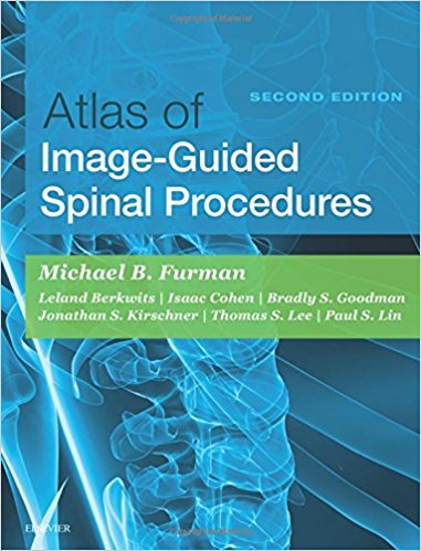
Atlas of Image-Guided Spinal Procedures, 2e 2nd Edition

[amazon_link asins=’0323401538′ template=’ProductAd’ store=’aishabano-20′ marketplace=’US’ link_id=’398a76f5-4276-11e8-a265-f5431a24bf92′]

The second edition of Atlas of Image-Guided Spinal Procedures features a highly visual atlas format to illustrate exactly how to perform each technique. This medical reference walks you through each procedure, step-by-step, to safely and efficiently relieve patients’ pain. This book presents an algorithmic, image-guided approach for each technique; trajectory view (demonstrates fluoroscopic “setup”); multiplanar confirmation views (AP, lateral, oblique); and “safety view” (what should be avoided during injection), along with optimal and suboptimal contrast patterns. Each fluoroscopic and ultrasound chapter also has the same “voice” so it is easy to follow.


  • Safely and efficiently relieve your patients’ pain with consistent, easy-to-follow chapters that guide you through each technique.
  •  Presents an algorithmic, image-guided approach for each technique: trajectory view (demonstrates fluoroscopic “setup”); multiplanarconfirmation views (AP, lateral, oblique); and “safety view” (what should be avoided during injection), along with optimal and suboptimal contrast patterns.
  • Special chapters on Needle Techniques, Procedural Safety, Fluoroscopic and Ultrasound Imaging Pearls, Radiation Safety, and L5-S1 Disc Access provide additional visual instruction.
  • View drawings of radiopaque landmarks and key radiolucent anatomy that cannot be viewed fluoroscopically.
  • Videos including procedural “set-up” and optimal and suboptimal constrast flow are available in the Expert Consult eBook version.
  • Includes new unique and diagrams demonstrating cervical, thoracic and lumbar radiofrequency probe placement and treatment zones on multiplanar views, as well as new unique tables and examplesdifferentiating between optimal and suboptimal epidural contrast flow
  • Features new coverage of ultrasound techniques, as well as new presentation of procedures for “spine masqueraders” such as the hip and shoulder.
  • Expert Consult eBook version included with purchase. This enhanced eBook experience allows you to access and search all of the text, figures, images, videos, and references from the book on a variety of devices

DOWNLOAD THIS BOOK FREE HERE

https://upsto.re/3tuuqdN

Atlas of Pediatric Dermatoscopy 1st ed

[amazon_link asins=’3319711679′ template=’ProductAd’ store=’aishabano-20′ marketplace=’US’ link_id=’8ccdf178-3cee-11e8-9b13-3d64dee5e969′]

About the Author

Atlas of Minimally Invasive Surgery for Lung and Esophageal Cancer 1st ed

[amazon_link asins=’9402408339′ template=’ProductAd’ store=’aishabano-20′ marketplace=’US’ link_id=’3ce825a9-3cee-11e8-8382-0b2408ab7e3b’]

This Atlas presents a state-of-the-art review of VATS and robotic approaches to managing lung and esophageal cancers. It discusses cancer staging, physiological evaluation of patients, and patient selection for minimally invasive surgery. The atlas offers detailed descriptions of individual operations accompanied by anatomic drawings, intraoperative images, and 3-dimensional anatomic reconstructions. Written by recognized experts in the field, it provides readers with an unparalleled resource for advancing their skills in managing these cancers.

It is a valuable reference work for thoracic surgeons in training as well as in practice who want to pursue minimally invasive surgery. It is unique in offering fully illustrated, step-by-step descriptions of the operative procedures.

DOWNLOAD THIS BOOK FREE HERE

https://upsto.re/3tR634Z

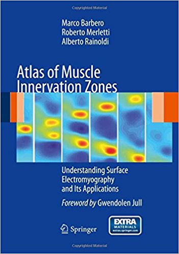
Atlas of Muscle Innervation Zones: Understanding Surface Electromyography and Its Applications

[amazon_link asins=’8847024625′ template=’ProductAd’ store=’aishabano-20′ marketplace=’US’ link_id=’57aa903c-3b6d-11e8-826b-5b51c0c83ea7′]

Invasive electromyography is a well-established diagnostic tool that has been used for decades by neurologists. Recently, new and alternative devices have increasingly become available that permit diagnosis without the use of needles. This developing area of science and the new tools have not, however, been sufficiently investigated in academic training. Consequently a gap exists between what science is making possible and the competence acquired during graduate studies. This handy volume has the aim of filling this gap by providing the information required by medical practitioners in rehabilitation, sports, and occupational health as well as by rehabilitation therapists, ergonomists, and sport coaches. The techniques that are presented and explained will help in monitoring and recording changes, evaluating the effectiveness of treatments and training, evaluating work stations, and preventing and documenting the evolution of occupational disorders of the neuromuscular system.

DOWNLOAD THIS BOOK FREE HERE

https://upsto.re/32H2SCF

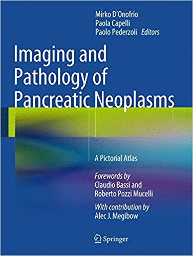
Imaging and Pathology of Pancreatic Neoplasms: A Pictorial Atlas

[amazon_link asins=’8847056772′ template=’ProductAd’ store=’aishabano-20′ marketplace=’US’ link_id=’855ae3aa-3744-11e8-b216-a3c77b66a078′]

Although interest in pancreatic pathology is very high in the radiological and gastroenterological communities, it is still the case that less is known about pathology of the pancreas than about liver pathology, for example. Diagnosis depends on the structure of the pancreatic lesion, which can be directly visualized on US, CT or MR images. This atlas, which encompasses both the imaging and the pathology of pancreatic neoplasms, will therefore be invaluable in enabling radiologists and sonographers to understand the underlying pathology and in allowing pancreatic pathologists to understand the imaging translation.

The emphasis in the atlas is very much on the pathological and imaging appearances, with most of the text concentrated at the beginning of the chapters. A comprehensive overview is provided of typical and atypical presentations and diverse aspects of common and uncommon pancreatic neoplasms, including ductal adenocarcinoma, neuroendocrine neoplasms, intraductal papillary mucinous neoplasms, cystic neoplasms, metastases and lymphoma.

DOWNLOAD THIS BOOK FREE HERE

https://upsto.re/3d2vHnf

Atlas of Imaging in Infertility: A Complete Guide Based in Key Images 1st ed

[amazon_link asins=’3319138928′ template=’ProductAd’ store=’aishabano-20′ marketplace=’US’ link_id=’b6d9417d-294a-11e8-9a2d-d729ddbe432d’]

This atlas gathers the most frequent imaging findings concerning alterations that cause infertility in both males and in females. Also, it discusses how the images should be analyzed and described, to serve as a guide for the physicians who request and perform these imaging tests.
Infertility is a highly prevalent condition – about 10% of couples are infertile with males and females equally affected. This book offers professionals dealing with infertility, including endocrinologists, gynecologists and urologists, a useful guide to the diagnostic imaging tests that are essential in treating these patients. The Atlas of Imaging in Infertility is a valuable resource for physicians from different specialties managing cases of infertility.
DOWNLOAD THIS BOOK FREE HERE
1 7 8 9 10 11 19