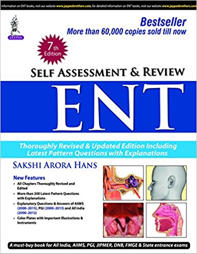
Self Assessment and Review ENT
[amazon_link asins=’9385999532′ template=’ProductAd’ store=’aishabano-20′ marketplace=’US’ link_id=’d0a2156b-3b6c-11e8-93ae-6b78f706ab80′]
Medical Books Library for Doctors, Physicians, Surgeons, Dentists, Intensivists, Physician Assistants, Nurses, Medical Technicians and Medical Students
Medical books library


[amazon_link asins=’9385999532′ template=’ProductAd’ store=’aishabano-20′ marketplace=’US’ link_id=’d0a2156b-3b6c-11e8-93ae-6b78f706ab80′]
[amazon_link asins=’3319673130′ template=’ProductAd’ store=’aishabano-20′ marketplace=’US’ link_id=’2f3cbee7-292c-11e8-abf8-8b719f87a8c4′]
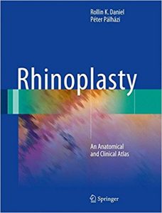
In this atlas, sequential anatomical dissections are presented which show each component of the nose in unprecedented meticulous detail. Anatomical photographs are often paired with anatomical drawings and even intraoperative clinical photographs to illustrate each part of the nose.
Rhinoplasty: An Anatomical and Clinical Atlas, provides an in-depth understanding of nasal anatomy and a wide variety of operative techniques. In rhinoplasty surgery, the surgeon must understand the tight linkage between surface aesthetics, underlying anatomy, and selection of operative techniques. The underlying anatomy is only revealed to a limited degree at the time of surgery and the surgeon must then adapt the operative plan to fit the actual anatomy observed in the operating room to achieve the patient’s desired aesthetic result. Ultimately, the goal of this atlas is to allow the surgeon to see the operative techniques in both cadavers and clinical cases which represents the best possible learning approach.
DOWNLOAD THIS BOOK FREE HERE

[amazon template=iframe image2&asin=3319480537]

[amazon template=image&asin=3131319127]
[amazon template=image&asin=3319533177]

[amazon template=image&asin=3319503936]
[amazon template=image&asin=160547715X]
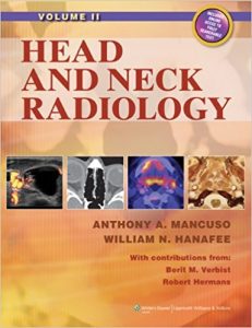
Written and edited by acknowledged masters in the field, this two-volume full-color text is the most comprehensive and current reference on head and neck radiology. It features more than 10,000 images and covers every disorder in every region of the head and neck.
The first two sections cover applied imaging fundamentals and general pathology, pathophysiology, patterns of disease, and natural history of head and neck disorders correlated with imaging appearance. Subsequent sections focus on specific anatomic regions: the eye, orbit, visual pathways, and cranial nerves III, IV, and VI; sinonasal and craniofacial region including cranial nerve V; temporal bone, posterior skull base, posterior fossa, and cranial nerves VII-XII; infrahyoid neck and cervico-thoracic junction (thoracic inlet); thyroid and parathyroid glands; major salivary glands; nasopharynx; oropharynx; oral cavity and floor of the mouth; larynx, hypopharynx, and cervical esophagus; trachea; hypopharynx; and cervical esophagus. The text covers all current imaging modalities, including plain film, MRI, CT, ultrasound, and nuclear medicine including PET.
A companion website will offer the fully searchable text and images. The first two sections will be online only.

[amazon template=image&asin=1444117955]
This innovative and accessible guide to ear, nose and throat medicine delivers everything you need to know to develop a thorough grounding in the subject, acting as an essential course companion and an ideal revision aid.
Written in a lively and accessible manner reflecting the author’s popular lecturing style, the content has been carefully matched to meet the demands of both the undergraduate curriculum and the core competencies for foundation study. Topics are approached in a problem-oriented manner, with basic science and clinical information integrated throughout in line with current teaching practice.
Essential ENT, praised in the first edition for its brevity, style and illustrations, remains an essential text for undergraduate medical students and foundation doctors, an excellent aide memoire for those embarking upon a career in ENT and studying for the Diploma in Laryngology and Otology, and a reliable companion in clinical practice for general practitioners and staff in the emergency department.

[amazon template=iframe image2&asin=0387244476]
Recognizing the clinician’s need for quick access to a comprehensive and immediately useful presentation of evidence-based material, the authors and editors have condensed the research on the most common otorhinolaryngological complaints into this indispensable volume. Their unique approach color-codes the level of research backing each set of evidence in order to make assessment of the evidence as quick and useful as possible. Each clinical problem is presented with a “color key,” letting the physician know the level of evidence available: green (high-level evidence), yellow (low–moderate levels of evidence), or red (major disagreement or only minimal low-level evidence). The content of each chapter is structured in the same manner so the reader quickly becomes accustomed to finding precisely the information needed for each new case.
Featuring sections on general otolaryngology, head and neck surgery, pediatrics, and otology, Evidence-Based Otolaryngology not only presents the research, but gives the clinician immediately applicable recommendations for patient treatment.
[amazon template=iframe image2&asin=0387890270]
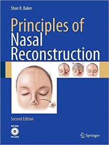
Skin cancer is among the most commonly occurring cancers, with incidence rates climbing among patients of all ages. The nose is the most common site for these cases. The vast majority of skin cancers of the nose are treated surgically by plastic surgeons, dermatologists, and otolaryngologists. Surgical excision requires reconstruction to one degree or another and Principles of Nasal Reconstruction will prove extremely helpful to any surgeon contemplating reconstruction of defects resulting from skin cancer removal. This book offers multiple guided surgical techniques and references to provide insight and practical guidance to the surgeon and trainee performing nasal reconstructions