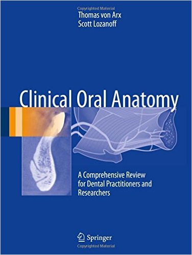
Clinical Oral Anatomy: A Comprehensive Review for Dental Practitioners and Researchers 1st ed. 2017 Edition
[amazon template=iframe image2&asin=3319419919]

This superbly illustrated book presents the most current and comprehensive review of oral anatomy for clinicians and researchers alike. In 26 chapters, the reader is taken on a unique anatomical journey, starting with the oral fissure, continuing via the maxilla and mandible to the tongue and floor of the mouth, and concluding with the temporomandibular joint and masticatory muscles. Each chapter offers a detailed description of the relevant anatomical structures and their spatial relationships, provides quantitative morphological assessments, and explains the relevance of the region for clinical dentistry. All dental health care professionals require a sound knowledge of anatomy for the purposes of diagnostics, treatment planning, and therapeutic intervention. A full understanding of the relationship between anatomy and clinical practice is the ultimate objective, and this book will enable the reader to achieve such understanding as the basis for provision of the best possible treatment for each individual patient as well as recognition and comprehension of unexpected clinical findings.

DOWNLOAD THIS BOOK FREE HERE

http://upsto.re/vuRSdLm

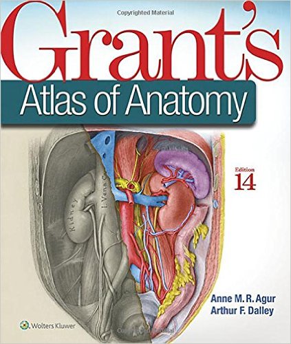
Grant’s Atlas of Anatomy (Grant, John Charles Boileau//Grant’s Atlas of Anatomy) 14th Edition

[amazon template=iframe image2&asin=1469890682]

Illustrations drawn from real specimens, presented in surface-to-deep dissection sequence, set Grant’s Atlas of Anatomy apart as the most accurate reference available for learning human anatomy. These realistic representations provide students with the ultimate lab resource.

For more than seventy years, Grant’s Atlas of Anatomy has continually adapted to meet the needs of each generation of students, while maintaining the Grant’s tradition of excellence. The meticulously updated fourteenth edition is a visually stunning reference that delivers the accuracy, pedagogy, and clinical relevance expected of this classic atlas, with new features that make it even more practical and user-friendly:

  • Recolorization of the original Grant’s Atlas images from high-resolution scans add a new level of organ luminosity and tissue transparency, and enable the formation of 3D constructs for each body region. This lavish visual enhancement ensures a vastly improved experience in learning the structure and function of the human body. See the enhanced color and detail here:  https://youtu.be/KdLzQUoG54Y
  • Schematic illustrations now feature a uniform style and consistent coloring, clearly showing the relationships of structures and clarifying anatomical concepts.
  • Newly revised legends with highlighted clinical applications describe the anatomic features and provide context for health care practice.
  • Enhanced medical imaging includes more than 100 clinically significant MRIs, CT images, ultrasound scans, and corresponding orientation drawings.
  • Updated and improved tables help organize muscles, vessels, and other anatomic information in an easy-to-use format ideal for review and study.

DOWNLOAD THIS BOOK FREE HERE

PART I http://upsto.re/v4RW8aM

PART II http://upsto.re/v4K7rJf

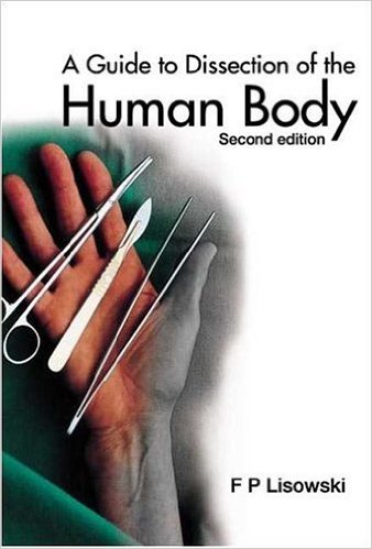
A Guide to Dissection of the Human Body 2nd ed. Edition

[amazon template=iframe image2&asin=9812388176]

In this second edition of A Guide to Dissection of the Human Body, certain dissecting instructions have been revised to increase clarity. Methodical and comprehensive, the guide complements various anatomy courses. The terminology has been checked and brought up to date, in accordance with the latest version of Terminologia Anatomica (1998).The major aim of this guide remains — to provide a well-rounded dissecting manual that reinforces, but does not replace, a textbook of human anatomy. The details of human anatomy covered here are of interest and importance primarily in a medical context. To this end the guide has been designed for medical, dental, osteopathy and physiotherapy students, and for students of alternative medicine where dissection of the human body is required. It has also been planned for postgraduate students proceeding to specialise in the various clinical (surgical, radiological, emergency medical, and gynaecological) sciences and thus need to revise their anatomical knowledge through dissection.It has to be stressed, learning anatomy is most efficient and retention is highest when didactic study is combined with the experience of dissection. In using this method one observes, palpates and moves parts of the body. It is a three-dimensional and visuo-tactile approach to the examination of the human body. Undoubtedly dissection reinforces and expands the knowledge gained from the textbook and from atlases and computerized-anatomy programs, The Guide is flexible enough for use in long as well as short courses, and is thus structured in such a way that the dissection of the body can be completed in 100 to 160 hours. It provides a link to real, living and variable anatomy.

DOWNLOAD THIS BOOK FREE HERE

http://upsto.re/v4Qg9xz

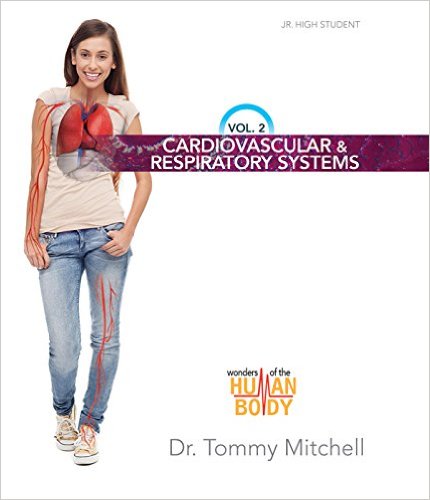
Wonders of the Human Body: Cardiovascular & Respiratory Systems

41-ktbdn2dl-_sx428_bo1204203200_-1

[amazon template=iframe image2&asin=0890519285]

There is absolutely no possibility that you were simply the process of random chance!

In Volume 2 of the Wonders of the Human Body series, Dr. Tommy Mitchell covers the intricate design of both the cardiovascular system, consisting of the blood, blood vessels, and heart, as well as the respiratory system that focuses on the transportation of oxygen through the body. From the level of the cells to the organs themselves, you will examine these systems in depth.

In the Cardiovascular & Respiratory Systems, prepare to discover the incredible design of the human heart, including

  • How blood moves through an incredible network of arteries and veins
  • What “blood pressure” is and the marvelous systems that help regulate it
  • How the respiratory system allows us to get the “bad air out” and the “good air in!”

Along the way, you will see what happens when things go wrong, but we made sure that you’ll find suggestions to help keep your heart and lungs healthy. Although the world insists that our bodies are merely the result of time and chance, as you examine your human body closely, you will see that it cannot be an accident. You can only be the living creation of our Master Designer!

DOWNLOAD THIS BOOK FREE HERE

http://upsto.re/vLG4RjE

Anatomy & Physiology

41iztyohhgl-_sx385_bo1204203200_

[amazon template=iframe image2&asin=1938168135]

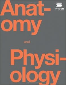
Anatomy and Physiology is a dynamic textbook for the yearlong Human Anatomy and Physiology course taught at most two- and four-year colleges and universities to students majoring in nursing and allied health. A&P is 29 chapters of pedagogically effective learning content, organized by body system, and written at an audience-appropriate level. The lucid text, strategically constructed art, inspiring career features, and links to external learning tools address the critical teaching and learning challenges in the course. Color is used for pedagogical effect in A&P. Most art will consist of elegant black line, with the strongest line illustrating the most important structure(s) and shading used to show dimension and shape. Color (used only when needed) highlights and clarifies the primary anatomical or functional point of the illustration. Student focus is drawn to the most important learning point in each illustration, without distraction from excessive gradients, shadows, and loud highlights. The online book provides students with links to surgical videos, histology, interactive diagrams, and cadaver imagery at critical junctures. This book is available online for free at openstaxcollege dot org

DOWNLOAD THIS BOOK FREE HERE

PART I http://upsto.re/vSpSr2o

PART II http://upsto.re/vSpYZE9

Surgical Anatomy Around the Orbit: The System of Zones

51tb6wljbhl-_sx471_bo1204203200_

[amazon template=iframe image2&asin=0781750814.pdf]

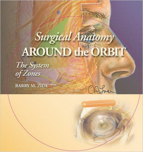
This full-color surgical atlas provides a detailed, true-to-life view of periorbital anatomy as seen during an operative procedure. More than 130 photographs of fresh cadaver dissections and more than 100 vivid drawings by an award-winning ophthalmic artist enable surgeons to thoroughly preview this complex anatomy prior to surgery.

Using his popular concept of facial zones, Dr. Zide depicts the fine anatomic details necessary for today’s advanced cosmetic and reconstructive procedures. A chapter on the seventh cranial nerve shows what tissue is innervated and delineates the exact zone of safety. A chapter on anatomy for facial nerve blocks is included.

DOWNLOAD THIS BOOK FREE HERE

http://upsto.re/vnkS2ko

Handbook of Anthropometry: Physical Measures of Human Form in Health and Disease 2012th Edition

41iopf8j-gl-_sx350_bo1204203200_

[amazon template=iframe image2&asin=144191787X]

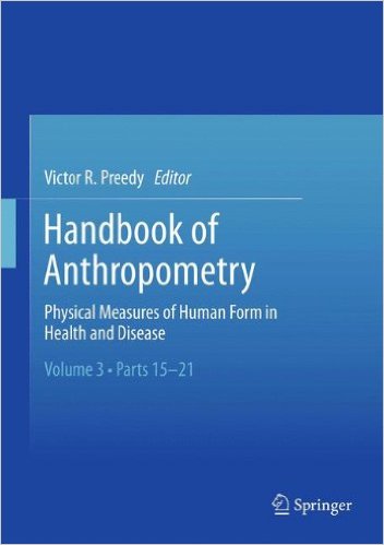
Although its underlying concept is a relatively simple one―the measurement of the human body and its parts―anthropometry employs a myriad of methods and instruments, and is useful for a variety of purposes, from understanding the impact of disease on individuals to tracking changes in populations over time. The first interdisciplinary reference on the subject, the Handbook of Anthropometry brings this wide-ranging field together: basic theory and highly specialized topics in normal and abnormal anthropometry in terms of health, disease prevention, and intervention. Over 140 self-contained chapters cover up-to-date indices, the latest studies on computerized methods, shape-capturing systems, and bioelectrical impedance, data concerning single tissues and whole-body variables, and reports from different areas of the world. Chapters feature helpful charts and illustrations, cross-references to related chapters are included, and key points are presented in bullet form for ease of comprehension. Together, the Handbook’s thirteen sections entail all major aspects of anthropometrical practice and research, including: Tools and techniques. Developmental stages, from fetus to elder. Genetic diseases, metabolic diseases, and cancer. Exercise and nutrition. Ethnic, cultural, and geographic populations. Special conditions and circumstances. The Handbook of Anthropometry is an invaluable addition to the reference libraries of a broad spectrum of health professionals, among them health scientists, physicians, physiologists, nutritionists, dieticians, nurses, public health researchers, epidemiologists, exercise physiologists, and physical therapists. It is also useful to college-level students and faculty in the health disciplines, as well as to policymakers and ergonomists.

DOWNLOAD THIS BOOK FREE HERE
http://upsto.re/vyE2vhb

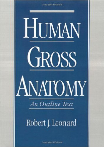
Human Gross Anatomy: An Outline Text 1st Edition

51vvy9kf2sl-_sx351_bo1204203200_

[amazon template=iframe image2&asin=0195090039]

This text presents gross anatomy in an expanded outline format. Its conciseness and logical organization permit easy previewing of the subject matter before a lecture or laboratory as well as efficient learning and reviewing of the material before an examination. Notwithstanding its succinct style, the text’s coverage of gross anatomy is comprehensive and is written at a level of detail appropriate for the medical student.
The book begins with an introduction, setting forth basic principles and terminology that will serve the student throughout the remainder of the text. Overviews of the nervous, muscular, skeletal, and vascular systems are also included in the introduction. The main part of the text is organized in the following sequence: The Back; The Thorax; The Abdomen; The Perineum and Pelvis; The Head and Neck; The Upper Limb; and The Lower Limb. Clinical notes that highlight anatomy relevant to current medical practice appear throughout these chapters. Since each regional anatomy chapter is complete in itself, the order in which they are studied is optional.

DOWNLOAD THIS BOOK FREE HERE
http://upsto.re/vyEwLM3

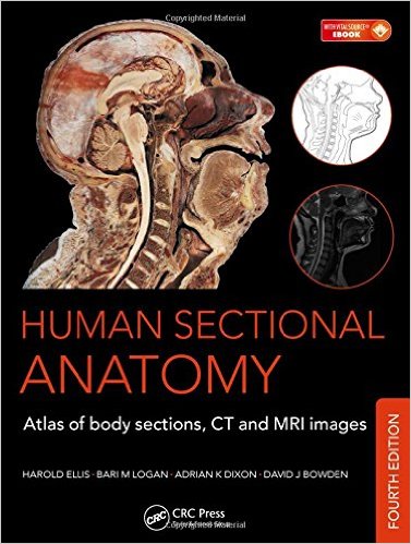
Human Sectional Anatomy: Atlas of Body Sections, CT and MRI Images, Fourth Edition 4th Edition

51sjhkrat7l-_sx375_bo1204203200_

[amazon template=iframe image2&asin=1498703607]

First published in 1991, Human Sectional Anatomy set new standards for the quality of cadaver sections and accompanying radiological images. Now in its fourth edition, this unsurpassed quality remains and is further enhanced by the addition of new material.

The superb full-colour cadaver sections are compared with CT and MRI images, with accompanying, labelled, line diagrams. Many of the radiological images have been replaced with new examples for this latest edition, captured using the most up-to date imaging technologies to ensure excellent visualization of the anatomy. The photographic material is enhanced by useful notes with details of important anatomical and radiological features.

Beautifully presented in a generous format, Human Sectional Anatomy continues to be an invaluable resource for all radiologists, radiographers, surgeons and medics, in training and in practice, and an essential component of departmental and general medical library collections.

DOWNLOAD THIS BOOK FREE HERE
http://upsto.re/vrqJ3VZ

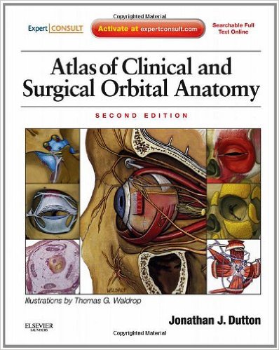
Atlas of Clinical and Surgical Orbital Anatomy: Expert Consult: Online and Print, 2e 2nd Edition

51mgwsmipil-_sx396_bo1204203200_

[amazon template=iframe image2&asin=1437722725]

Atlas of Clinical and Surgical Orbital Anatomy, by Dr. Jonathan Dutton, demonstrates the complex area of orbital anatomy through unique illustrations and comprehensive coverage that goes from embryology to adult anatomy. This completely updated and revised new edition features a new chapter on the cavernous sinus, illustrations modified to reflect recent anatomic findings, and new sections covering clinical correlations. With access to the fully searchable text and a downloadable image gallery online at www.expertconsult.com, you’ll have the detailed visual guidance you need to perform successful orbital surgery.

  • Clearly see the nuances of each anatomic system with layered illustrations that use multiple artworks to display relevant structures and highlight key intricacies.
  • Visualize each system three-dimensionally through depictions from frontal, lateral, and superior angles.
  • Apply a comprehensive approach to common orbital diseases using coverage of clinical correlations from embryology to adult anatomy.
  • Access the fully searchable text online at www.expertconsult.com, along with a downloadable image gallery.
  • Master the anatomy-disease-surgery relationship thanks to new chapter sections on clinical correlations.
  • Get a more complete understanding of orbital disease through a new chapter on the cavernous sinus and illustrations modified to reflect recent anatomic findings.
  • Stay current on the newest research data with completely revised and updated chapters and references.

Completely understand the complex area of orbital anatomy with this uniquely illustrated guide

DOWNLOAD THIS BOOK FREE HERE
http://upstore.net/vrKiYF4

1 16 17 18 19 20 22