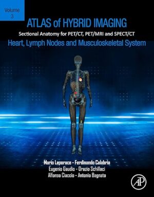
Atlas of Hybrid Imaging Sectional Anatomy for PET/CT, PET/MRI and SPECT/CT Vol. 3: Heart, Lymph Node and Musculoskeletal System

Atlas of Hybrid Imaging Sectional Anatomy for PET/CT, PET/MRI and SPECT/CT Vol. 3: Heart, Lymph Node and Musculoskeletal System

Atlas of Hybrid Imaging of the Heart, Lymph Nodes and Musculoskeletal System, Volume Three: Sectional Anatomy for PET/CT, PET/MRI and SPECT/CT provides a guide for interpreting PET and SPECT in relation to co-registered CT and/or MRI. In this atlas, exclusively dedicated to heart, lymph nodes and musculoskeletal system, nuclear physicians and radiologists cover hybrid nuclear medicine based on their own case studies. The practical structure in two-page unit offers readers a navigational tool based on anatomical districts, with labeled and explained low-dose multiplanar CT or MRI views merged with PET fusion imaging on one side and enhanced CT or MRI on the other.
This new format enables the rapid identification of hybrid nuclear medicine findings which are now routine at leading medical centers. Each chapter begins with three-dimensional CT and/or MRI views of the evaluated anatomical region, bringing forward sectional tables. Clinical cases, tricks and pitfalls linked to several PET or SPECT radiopharmaceuticals help introduce the reader to peculiar molecular pathways and improve confidence in cross-sectional imaging that is vital for accurate diagnosis and treatments.

– Presents a compact, comprehensive, easy-to-read guide on sectional imaging and multiplanar evaluation of hybrid PET and SPECT
– Includes more than 200 fully colored, labeled, high quality original images of axial, coronal and sagittal CT, contrast enhanced CT, PET/CT and/or PET/MRI
– Displays clinical cases that showcase both common and unusual findings that nuclear physicians and radiologists could encounter in their clinical practice
– Provides specific text boxes that explain anatomical variants, radiological advices and physiological findings linked to tracer bio-distribution

DOWNLOAD THIS MEDICAL BOOK 

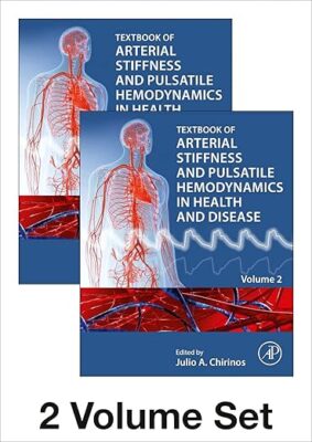
Textbook of Arterial Stiffness and Pulsatile Hemodynamics in Health and Disease 1st Edition

Textbook of Arterial Stiffness and Pulsatile Hemodynamics in Health and Disease 1st Edition

Textbook of Arterial Stiffness and Pulsatile Hemodynamics in Health and Disease, Two Volume Set covers the principles, physiology, biologic pathways, clinical implications and therapeutics surrounding arterial stiffness and pulsatile hemodynamics, along with a thorough overview of the field. The book presents complex engineering concepts in a way that those in science and medicine can more easily understand. It includes detailed illustrations. Additionally, it presents advanced bioengineering concepts in boxes for readers who wants more in-depth biophysical knowledge. This is a must-have reference for students, researchers and clinicians interested in learning more about this field.

  • Incorporates case studies and calculations/worked examples with mathematical principles explained in a conceptual manner without complicated formulas
  • Features chapter contributions from leading international researchers and clinicians
  • Covers principles, physiology, biologic pathways, clinical implications and therapeutics

DOWNLOAD THIS MEDICAL BOOK 

Moore’s Clinically Oriented Anatomy

Moore’s Clinically Oriented Anatomy 

Renowned for its comprehensive coverage and engaging, storytelling approach, the bestselling Moore’s Clinically Oriented Anatomy, 9th Edition, guides students from initial anatomy and foundational science courses through clinical training and practice. A popular resource for a variety of programs, this proven text serves as a complete reference, emphasizing anatomy that is important in physical diagnosis for primary care, interpretation of diagnostic imaging, and understanding the anatomical basis of emergency medicine and general surgery. The 9th Edition reflects the latest changes in the clinical application of anatomy as well as preparation for the USMLE while maintaining the highest standards for scientific and clinical accuracy.

DOWNLOAD THIS MEDICAL BOOK 

Netter’s Concise Radiologic Anatomy: With STUDENT CONSULT Online Access (Netter Basic Science) 2nd Edition

Netter’s Concise Radiologic Anatomy: With STUDENT CONSULT Online Access (Netter Basic Science) 2nd Edition

Designed to make learning more interesting and clinically meaningful, Netter’s Concise Radiologic Anatomy, 2nd Edition matches radiologic images―from MR and ultrasound to CT and advanced imaging reconstructions―to the exquisite artwork of master medical illustrator Frank H. Netter, MD. As a companion to the bestselling Netter’s Atlas of Human Anatomy, this updated medical textbook begins with the anatomy and matches radiologic images to the anatomic images; the result is a concise, visual guide that shows how advanced diagnostic imaging is an amazing “dissection tool” for viewing human anatomy in the living patient!

“Overall I feel that this textbook is a good overview of radiology, and perfectly adequate for medical students as we are not expected to have a very in depth knowledge of radiology past chest and abdominal x rays. It may well prove valuable for the Netter-collectors out there, and provides an excellent quick guide to radiology.” Reviewed by: TheMedicalStudent.co.uk Date: July 2014

  • Quickly review key information with a concise, user-friendly format that is organized and color-coded to be in-line with Netter’s Atlas of Human Anatomy, 6th Edition.
  • View direct, at-a-glance comparisons between idealized anatomic illustrations and real-life medicine with side-by-side radiology examples of normal anatomy and common variants with corresponding anatomy illustrations.
  • Improve upon your knowledge with a brief background in basic radiology, including reconstructions and a list of common abbreviations for the images presented.
  • Broaden your visual comprehension with the help of 30 brand-new ultrasound images.
  • Access the complete contents online at Student Consult.

DOWNLOAD THIS MEDICAL BOOK 

Netter Atlas of Human Anatomy: A Systems Approach: paperback + eBook (Netter Basic Science) 8th Edition

Netter Atlas of Human Anatomy: A Systems Approach: paperback + eBook (Netter Basic Science) 8th Edition

For students and clinical professionals who are learning anatomy, participating in a dissection lab, sharing anatomy knowledge with patients, or refreshing their anatomy knowledge, the Netter Atlas of Human Anatomy illustrates the body, system by system, in clear, brilliant detail from a clinician’s perspective. Unique among anatomy atlases, it contains illustrations that emphasize anatomic relationships that are most important to the clinician in training and practice. Illustrated by clinicians, for clinicians, it contains more than 550 exquisite plates plus dozens of carefully selected radiologic images for common views.

  • Presents world-renowned, superbly clear views of the human body from a clinical perspective, with paintings by Dr. Frank Netter as well as Dr. Carlos A. G. Machado, one of today’s foremost medical illustrators.
  • Content guided by expert anatomists and educators: R. Shane Tubbs, Paul E. Neumann, Jennifer K. Brueckner-Collins, Martha Johnson Gdowski, Virginia T. Lyons, Peter J. Ward, Todd M. Hoagland, Brion Benninger, and an international Advisory Board.
  • Offers system-by-system coverage, including quick reference notes on structures with high clinical significance in common clinical scenarios and a muscle table appendix.
  • Contains new illustrations by Dr. Machado including clinically important areas such as the pelvic cavity, temporal and infratemporal fossae, nasal turbinates, and more.
  • Features new nerve tables devoted to the cranial nerves and the nerves of the cervical, brachial, and lumbosacral plexuses.
  • Uses updated terminology based on the second edition of the international anatomic standard, Terminologia Anatomica, and includes common clinically used eponyms.
  • Enhanced eBook version included with purchase. Your enhanced eBook allows you to access all of the text, figures, and references from the book on a variety of devices.
  • Provides access to extensive digital content: every plate in the Atlas―and over 100 bonus plates including illustrations from previous editions―is enhanced with an interactive label quiz option and supplemented with “Plate Pearls” that provide quick key points of the major themes of each plate. Digital content also includes over 300 multiple choice questions and other learning tools.

Also available:

• Netter Atlas of Human Anatomy: Classic Regional Approach―Same content as the systems approach, but organized traditionally, body region by body region.

Both options contain the same table information and same 550+ illustrated plates painted by clinician artists, Frank H. Netter, MD, and Carlos Machado, MD.

DOWNLOAD THIS MEDICAL BOOK 

Grant’s Atlas of Anatomy (Lippincott Connect) 15th Edition

Grant’s Atlas of Anatomy (Lippincott Connect) 15th Edition

Selected as a Doody’s Core Title for 2021 and 2023!

For more than seventy-five years, 
Grant’s Atlas of Anatomy has maintained a tradition of excellence while continually adapting to meet the needs of each generation of students. The updated fifteenth edition is a visually stunning reference that delivers the accuracy, pedagogy, and clinical relevance expected of this classic atlas, with new and enhanced features that make it even more practical and user-friendly.
 
Illustrations drawn from real specimens, presented in surface-to-deep dissection sequence, set 
Grant’s Atlas of Anatomy apart as the most accurate reference available for learning human anatomy. These realistic representations bring structures to life and provide students with the ultimate lab resource.

  • NEW Illustration overviews highlight the autonomic nerves to clarify nerve and muscle innervations.
  • NEW and updated images reflect the latest clinical insights through:
    • 15 NEW Illustrations
    • 160 revised figures
    • NEW and updated surface anatomy images
    • NEW pulses photos
  • Renowned, high-resolution, dynamically colored illustrations organized in dissection sequence enable the formation of 3-D constructs for each body region and provide detailed, realistic reference during dissection.
  • Tables detail muscles, vessels, and other anatomic information in an easy-to-use format ideal for review and study.
  • Enhanced medical imaging includes more than 100 clinically significant MRIs, CT images, ultrasound scans, and corresponding orientation drawings to help students confidently apply the laboratory experience to clinical rotations.
  • Color schematic illustrations reinforce the relationships of structures and anatomical concepts in vibrant detail.

DOWNLOAD THIS MEDICAL BOOK 

Skandalakis’ Surgical Anatomy: The Embryologic and Anatomic Basis of Modern Surgery First Edition, First ed.

Skandalakis’ Surgical Anatomy: The Embryologic and Anatomic Basis of Modern Surgery First Edition, First ed.

Brief facts of embryogenesis are included in this book because embryology leads the student to a more thorough understanding of human anatomy. Applied, surgically oriented anatomy emphasizing both surgical applications and ways to avoid anatomic complications have been included. Because minimally invasive and robotic surgery is definitely the surgery of today and tomorrow, the modern surgeon must now know this other type of anatomy, the “”non-touch, non-see”” anatomy. In other words, the surgeon must know anatomy very thoroughly indeed.

DOWNLOAD THIS MEDICAL BOOK 

Gray’s Surgical Anatomy

Gray’s Surgical Anatomy 

Written and edited by expert surgeons in collaboration with a world-renowned anatomist, this exquisitely illustrated reference consolidates surgical, anatomical and technical knowledge for the entire human body in a single volume. Part of the highly respected Gray’s ‘family,’ this new resource brings to life the applied anatomical knowledge that is critically important in the operating room, with a high level of detail to ensure safe and effective surgical practice. Gray’s Surgical Anatomy is unique in the field: effectively a textbook of regional anatomy, a dissection manual, and an atlas of operative procedures – making it an invaluable resource for surgeons and surgical trainees at all levels of experience, as well as students, radiologists, and anatomists.

  • Brings you expert content written by surgeons for surgeons, with all anatomical detail quality assured by Lead Co-Editor and Gray’s Anatomy Editor-in-Chief, Professor Susan Standring.

  • Features superb colour photographs from the operating room, accompanied by detailed explanatory artwork and figures from the latest imaging modalities – plus summary tables, online videos (intra-operative and cadaveric), self-assessment questions, and case-based scenarios – making it an ideal reference and learning package for surgeons at all levels.

  • Reflects contemporary practice with chapters logically organized by anatomical region, designed for relevance to surgeons across a wide range of subspecialties, practice types, and clinical settings – and aligned to the requirements of current trainee curricula.

    • Maximizes day-to-day practical application with references to core surgical procedures throughout, as well as the ‘Tips and Anatomical Hazards’ from leading international surgeons.

    • Demonstrates key anatomical features and relationships that are essential for safe surgical practice – using brand-new illustrations, supplemented by carefully selected contemporary artwork from the most recent edition of Gray’s Anatomy and other leading publications.

    • Integrates essential anatomy for robotic and minimal access approaches, including laparoscopic and endoscopic techniques.

      • Features dedicated chapters describing anatomy of lumbar puncture, epidural anaesthesia, peripheral nerve blocks, echocardiographic anatomy of the heart, and endoscopic anatomy of the gastrointestinal tract – as well as a unique overview of human factors and minimizing error in the operating room, essential non-technical skills for improving patient outcomes and safety.
      • Enhanced eBook version included with purchase. Your enhanced eBook allows you to access all of the text, figures, and references from the book on a variety of devices – PLUS a wealth of BONUS content not found in the printed book.


DOWNLOAD THIS MEDICAL BOOK 

1 2 3 22