The Rules of Radiology

The Rules of Radiology

This book gets to the heart of what radiology is and what radiologists do. As a relatively young speciality, there is no guide for radiologists to act as a moral compass. Until now, that is. You will not find any dry technical matters in here. You will not find any clues about how to interpret images better. This book details the ‘other 50%’: the rest of the working week when a radiologist is not reading scans or performing procedures.

The essence of radiology is distilled and offered up to the reader. If you want a comfortable read that offers bland reassurances, look elsewhere. If you want a book that questions everything and discusses uncomfortable truths, this is the book for you. Each of the Rules addresses an important part of professional practice. This book is a manifesto for all radiologists across the globe to raise their game, to be more effective and to serve their patients better.

DOWNLOAD THIS MEDICAL BOOK


Fast Facts for the Radiology Nurse 2nd Edition

Fast Facts for the Radiology Nurse 2nd Edition

The second edition of this pocket-sized resource for radiology nurses continues to demystify complex procedures by providing concise yet comprehensive information on the basics of radiology. As the complexity of healthcare grows, so too does imaging ability. Different modalities, technologies, and skills levels must all work in harmony to provide precision images. Radiology nurses must be proactive, patient-focused, and able to work with a diverse team of individuals to care for the widest range of patients.

Describing essential procedures and protocols in quick access style, Fast Facts for the Radiology Nurse, Second Edition covers over 50 different Interventional Radiology procedures. Woven throughout is an emphasis on interprofessional care and effective communication along with guidance on forming and maintaining a high-performing team. Abundant Clinical Pearls disseminate the hard-earned wisdom of expert radiology nurses. Extensively updated with the most current guidelines and protocols, the second edition presents four completely new chapters on Legal Issues Affecting Radiology, Strategies for Working with Difficult People, Safety and Quality in the Radiology Setting, and Emergency Management and Catastrophe Response.

DOWNLOAD THIS MEDICAL BOOK


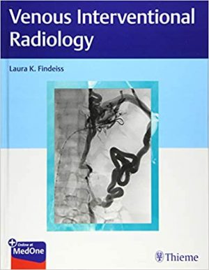
Venous Interventional Radiology

Venous Interventional Radiology

The diagnosis and management of venous disorders is a core competency of interventional radiology, comprising up to 75% of an interventional radiologist’s caseload in some institutions. Venous Interventional Radiology by prominent interventional radiologist Laura Findeiss and an impressive group of contributors is a highly practical vascular interventional radiology reference that covers a full spectrum of venous disease. The richly illustrated book starts with an opening chapter on venous anatomy, physiology, and epidemiology.

Disease-specific chapters cover common to complex disorders, from varicose veins and venous ulceration, to thromboembolic disease and pulmonary embolism. Each chapter presents the latest minimally invasive image-guided interventions for the diagnosis and management of a specific disease. Clinical cases provide discussion of patient presentation/symptoms, clinical evaluation, procedural and non-invasive management strategies, and follow-up, all of which are key to delivering comprehensive patient care.

DOWNLOAD THIS MEDICAL BOOK


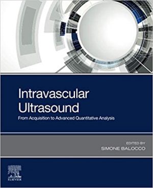
Intravascular Ultrasound: From Acquisition to Advanced Quantitative Analysis

Intravascular Ultrasound: From Acquisition to Advanced Quantitative Analysis

Intravascular Ultrasound: From Acquisition to Advanced Quantitative Analysis covers topics of the whole imaging pipeline, ranging from the definition of the clinical problem and image acquisition systems to image processing and analysis, including the assisted clinical-decision making procedures and treatment planning (stent deployment and follow up). Atherosclerosis, a disease of the vessel wall that produces vessel narrowing and obstruction, is the major cause of cardiovascular diseases, such as heart attack or stroke. This book covers all aspects of this imaging tool that allows for the visualization of internal vessel structures and the quantification and characterization of coronary plaque.

DOWNLOAD THIS MEDICAL BOOK


Felson’s Principles of Chest Roentgenology A Programmed 5th Edition
Felson’s Principles of Chest Roentgenology A Programmed 5th Edition

Easy to read, engaging, and highly interactive, Felson’s Principles of Chest Roentgenology: A Programmed Text, 5th Edition, has long been the go-to learning resource for medical students, residents, radiologists, and others who order and interpret chest x-rays. It offers a clear, self-directed tutorial on all aspects of chest imaging, including pathologies and anatomic challenges. You’ll find essential, accessible explanations of basic science, image reading and interpretation, and key terminology, along with hundreds of high-quality radiographs and interactive quizzes that have made this best-selling title the must-have primer of chest radiology.

Presents essential concepts in a straightforward, logically sequenced manner, with one chapter building on the next.

Emphasizes basic radiographic anatomy and signs of disease seen in everyday practice on the chest x-ray, helpfully presented from various points of view.

Includes more than 550 radiographs (many are new!) with correlative PET, CT, and MR images as appropriate―all presented with humor and insightful comments that provide a uniquely engaging self-directed learning experience for clinical application or board review.

Keeps you up to date with the latest thoracic imaging topics, including pleuroparenchymal fibroelastosis, combined pulmonary fibrosis with emphysema (CPFE), age-related lung changes, interstitial lung disease (ILD), lung cancer screening and tumor classification, and lower radiation dosing and safety considerations.

Provides numerous multiple-choice questions and quizzes throughout, along with answers, annotated x-rays, line drawings, cartoons, and engaging clinical tips.

Includes access to robust interactive offerings online, such as easy-to-access quizzes and board review questions.

Enhanced eBook version included with purchase. Your enhanced eBook allows you to access all of the text, figures, and references from the book on a variety of devices.

DOWNLOAD THIS MEDICAL BOOK


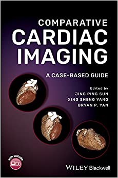
Comparative Cardiac Imaging: A Case-based Guide

Comparative Cardiac Imaging: A Case-based Guide

Comparative Cardiac Imaging: A Case-based Guide

Most books currently available on cardiac imaging focus on just modality. As the number of imaging options has increased, the choice of procedure has become more complicated. This comprehensive book will guide the practitioner in choosing the most appropriate test when confronted by various cardiac symptoms and diseases and to understand the benefits and limitations of each imaging modality. It demonstrates the advantages and disadvantages of various imaging modalities such as echocardiography, computer tomography, MRI, and nuclear cardiology in the evaluation of various disease states both commonly and infrequently seen in a standard practice.

Comparative Cardiac Imaging―A Case-based Guide utilizes actual case examples to demonstrate the state of the art in comparative cardiac imaging. It offers in-depth chapter coverage of Aortic Diseases; Aortic Valvular Diseases; Mitral Valvular Diseases; Prosthetic Valves; Coronary Artery Disease; Pulmonary Artery Diseases; Congenital Heart Disease; Cardiac Tumor; Infective Disease; Cardiomypathy; and Cardiac Trauma.

DOWNLOAD THIS MEDICAL BOOK


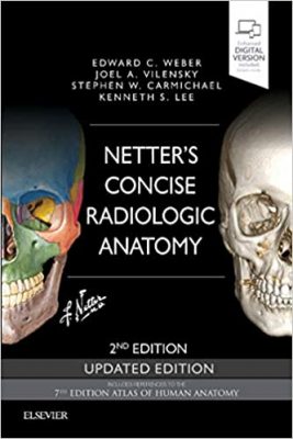
Netter’s Concise Radiologic Anatomy Updated Edition 2nd Edition

Netter’s Concise Radiologic Anatomy Updated Edition 2nd Edition

Designed to make learning more interesting and clinically meaningful, Netter’s Concise Radiologic Anatomy matches radiologic images?from MR and ultrasound to CT and advanced imaging reconstructions?to the exquisite artwork of master medical illustrator Frank H. Netter, MD. As a companion to the bestselling Netter’s Atlas of Human Anatomy, this updated medical textbook begins with the anatomy and matches radiologic images to the anatomic images; the result is a concise, visual guide that shows how advanced diagnostic imaging is an amazing “dissection tool” for viewing human anatomy in the living patient!

View direct, at-a-glance comparisons between idealized anatomic illustrations and real-life medicine with side-by-side radiology examples of normal anatomy and common variants with corresponding anatomy illustrations.

Improve upon your knowledge with a brief background in basic radiology, including reconstructions and a list of common abbreviations for the images presented.

Broaden your visual comprehension with the help of 30 brand-new ultrasound images.

Access the complete contents online at Student Consult.

DOWNLOAD THIS MEDICAL BOOK


Weir & Abrahams’ Imaging Atlas of Human Anatomy 6th Edition

Weir & Abrahams’ Imaging Atlas of Human Anatomy 6th Edition

Imaging is ever more integral to anatomy education and throughout modern medicine. Building on the success of previous editions, this fully revised sixth edition provides a superb foundation for understanding applied human anatomy, offering a complete view of the structures and relationships within the whole body, using the very latest imaging techniques.

FOR MORE BOOKS VISIT EDOWNLOADS.ME

All relevant imaging modalities are included, from plain radiographs to more advanced imaging of ultrasound, CT, MRI, functional imaging and angiography. Coverage is further enhanced by a carefully selected range of BONUS electronic content, including clinical photos and cases, ultrasound videos, labelled radiograph ‘slidelines’, cross-sectional imaging stacks and test-yourself materials. Uniquely, key syllabus image sets are now highlighted throughout to aid efficient study, as well as the most common, clinically important anatomical variants that you should be aware of.

This superb package is ideally suited to the needs of medical students, as well as radiologists, radiographers and surgeons in training. It will also prove invaluable to the range of other students and professionals who require a clear, accurate, view of anatomy in current practice.

DOWNLOAD THIS MEDICAL BOOK


Musculoskeletal MRI 3rd Edition

Musculoskeletal MRI 3rd Edition

Ideal for residents, practicing radiologists, and fellows alike, this updated reference offers easy-to-understand guidance on how to approach musculoskeletal MRI and recognize abnormalities. Concise, to-the-point text covers MRI for the entire musculoskeletal system, presented in a highly templated format. Thoroughly revised and enhanced with full-color artwork throughout, this resource provides just the information you need to perform and interpret quality musculoskeletal MRI.

Includes the latest protocols, practical advice, tips, and pearls for diagnosing conditions impacting the temporomandibular joint, shoulder, elbow, wrist/hand, spine, hips and pelvis, knee, and foot and ankle.

Follows a quick-reference format throughout, beginning with basic technical information on how to obtain a quality examination, followed by a discussion of the normal appearance and the abnormal appearance for each small unit that composes a joint.

Depicts both normal and abnormal anatomy, as well as disease progression, through more than 600 detailed, high-quality images, most of which are new to this edition.

Features key information boxes throughout for a quick review of pertinent material.

Expert Consult™ eBook version included with purchase. This enhanced eBook experience allows you to search all of the text, figures, and references from the book on a variety of devices.
DOWNLOAD THIS MEDICAL BOOK


Radiology Structured Reporting Handbook

Radiology Structured Reporting Handbook

Multiple studies show that referring physicians have a clear preference for structured radiology reports due to clarity and ease of interpretation, yet a one-size-fits all approach does not address disease complexities. Concurrently, the use of structured radiology templates has increased, driven in part by the need to comply with big data and artificial intelligence as well as reimbursement. Standardization of reporting is one of the first essential steps in the transformation of radiology from “the art of imaging” to a robust data science.

Radiology Structured Reporting Handbook: Disease-Specific Templates and Interpretation Pearls by Professors Olga R. Brook, Wieland H. Sommer, and esteemed colleagues is a highly practical guide on structured reporting for every major area of radiology. Featuring disease-specific templates, the book is organized in six sections and 53 chapters. Section one covers core foundation topics, from different definitions of structured reporting and pros and cons to change management and how to build templates. Five disease-specific sections encompass specific cancers and a variety of abdominal, thoracic, neurological, and cardiovascular diseases and conditions.

Key Highlights

  • Downloadable disease-specific templates for a variety of clinical entities including cardiovascular, thoracic, abdominal, oncological, and neuroradiology
  • Essential interpretation pearls for specific diseases from top experts in a bullet format, accompanied by relevant figures and tables


Together, the templates and pearls provide an essential and unique practice resource for optimal and clinically relevant reporting. The book also serves as a succinct educational tool for radiology trainees and practicing radiologists who may not interpret specific highly specialized types of studies on a daily basis.

DOWNLOAD THIS BOOK


1 6 7 8 9 10 62