The Final FRCR: Self-Assessment

The Final FRCR: Self-Assessment
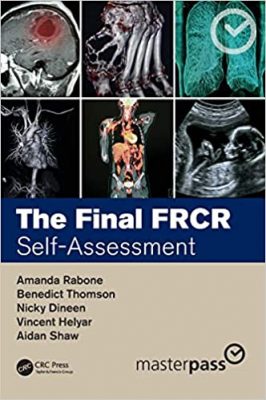
The Final FRCR: Self-Assessment

This is an SBA question medical book aimed at the post-graduate radiology market, specifically those taking the Fellowship of the Royal College of Radiology (FRCR) part 2 (‘final’) exams. This is a complementary title to The Final FRCR: Complete Revision Notes, which published in 2018.

Part 2 of the FRCR is itself composed of two elements. Part 2a is a series of six multiple choice exams covering the major body systems: musculoskeletal & trauma, gastrointestinal, genitourinary, head and neck, pediatrics and chest. Part 2b involves a written exam and an oral viva and is typically taken at the beginning of the fourth year of specialty training. Approximately 700-1000 trainees sit the exam each year. The SBAs would also be applicable for those studying for other exams or coming to the UK to sit the UK exams from Asia and the Middle East.

FOR MORE BOOKS VISIT EDOWNLOADS.ME

Key Features

Resource designed for those taking the final FRCR (UK exam) designed to be a complementary product to The Final FRCR: Complete Revision Notes.

Templated question format across the six major body systems.

Written by recent graduates of the FRCR exams who know how best to approach the topic.

Reviewed by senior advisors.

DOWNLOAD THIS MEDICAL BOOK


FRCR Physics Notes: Medical imaging physics for the First FRCR examination

FRCR Physics Notes: Medical imaging physics for the First FRCR examination

FRCR Physics Notes: Medical imaging physics for the First FRCR examination

This superb medical book is suited to those sitting the first FRCR physics examination in the UK and covers the scope of the Royal College of Radiologists syllabus. Although aimed at UK radiology trainees, it is also suitable for international residents taking similar examinations, postgraduate medical physics students and radiographers. The notes provide an excellent overview for anyone interested in the physics of radiology or just refreshing their knowledge.

FOR MORE MEDICAL BOOKS VISIT EDOWNLOADS.ME

The notes cover all of radiology physics, including:

  • Basic science
  • X-ray imaging
  • CT imaging
  • Ultrasound imaging
  • MR imaging (MRI)
  • Molecular imaging
  • Radiation dosimetry, protection and legislation


This third edition includes updates to reflect new legislation and many new illustrations, added sections, and removal of content no longer relevant to the FRCR physics exam. This edition has gone through strict critique and evaluation by physicists and other specialists to provide an accurate, understandable and up-to-date resource. The book summarises and pulls together content from the FRCR Physics Notes at Radiology Cafe and delivers it as a paperback or eBook for you to keep and read anytime.

There are 7 main chapters, which are further subdivided into 60 sub-chapters so topics are easy to find, and a comprehensive appendix and index have been included at the back of the book.

DOWNLOAD THIS MEDICAL BOOK


AI Innovation in Medical Imaging Diagnostics

AI Innovation in Medical Imaging Diagnostics

AI Innovation in Medical Imaging Diagnostics

Recent advancements in the technology of medical imaging, such as CT and MRI scanners, are making it possible to create more detailed 3D and 4D images. These powerful images require vast amounts of digital data to help with the diagnosis of the patient. Artificial intelligence (AI) must play a vital role in supporting with the analysis of this medical imaging data, but it will only be viable as long as healthcare professionals and AI interact to embrace deep thinking platforms such as automation in the identification of diseases in patients.

FOR MORE BOOKS VISIT EDOWNLOADS.ME

AI Innovation in Medical Imaging Diagnostics is an essential reference source that examines AI applications in medical imaging that can transform hospitals to become more efficient in the management of patient treatment plans through the production of faster imaging and the reduction of radiation dosages through the PET and SPECT imaging modalities. The book also explores how data clusters from these images can be translated into small data packages that can be accessed by healthcare departments to give a real-time insight into patient care and required interventions. Featuring research on topics such as assistive healthcare, cancer detection, and machine learning, this book is ideally designed for healthcare administrators, radiologists, data analysts, computer science professionals, medical imaging specialists, diagnosticians, medical professionals, researchers, and students.

DOWNLOAD THIS MEDICAL BOOK


Top 3 Differentials in Musculoskeletal Imaging: A Case Review

Top 3 Differentials in Musculoskeletal Imaging: A Case Review

Top 3 Differentials in Musculoskeletal Imaging: A Case Review

This medical book Top 3 Differentials in Musculoskeletal Imaging: A Case Review by Jasjeet Bindra, Robert D. Boutin, and expert contributors is one in a series of radiology case books mirroring the format of the highly acclaimed O’Brien classic, Top 3 Differentials in Radiology: A Case Review. The book is organized in 10 parts: trauma, bone tumors, upper extremity, lower extremity, arthropathies, infection, soft tissue tumors, metabolic musculoskeletal conditions, spine, and pediatric/developmental musculoskeletal conditions.

FOR MORE BOOKS VISIT EDOANLOADS.ME

In this medical book each case is formatted as a two-page unit. The left page features clinical images, succinctly captioned findings, and pertinent clinical history. The right page includes the key imaging gamut, differential diagnoses, additional diagnostic considerations, the diagnosis, clinical pearls, and suggested readings.

Key Features

  • A total of 146 cases, each focused on a specific imaging finding, including aggressive periosteal reaction, focal cortical thickening, diffusely increased bone density, focal periphyseal edema, acro-osteolysis, and more
  • A wealth of meticulously selected, high-quality radiographs, CTs, and MRIs enhance diagnostic skills
  • A list of differential diagnoses provides an ideal curriculum guide for trainees and educators alike


Radiology residents, fellows, and staff radiologists preparing for certification will greatly benefit from reading this text to prep for the radiology core and certifying exams. This is also an outstanding, day-to-day practice resource for practicing radiologists, clinicians, and orthopaedic surgeons involved in reviewing and interpreting musculoskeletal radiology studies.

DOWNLOAD THIS MEDICAL BOOK


Echocardiography in Pediatric and Congenital Heart Disease 3rd Edition

Echocardiography in Pediatric and Congenital Heart Disease 3rd Edition

Echocardiography in Pediatric and Congenital Heart Disease provides comprehensive guidance on the use of non-invasive ultrasound imaging in the diagnosis and treatment of pediatric cardiac conditions. Written by a team of experts from the world’s leading pediatric cardiology centers, this highly-illustrated, full-color reference covers anatomy, pathophysiology, ultrasound physics, laboratory setup, patient preparation and safety, pediatric echocardiogram protocols, quantitative methods of echocardiographic evaluation, and more.

Offering a wealth of additional material on state-of-the-art techniques and technologies in echocardiography, the thoroughly revised third edition features entirely new chapters on examination guidelines and standards, quality improvement in the laboratory, perioperative echocardiography, hemodynamic assessment of the neonate, early fetal echocardiography, and multimodality imaging. This edition offers updated and expanded discussion of the latest advances in echocardiography, particularly those related to speckle tracking and 3D echocardiography. An essential resource for all practitioners, instructors, and trainees in the field, Echocardiography in Pediatric and Congenital Heart Disease:

  • Provides up-to-date reference to ultrasound imaging of the hearts of fetuses, children, and adults with both acquired and congenital heart disease
  • Covers the echocardiographic examination of congenital cardiovascular abnormalities before, during, and after treatment
  • Describes quantitative methods of echocardiographic evaluation, including assessment of diastolic function, right ventricular function and assessment of the post-Fontan patient
  • Discusses intraoperative echocardiography, heart disease in pregnancy, and other special techniques and topics
  • Includes more than 1200 high-quality color images as well as a companion website with over 600 video clips

Echocardiography in Pediatric and Congenital Heart Disease, Third Edition, remains an essential textbook for cardiac sonographers, pediatric and adult cardiologists, echocardiography nurses and technicians, and adult cardiologists with interest in congenital heart disease.

DOWNLOAD THIS MEDICAL BOOK


Diagnostic Radiology Physics with MATLAB

Diagnostic Radiology Physics with MATLAB

Imaging modalities in radiology produce ever-increasing amounts of data which need to be displayed, optimized, analyzed and archived: a “big data” as well as an “image processing” problem. Computer programming skills are rarely emphasized during the education and training of medical physicists, meaning that many individuals enter the workplace without the ability to efficiently solve many real-world clinical problems.

This book provides a foundation for the teaching and learning of programming for medical physicists and other professions in the field of Radiology and offers valuable content for novices and more experienced readers alike.

It focuses on providing readers with practical skills on how to implement MATLAB® as an everyday tool, rather than on solving academic and abstract physics problems. Further, it recognizes that MATLAB is only one tool in a medical physicist’s toolkit and shows how it can be used as the “glue” to integrate other software and processes together. Yet, with great power comes great responsibility. The pitfalls to deploying your own software in a clinical environment are also clearly explained. This book is an ideal companion for all medical physicists and medical professionals looking to learn how to utilize MATLAB in their work.

Features


  • Encompasses a wide range of medical physics applications in diagnostic and interventional radiology
  • Advances the skill of the reader by taking them through real-world practical examples and solutions with access to an online resource of example code
  • The diverse examples of varying difficulty make the book suitable for readers from a variety of backgrounds and with different levels of programming experience.

DOWNLOAD THIS MEDICAL BOOK


Chapman & Nakielny’s Aids to Radiological Differential Diagnosis 7th Edition

Chapman & Nakielny’s Aids to Radiological Differential Diagnosis 7th Edition

Chapman & Nakielny’s Aids to Radiological Differential Diagnosis is a well-loved radiology resource, used by trainees and practitioners worldwide to hone their knowledge of radiological differential diagnosis for the most commonly encountered conditions throughout the body. It is an invaluable quick-reference companion in everyday practice, as well as an essential study tool when preparing for the FRCR or similar examinations.

First published 35 years ago Stephen Chapman and Richard Nakielny’s original aims remain as relevant today as when the book was originally conceived. However radiology has expanded rapidly in recent years and this Seventh Edition is the biggest revision this book has had in its long history with major changes incorporated into nearly every section.

    • Comprehensive lists of differential diagnoses to aid effective diagnoses
    • Closely aligned to the needs of current FRCR curriculum
  • Brief, to the point text and clear page format allows for rapid access to key information
    • Part 2 of the book has been restructured to focus on multisystem disorders which cannot be fully covered in the individual chapters in Part 1.
    • A new chapter on Nuclear Medicine has been added to reflect its importance in modern medical imaging.
    • The chapter on head and neck conditions has been significantly expanded.
    • Important discriminating features have been added to nearly every differential to aid the reader in developing a strategy for reaching a diagnosis.
  • The top differentials in each list which are considered important for radiology trainees to learn for exams are underlined.

DOWNLOAD THIS MEDICAL BOOK


Core Radiology: A Visual Approach to Diagnostic Imaging 2nd Edition

Core Radiology: A Visual Approach to Diagnostic Imaging 2nd Edition

Embodying the principle of ‘everything you need but still easy to read’, this fully updated edition of Core Radiology is an indispensable aid for learning the fundamentals of radiology and preparing for the American Board of Radiology Core exam. Containing over 2,100 clinical radiological images with full explanatory captions and color-coded annotations, streamlined formatting ensures readers can follow discussion points effortlessly. Bullet pointed text concentrates on essential concepts, with text boxes, tables and over 400 color illustrations supporting readers’ understanding of complex anatomic topics.

Real-world examples are presented for the readers, encompassing the vast majority of entitles likely encountered in board exams and clinical practice. Divided into two volumes, this edition is more manageable whilst remaining comprehensive in its coverage of topics, including expanded pediatric cardiac surgery descriptions, updated brain tumor classifications, and non-invasive vascular imaging. Highly accessible and informative, this is the go-to introductory textbook for radiology residents worldwide.

DOWNLOAD THIS MEDICAL BOOK


Webb, Müller and Naidich’s High-Resolution CT of the Lung Sixth Edition

Webb, Müller and Naidich’s High-Resolution CT of the Lung Sixth Edition

Over the past 30 years high-resolution CT (HRCT) has matured to become an integral part of the multidisciplinary evaluation in diffuse lung disease. In this regard, Webb, Muller and Naidich’s High-Resolution CT of the Lung, 6th Edition, is a ‘gold standard’ reference that aims to keep radiologists and pulmonologists alike at the cutting edge of the ever-evolving field of thoracic imaging. The new US-European author team continues the tradition of excellence which readers have come to expect while the underlying layout and ethos — established by the ‘founding’ author team — remain. The new edition aims to brings readers up to date not only with recent advances but also with the important conceptual changes in thinking in various fields of thoracic imaging. Also featured in this updated edition is authoritative guidance on HRCT findings and differential diagnosis, as well as the characteristics of the common lung diseases assessed using HRCT, all enhanced by a multitude of new images and updated content throughout.

Covers the physical and anatomic principles governing HRCT imaging and the importance of the ‘pattern-based’ approach to HRCT interpretation.

Updates to chapters on interstitial lung diseases and airways diseases & connective tissue diseases reflect the dramatic changes in thinking in these fields.

Includes new and improved features including key HRCT findings in separate tables in relevant sections, where relevant, and new and exciting topics including artificial intelligence and machine learning. [Reword to: Includes improved features such as key HRCT findings in separate tables, and new topics including artificial intelligence and machine learning. ?]

Contains full-color illustrations of histologic findings in lung disease linked with HRCT manifestations.

Provides diagnostic algorithms, an illustrated quick reference guide at the front of the text, and helpful tables and line drawings.

DOWNLOAD THIS MEDICAL BOOK


1 5 6 7 8 9 62