Musculoskeletal Ultrasound in Rheumatology Review 1st ed
[amazon template=iframe image2&asin=3319323652]

This book provides a comprehensive clinical review of diagnostic and interventional applications of musculoskeletal ultrasound at the point-of-care. As more rheumatologists and other musculoskeletal providers in training and in practice learn the skill of musculoskeletal ultrasound, an increasing number of them will seek study materials for exam preparation and practical knowledge that apply to their clinical practice. Each chapter covers a standardized protocol of joint images with probe placement, and includes numerous examples of common ultrasound pathologies, clearly addressing what kind of pathology to look for with specific ultrasound image views. Review topics are emphasized, and study tools such as key-concept overviews, lists of important studies in the field, and extensive questions for self-assessment are included throughout. Because ultrasound training is moving toward becoming a mandatory part of rheumatology fellowship and has become mandatory in physical medicine and rehabilitation residencies, this book is a valuable educational resource for rheumatologists, physiatrists, and musculoskeletal providers seeking a practical review guide for preparation of certification exams and use in clinical practice.

DOWNLOAD THIS BOOK FREE HERE

http://upsto.re/FJDDvce

Perinatal Neuroradiology: From the Fetus to the Newborn 1st ed

[amazon template=iframe image2&asin=8847053242]

The novel aim of this book is to illustrate the MR imaging features of the fetal and the neonatal brain by matching prenatal and postnatal images for a wide range of neurological abnormalities. The focus is on both conventional and advanced MR imaging techniques, including high-resolution MR autopsy of the fetal brain.

During the past ten years, neuroradiological evaluation of the neonatal and the prenatal brain has advanced tremendously. However, even though they are intrinsically related, these two critical stages in brain development are usually studied and presented separately. In order to have a sound understanding of neonatal brain diseases, detailed knowledge of prenatal brain pathology is immensely helpful; conversely, knowledge of neonatal brain disease is a prerequisite for understanding many fetal brain lesions. Written by experts in the field, Perinatal Neuroradiology will be of value for neuroradiologists and pediatric radiologists, as well as obstetricians and neonatologists.

DOWNLOAD THIS BOOK FREE HERE

http://upsto.re/FJAdYf4

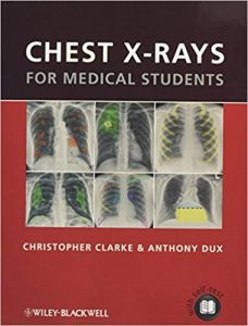
Chest X-rays for Medical Students 1st Edition

[amazon template=iframe image2&asin=0470656190]

Chest X-rays for Medical Students is a unique teaching and learning resource that offers students, junior doctors, trainee radiologists, nurses, physiotherapists and nurse practitioners a basic understanding of the principles of chest radiology.

  • Provides a memorable way to analyze and present chest radiographs – the unique ‘ABCDE’ system as developed by the authors
  • Explains how to recognize basic radiological signs, pathology and patterns associated with common medical conditions as seen on plain PA and AP chest radiographs
  • Presents each radiograph twice, side by side – once as would be seen in a clinical setting and again with the pathology clearly highlighted
  • Includes a section of self-assessment and presentation exercises to test knowledge and presentation technique

Ideal for study and clinical reference, this book will be the ideal companion for any medical student, junior doctor or trainee radiographer.

DOWNLOAD THIS BOOK FREE HERE

http://upsto.re/Fwp52zU

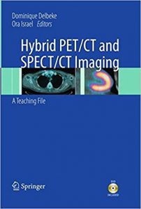
Hybrid PET/CT and SPECT/CT Imaging: A Teaching File

[amazon template=iframe image2&asin=1461400902]

Hybrid PET/CT and SPECT/CT Imaging: A Teaching File presents a complete book on state-of-the-art integrated PET/CT and SPECT/CT imaging systems. Edited by Drs. Dominique Delbeke and Ora Israel, this practical guide includes in-depth cases that will advance the skills of nuclear physicians and radiologists while also preparing residents for the field of nuclear medicine and radiology. Internationally-recognized contributors provide the reader with chapters covering the technical and clinical aspects of hybrid imaging, including basic principles, physics and instrumentation, the normal distribution of radiopharmaceuticals, and protocols critical to the field. A comprehensive review of nuclear oncology cases found in everyday practice are addressed, including among other clinical applications head and neck tumors, breast cancer, lung neoplasms, colorectal cancer, pancreatic cancer, and genitourinary tumors. Additional chapters examine the use of these modalities in pediatric patients, benign bone diseases and infection, and cardiac hybrid imaging. A wealth of illustrations reinforces the key teaching points discussed throughout the book.

A beneficial feature is the accompanying CD-ROM containing DICOM images from selected cases showing views in all planes, including the fusion images, which simulates an actual workstation used in a clinical scenario. The CD-ROM has been provided by Hermes Medical Solutions, Inc.

DOWNLOAD THIS BOOK FREE HERE

http://upsto.re/FwbNacb

Diagnostic Neuroradiology

[amazon template=iframe image2&asin=3540756523]

In this monograph, the authors summarize their findings in complex neuroimaging work (cranio-, spondylo-, myelo- and angiography as well as CT and MR imaging of the brain and spine) during their longstanding experience at the N. Burdenko Neurosurgical Institute in Moscow. The book begins with a review of modern neuroimaging techniques: CT and MR angiography, perfusion and diffusion imaging, tractography, spectroscopy and functional MR imaging.

The problems and various other aspects of diagnosis of intra- and extra-axial brain tumors (more than 30,000 verified cases) as well as of cerebrovascular, infectious, demyelinating, degenerative and traumatic brain and spine lesions are discussed. The volume is well illustrated with angiographic, CT and MR images of complex diagnostic studies. The numerous images represent a “visual text,” which can be used as an atlas by practical clinicians.

This book is a comprehensive reference manual for neurologists, neurotraumatologists and radiologists. It may also be of interest to technicians, medical physicists, students and other specialists interested in neurovisualization and diagnostic imaging

Valery N. Kornienko, MD, PhD, head of the Neuroimaging Dept. of the Burdenko Neurosurgical Institute RAMS in Moscow, Russia. Doctor of medical science, professor, honorary scientist of the Russian Federation, author of 12 monographs on neuroradiology and more than 500 scientific publications.

Igor N. Pronin, MD, PhD, professor at the Neuroimaging Dept. of the Burdenko Neurosurgical Institute RAMS in Moscow, Russia. Author of 2 monographs on neuroradiology and 220 scientific publications

DOWNLOAD THIS BOOK FREE HERE

http://upsto.re/FwaHbJs

Hemodynamic Monitoring Using Echocardiography in the Critically Ill

[amazon template=iframe image2&asin=3540879544]

The hemodynamic evaluation of patients with acute circulatory failure and respiratory failure has in the past usually been performed using invasive procedures but in recent years less invasive monitoring devices have been introduced. Echocardiography can be used for both the diagnosis and the management of circulatory and respiratory failure. This book provides all the essential information required by readers in order to perform optimal hemodynamic management of the critically ill based on echocardiographic guidance. After an introductory section on basic principles, hemodynamic assessment using echocardiography is discussed in detail. The diagnosis and management of all types of circulatory and acute respiratory failure by means of echocardiography are then rigorously considered, and specific situations such as thoracic trauma and acute aortic syndrome are examined. The final section is devoted to future issues and applications.

DOWNLOAD THIS BOOK FREE HERE

http://upsto.re/FwaauFj

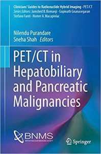
PET/CT in Hepatobiliary and Pancreatic Malignancies (Clinicians’ Guides to Radionuclide Hybrid Imaging) 1st ed

[amazon template=iframe image2&asin=3319605062]

About the Author

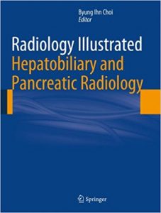
Radiology Illustrated: Hepatobiliary and Pancreatic Radiology

[amazon template=iframe image2&asin=3642358241]

Radiology Illustrated: Hepatobiliary and Pancreatic Radiology is the first of two volumes that will serve as a clear, practical guide to the diagnostic imaging of abdominal diseases. This volume, devoted to diseases of the liver, biliary tree, gallbladder, pancreas, and spleen, covers congenital disorders, vascular diseases, benign and malignant tumors, and infectious conditions. Liver transplantation, evaluation of the therapeutic response of hepatocellular carcinoma, trauma, and post-treatment complications are also addressed.

The book presents approximately 560 cases with more than 2100 carefully selected and categorized illustrations, along with key text messages and tables, that will allow the reader easily to recall the relevant images as an aid to differential diagnosis. At the end of each text message, key points are summarized to facilitate rapid review and learning. In addition, brief descriptions of each clinical problem are provided, followed by both common and uncommon case studies that illustrate the role of different imaging modalities, such as ultrasound, radiography, CT, and MRI.

DOWNLOAD THIS BOOK FREE HERE

http://upsto.re/FhzjRBg

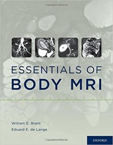
Essentials of Body MRI 1st Edition

[amazon template=iframe image2&asin=0199738491]

Essentials of Body MRI extensively covers the field, offering clear and detailed guidance on MRI as an invaluable tool for the primary diagnosis and problem solving of diseases of the body, including the abdomen, liver, pancreas, pelvis, heart, urinary tract, and great vessels. The beginning chapters focus on the physics, pulse sequences, and other practical considerations related to body MR imaging, explained in an easy to understand way, to help the reader fully comprehend the imaging appearance of clinical disease. The remaining chapters discuss clinical applications, with topics spanning from the normal anatomic structures and diagnosis of abdominal, pelvic, cardiac, and vascular diseases to the modality’s role as a tool for solving diagnostic problems. The key points of each chapter are boxed as Essentials to Remember for rapid review and learning. Written in clear, accessible text, and featuring 887 figures and numerous tables, Essentials of Body MRI is a resource that radiology residents, fellows, and anyone else who wants to learn about Body MRI, will turn to again and again.

DOWNLOAD THIS BOOK FREE HERE

http://upsto.re/FZbXfd9

1 38 39 40 41 42 62