Medical Imaging Technology (SpringerBriefs in Physics)
[amazon template=image&asin=1461470722]

From the Back Cover

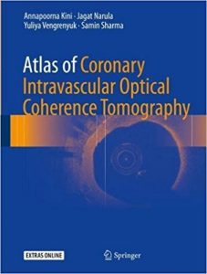
Atlas of Coronary Intravascular Optical Coherence Tomography 1st ed. 2018 Edition

[amazon template=image&asin=3319626647]

About the Author

Jagat Narula, MD, PhD is a world-renowned physician-scientist in cardiovascular medicine and imaging and leads the cardiac services at Mount Sinai St. Luke’s and Mount Sinai Roosevelt hospital campuses of the Mount Sinai Health System. He is involved in clinical and basic research in the fields of heart failure and atherosclerosis, with major emphasis on development of novel noninvasive imaging techniques. He has made contributions to the imaging of apoptotic cell death in heart muscle, and to the imaging of atherosclerotic plaques vulnerable to rupture.

Annapoorna Kini serves as a Professor of Medicine, Director of Cardiac Catheterization Laboratory, Director of Interventional Structural Heart Disease Program, and Director of Interventional Cardiology Fellowship at The Mount Sinai Medical Center, where she was named Zena and Michael A. Wiener Medicine Professor in 2016. She researches on the field of percutaneous coronary intervention and heart valve therapy. Kini co-established, with Dr Sharma, the Live Symposium of Complex Coronary and Vascular Cases in 1998 and has served as Director of the Annual Live Symposium of Complex Coronary & Vascular Cases at The Mount Sinai Medical Center. She has been among the first interventional cardiologists in the country to use the TAVI procedure in the treatment of inoperable patients with critical aortic stenosis.

Dr Yuliya Vengrenyuk is an instructor at Mount Sinai Hospital and the Icahn School of Medicine at Mount Sinai.

Dr Samin K. Sharma is an interventional cardiologist holding the American record for the highest number of complex coronary interventions performed annually, along with the highest angioplasty success rate in New York State. Dr. Sharma is responsible for operational issues related to Cardiology at the main campus and in the Mount Sinai network. He also increases Mount Sinai’s network of affiliated cardiologists in the tri-state area. As Dean for International Clinical Affiliations, Dr. Sharma serves as Mount Sinai’s clinical representative to develop relationships and affiliations worldwide.

DOWNLOAD THIS BOOK FREE HERE

http://upsto.re/Fj4gVFx

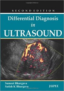
Differential Diagnosis in Ultrasound 2nd Edition

[amazon template=image&asin=9350259990]

Differential diagnosis is the process of weighing the probability of one disease versus that of other diseases possibly accounting for a patient’s illness (MedicineNet.com) This book is a practical reference providing numerous ultrasound images with detailed descriptions of various disease entities or systems of the body to help radiologists and sonologists determine a correct diagnosis. With more than 650 line diagrams and illustrations, each chapter discusses conditions and diseases in a different part of the body. Key Points * Practical guide to differential diagnosis in ultrasound * Features numerous ultrasound images with detailed descriptions * Covers diseases and conditions in all different parts of the body * Includes more than 650 line diagrams and illustrations * Previous edition published 2007

DOWNLOAD THIS BOOK FREE HERE

http://upsto.re/Fqvrvdt

Medical Image Understanding and Analysis: 21st Annual Conference, MIUA 2017, Edinburgh, UK, July 11–13, 2017, Proceedings (Communications in Computer and Information Science) 1st ed. 2017 Edition

[amazon template=image&asin=3319609637]

This book constitutes the refereed proceedings of the 21st Annual Conference on Medical Image Understanding and Analysis, MIUA 2017, held in Edinburgh, UK, in July 2017.
The 82 revised full papers presented were carefully reviewed and selected from 105 submissions. The papers are organized in topical sections on retinal imaging, ultrasound imaging, cardiovascular imaging, oncology imaging, mammography image analysis, image enhancement and alignment, modeling and segmentation of preclinical, body and histological imaging, feature detection and classification.

The chapters ‘Model-Based Correction of Segmentation Errors in Digitised Histological Images’ and ‘Unsupervised Superpixel-Based Segmentation of Histopathological Images with Consensus Clustering’ are open access under a CC BY 4.0 license.

DOWNLOAD THIS BOOK FREE HERE

http://upsto.re/FjZRMvV

Women’s Health in Interventional Radiology

[amazon template=image&asin=1441958754]

Written as a concise, clinical guide to major interventional procedures impacting upon women’s health, Women’s Health in Interventional Radiology focuses on a wide range of vascular and non-vascular interventions commonly performed in daily practice, including those related to the pelvis, fallopian tube, spine, and lower extremities. Chapters provide key clinical background information on each intervention, including pathophysiology, clinical manifestations, basic anatomy, and imaging features, before moving on to step-by-step explanations of the techniques. Accompanied by high-quality illustrations and images throughout, chapters also include technical hints, pearls, and pitfalls, postoperative care guidelines, outcomes data, and complications. Written by an expert group of experienced interventionalists, this book promises the reader a comprehensive overview of interventions currently performed to treat problems affecting the health of women and is a valuable resource for both practicing physicians and those in training.

DOWNLOAD THIS BOOK FREE HERE

http://upsto.re/FjQUkoG

Interventional Radiology for Medical Students 1st ed. 2018 Edition

[amazon template=image&asin=3319538527]

This book provides easily accessible technique specific information on interventional radiology (IR) procedures at a basic level for medical students and junior doctors. It covers subject topics in both vascular and non-vascular interventional radiology appropriate for a non-specialist audience. Case based contents help to introduce IR techniques and demonstrate the role of minimally invasive therapy in the overall clinical management. Pre and post-procedural patient assessments are also included in the chapters to enhance patient safety and early detection of complications.

Interventional Radiology treatments now play a major role in many disease processes and continues to grow with new procedures added to the armamentarium of the interventional radiologist, almost on a yearly basis.  Interventional radiology because of its pivotal role in medical care, requires its own teaching curriculum.

Recognising this, the Cardiovascular and Interventional Radiology Society of Europe (CIRSE) devised an undergraduate curriculum in IR, specifically designed for teaching medical students.

This book will be primarily of benefit to medical students.  In addition, it will also be of help to non-radiology junior hospital doctors and general practitioners.

DOWNLOAD THIS BOOK FREE HERE

http://upsto.re/FxcBoPp

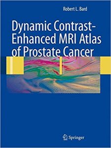
Dynamic Contrast-Enhanced MRI Atlas of Prostate Cancer 2009th Edition

[amazon template=image&asin=3540784225]

MRI Atlas of Prostate Cancer analyses high-resolution MRI scanning and dynamic contrast-enhanced (DCE) MRI. This combination improves the diagnosis and staging of prostate cancer and may soon replace PSA testing and digital rectal examination. The first two chapters focus on normal anatomy, anatomic variations, benign disease and intraprostatic tumors. The subsequent chapters on MRI of extracapsular disease create a useful atlas of pathologic anatomy. This is the first text of its kind to show color-coded DCE-MRI scans of prostate cancer and to correlate these imaging findings with tumor grading. The chapters on the post-treatment prostate clearly display the increasing incidence of post-therapy recurrences. This book is intended for internists, radiologists, radiotherapists, oncologists, urologists, family practitioners, and general surgeons. Ultrasound, MRI, and radiotherapy technicians will find it extremely useful as a reference guide.

DOWNLOAD THIS BOOK FREE HERE

http://upsto.re/FxgXBzW

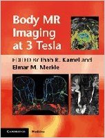
Body MR Imaging at 3 Tesla (Cambridge Medicine (Hardcover)) 1st Edition

[amazon template=image&asin=0521194865]

Body MR Imaging at 3.0 Tesla is a practical text enabling radiologists to maximise the benefits of high field 3T MR systems in a range of body applications. It explains the physical principles of MR imaging using 3T magnets, and the differences between 1.5T and 3T when applied extracranially. The book’s organ-based approach focuses on optimized techniques, providing recommended protocols for the main vendors of 3T MRI systems. All major thoracic and abdominal organs are covered, including breast, heart, liver, pancreas, the GI tract, kidneys, prostate and female pelvic organs. Abdominal and pelvic MR angiography and MRCP are also discussed. Protocol optimization, appearance of artifacts and novel applications using 3T are emphasized. Written and edited by experts in the field, Body MR Imaging at 3.0 Tesla guides radiologists in optimizing imaging protocols for 3T MR systems, reducing artifacts and identifying the advantages of using 3T in body applications.

DOWNLOAD THIS BOOK FREE HERE

http://upsto.re/FxiEMtb

Combined Scintigraphic and Radiographic Diagnosis of Bone and Joint Diseases Softcover reprint of hardcover 3rd ed

[amazon template=iframe image2&asin=3642061702]

The third edition of this book has been comprehensively rewritten and rearranged. In addition to the bone and joint diseases described in the two earlier editions, it now encompasses hitherto unpublished novel applications of pinhole scanning to the diagnosis of a broader spectrum of skeletal disorders. The book has been considerably expanded to discuss five new themes and is complemented by the addition of some 90 recently acquired cases.

DOWNLOAD THIS BOOK FREE HERE

http://upsto.re/FK3nuWo

Combined Scintigraphic and Radiographic Diagnosis of Bone and Joint Diseases: Including Gamma Correction Interpretation 4th ed

[amazon template=iframe image2&asin=3642251439]

In this fourth edition of Combined Scintigraphic and Radiographic Diagnosis of Bone and Joint Diseases, the text has been thoroughly amended, updated, and partially rearranged to reflect the latest advances. In addition to discussing the role of pinhole imaging in the range of disorders previously covered, the new edition pays detailed attention to the novel diagnostic use of gamma correction pinhole bone scan in a broad spectrum of skeletal disorders, including physical, traumatic, and sports injuries, infectious and non-infectious bone diseases, benign and malignant bone tumors, and soft tissue diseases. A large number of state of the art pinhole scans and corroborative CT, MRI, and/or ultrasound images are presented side by side. The book has been enlarged to encompass various new topics, including occult fractures; cervical sprain and whiplash trauma; bone marrow edema; microfractures of trabeculae; evident, gaping, and stress fractures; and differential diagnosis. This new edition will be essential reading for practitioners and researchers in not only nuclear medicine but also radiology, orthopedic surgery, and pathology.

DOWNLOAD THIS BOOK FREE HERE

http://upsto.re/FKCJ2cR

1 36 37 38 39 40 62