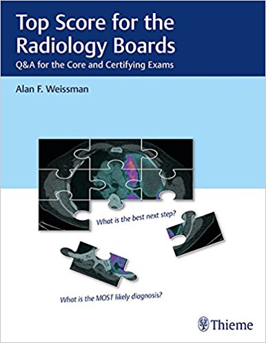
Top Score for the Radiology Boards: Q&A for the Core and Certifying Exams 1st Edition

[amazon_link asins=’1626234094′ template=’ProductAd’ store=’aishabano-20′ marketplace=’US’ link_id=’85e6c1a0-554f-11e8-b3fb-cb508be261e5′]

Top Score for the Radiology Boards: Q & A for the Core and Certifying Exams is the ideal diagnostic radiology board prep resource. Written by radiologist Alan Weissman, with contributions from dozens of leading experts at renowned institutions, Top Score has a simple ambition: to improve your test scores.

The book covers all exam categories, including non-interpretive skills (NIS), physics, safety, breast, cardiac, diagnostic radiology, gastrointestinal, genitourinary, interventional, musculoskeletal, neuroradiology, nuclear, pediatrics, thoracic, ultrasound/reproductive/endocrinology, vascular, and general radiology.

Chapters are composed of four types of test cases of varying focus and complexity, each on a two-page spread.

Essentials

starts with a patient presentation, followed by board-type multiple-choice questions. Details begins with a case presentation, followed by 10 rapid-fire questions, enabling brisk, high-volume learning. Image Rich presents multiple images that require accurate identification, enabling accelerated, high-volume image assessment practice. More Challenging follows the same format as Essentials but adds a higher degree of difficulty.

Key highlights

  • High-quality, board-type Q&A with detailed answer explanations
  • High yield “Top Tips” for each case
  • Special radiology artifacts section
  • Image Rich and Details sections aid in rapid and lasting topic mastery
  • Comprehensive review, covering all sections tested by the American Board of Radiology
  • Written by experienced, expert question writers
  • NIS chapter emphasizes proficiency in vital practice-related skills

This quintessential home-study guide will help radiology residents and fellows prep for and ace both the certifying and core exams.

DOWNLOAD THIS BOOK FREE HERE

https://upsto.re/3JMyeFV

Getting Started in Clinical Radiology: From Image to Diagnosis 1st Edition

[amazon_link asins=’3131403616′ template=’ProductAd’ store=’aishabano-20′ marketplace=’US’ link_id=’780b81c6-554e-11e8-a486-25ba08b28cc8′]

“I read the book for enjoyment and pleasure, as well as enlightenment. It was a delightful learning experience.” –Thomas Lee Bucky, MD

This book teaches radiology in a way that mimics a lively setting on the wards. To have fun in learning the
theoretical basis of imaging and the interpretation of radiographs and other
modalities, in the context of clinical examination and findings, this is the
book for you.

The basics of imaging are described using analogies from daily
life to make them as understandable and memorable as possible. The material of
radiology is described using actual cases; the most common differential
diagnoses are presented. A great amount of image material supports the learning
process.

A storyline runs through the book: four students in their final year of
medical school are involved in active discussion of the cases, so that the
reader also feels a part of the diagnostic process.

DOWNLOAD THIS BOOK FREE HERE

https://upsto.re/3JU8LpY

A-Z of Musculoskeletal and Trauma Radiology 1st Edition

[amazon_link asins=’0521700132′ template=’ProductAd’ store=’aishabano-20′ marketplace=’US’ link_id=’f8ad0511-5167-11e8-8cc1-e7f886282ec3′]

A-Z of Musculoskeletal and Trauma Radiology is an invaluable reference to the key aspects of imaging for all conditions of bones, muscles, tendons and ligaments. It provides the clinician with practical guidance on the key presenting characteristics, clinical features, diagnosis and management. The description of each condition is provided in a standard template of Characteristics, Clinical Features, Radiology and Management, enabling the reader to find the relevant information quickly. All diagnostic modalities are included and a separate section is dedicated to musculoskeletal trauma. Written by a multidisciplinary team of radiologists and an orthopaedic surgeon, A-Z of Musculoskeletal and Trauma Radiology is an invaluable resource for radiologists, orthopaedic surgeons, rheumatologists and all clinicians managing musculoskeletal conditions.

DOWNLOAD THIS BOOK FREE HERE

https://upsto.re/3wGk6A6

MDCT and MR Imaging of Acute Abdomen: New Technologies and Emerging Issues 1st ed. 2018 Edition

[amazon_link asins=’3319707779′ template=’ProductAd’ store=’aishabano-20′ marketplace=’US’ link_id=’93b837b4-4f0e-11e8-aebe-378bb4a858eb’]

This superbly illustrated book describes a comprehensive and modern approach to the imaging of abdominal and pelvic emergencies of traumatic and non-traumatic origin. The aim is to equip the reader with a full understanding of the roles of advanced cross-sectional imaging modalities, including dual-energy computed tomography (DECT) and magnetic resonance imaging (MRI). To this end, recent literature on the subject is reviewed, and current controversies in acute abdominal and pelvic imaging are discussed. Potential imaging and related pitfalls are highlighted and up-to-date information provided on differential diagnosis. The first two chapters explain an evidence-based approach to the evaluation of patients and present dose reduction strategies for multidetector CT imaging (MDCT).  The remaining chapters describe specific applications of MDCT, DECT, and MRI for the imaging of both common and less common acute abdominal and pelvic conditions, including disorders in the pediatric population and pregnant patients. The book will be of value to emergency and abdominal radiologists, general radiologists, emergency department physicians and related personnel, general and trauma surgeons, and trainees in all these specialties.

DOWNLOAD THIS BOOK FREE HERE

https://upsto.re/3hW8bEa

Imaging for Students Fourth Edition 4th Edition

[amazon_link asins=’1444121820′ template=’ProductAd’ store=’aishabano-20′ marketplace=’US’ link_id=’98086439-4cad-11e8-93eb-c3b6508118fa’]

Imaging for Students delivers step-by-step guidance to the range of imaging techniques available, providing a clear explanation of how each imaging modality actually works, and including information on the associated risks and hazards. Throughout, the importance of patient preparation and post-procedure observation is emphasized.

Taking information from evidence-based studies and published guidelines, in line with current clinical practice, the book takes a highly logical approach to the investigation of clinical scenarios, where possible indicating the “best first test”―vital to both appropriate clinical and cost-effective decision-making.

Drawing on the extensive clinical and teaching experience of its respected author, the fourth edition of Imaging for Students gives students and junior doctors everything they need to understand the advantages, disadvantages, and possible side effects of the imaging modalities available, and how to apply them appropriately in clinical practice.

DOWNLOAD THIS BOOK FREE HERE

https://upsto.re/3hdnB9e

Top 3 Differentials in Radiology: A Case Review 2nd Edition

[amazon_link asins=’1626232784′ template=’ProductAd’ store=’aishabano-20′ marketplace=’US’ link_id=’9d0ed196-4cac-11e8-bf91-ffbb41b35398′]

Praise for the first edition:

“Innovative…it would be difficult to find a better high-yield, high-quality textbook covering every subsection of the radiology…board examination.” – JAMA

“Extremely useful for practicing radiologists and radiology residents looking to…acquire the most pertinent knowledge in preparation for board exams, recertification, and general practice…This high-yield way of learning allows the rapid development of…differential diagnoses…for the full range of entities commonly encountered in practice and board exam settings.” – Academic Radiology

“One of the major strengths of this book is…it focuses on short succinct differentials and avoids the laundry-list differentials…The book…is a great value for those looking to develop short clear differentials for the imaging findings…” – Radiology

This fully revised second edition of Top 3 Differentials in Radiology provides a comprehensive core exam review of frequently encountered imaging gamuts in all major radiological subspecialties. Author William O’Brien utilizes the widely acclaimed format of his first edition, with 330 new and updated radiology cases organized into 12 core subspecialty sections. The last section, “Roentgen Classics,” includes cases from each of the previous core sections, with imaging findings characteristic of a single diagnosis.

This book reflects content included in the current radiology board examinations with accurate and concise descriptions of key differentials, which are integral to acing the boards. Each case is formatted as a two-page unit. The left page features clinical images, succinctly captioned radiographic findings, and pertinent clinical history. The right page includes the key imaging gamut, differential diagnoses rank-ordered by the Top 3, additional diagnostic considerations, and clinical pearls.

Key Features

  • More than 700 high quality images, including advanced imaging techniques
  • Rank ordered differentials organized into the Top 3 and additional diagnostic considerations
  • A high-yield review of important imaging and clinical manifestations for all entities on the list of differentials
  • Imaging pearls at the end of each case provide a quick review of key points

This outstanding resource provides radiologists and trainees with a solid foundation of core radiology topics and a wide spectrum of key imaging findings encountered in clinical practice. It is a must-have for radiology residents preparing for board examinations. Veteran radiologists looking for a comprehensive review of critical topics in radiology will also find this book invaluable.

DOWNLOAD THIS BOOK FREE HERE

https://upsto.re/3h2JpAR

Imaging Systems for Medical Diagnostics: Fundamentals, Technical Solutions and Applications for Systems Applying Ionizing Radiation, Nuclear Magnetic Resonance and Ultrasound 2nd Edition

[amazon_link asins=’3895782262′ template=’ProductAd’ store=’aishabano-20′ marketplace=’US’ link_id=’e4534a66-44a0-11e8-8a05-753ee4ec386b’]

Imaging Systems for Medical Diagnostics

The book provides a comprehensive compilation of basics, technical solutions and applications for medical imaging systems. It is intended as a handbook for students in biomedical engineering for medical physicists, and for engineers working on medical technologies, as well as for lecturers at universities and engineering schools. For qualified personnel at hospitals, and physicians working with these instruments it serves as a basic source of information. This also applies for service engineers and marketing specialists.

The book starts with the representation of the physical basics of image processing, implying some knowledge of Fourier transforms. After that, experts working in industry describe technical solutions and applications for imaging systems in medical diagnostics. The applications comprise the fields of X-ray diagnostics, computed tomography, nuclear medical diagnostics, magnetic resonance imaging, sonography, and molecular imaging. Considering the increasing importance of software based solutions, emphasis is also laid on the imaging software platform and hospital information systems.

DOWNLOAD THIS BOOK FREE HERE

https://upsto.re/36A242p

Atlas of Perioperative 3D Transesophageal Echocardiography: Cases and Videos 1st ed. 2016 Edition

[amazon_link asins=’9811005869′ template=’ProductAd’ store=’aishabano-20′ marketplace=’US’ link_id=’0f2b4466-44a0-11e8-9fdc-abc77f0e183c’]

This book presents a series of over 50 cases in 3D transesophageal echocardiography. In each case, background information is followed by a step-by-step approach to patient diagnosis and postoperative outcomes. The book’s most outstanding highlight consists in over 600 illustrations, which mainly focus on 3D transesophageal echocardiography in combination with 2D transesophageal echocardiography, X-ray fluoroscopy and computed tomography to demonstrate the various cardiovascular pathologies. Since the echo images obtained in clinic practice are moving images, it also includes more than 450 videos, which effectively supplement the static illustrations.

The atlas is divided into 11 chapters. Chapters 1 through 3 describe abnormalities affecting mitral, aortic, tricuspid and pulmonary valves, while prosthetic valves are discussed in Chapter 4. Chapter 5 details abnormalities of the aorta. Chapters 6-8 examine coronary artery disease, congenital heart disease and cardiomyopathies, while Chapter 9 explores cases of infective endocarditis. Chapter 10 deals with tumor and mass lesions, and the other cases are collectively addressed in Chapter 11. Offering a valuable new resource, the book will enhance the usefulness of echocardiography during operations as a “third eye” for surgeons.

DOWNLOAD THIS BOOK FREE HERE

https://upsto.re/36FFTHP

Callen’s Ultrasonography in Obstetrics and Gynecology, 6e 6th Edition

[amazon_link asins=’0323328342′ template=’ProductAd’ store=’aishabano-20′ marketplace=’US’ link_id=’364f668c-4275-11e8-b4c1-91c62b3b2af5′]

Get outstanding guidance from the world’s most trusted reference on OB/GYN ultrasound. Now brought to you by lead editor Dr. Mary Norton, Callen’s Ultrasonography in Obstetrics and Gynecology has been completely and exhaustively updated by a team of obstetric, gynecologic, and radiology experts to reflect the most recent advances in the field. It addresses the shift in today’s practice to a collaborative effort among radiologists, perinatologists, and OB/GYNs, with new emphasis placed on genetics and clinical management. This must-have resource covers virtually all aspects of fetal, obstetric and gynecologic ultrasound ― from the common to the rare ― in one essential clinical reference, allowing you to practice with absolute confidence.

  • Highly templated, full-color format allows you to locate information more quickly.
  • Full-color medical illustrations present key anatomic details in a clear manner.
  • Thousands of digital-quality images depict the complete range of normal and abnormal imaging presentations.


  • Expert Consult eBook version included with purchase. This enhanced eBook experience allows you to search all of the text, figures, images, and references from the book on a variety of devices. You’ll also access 20 real-time ultrasound videos of the fetal heart and other structures.
  • Provides extensive updates of text and images, including the latest in imaging, Doppler techniques, genetic testing, and clinical management.
  • Brand new chapters provide up-to-date, comprehensive coverage of topics relevant to current practice:

-First Trimester Fetal Anatomy

-Obstetric Ultrasound and the Obese Patient

-Evaluation of Pelvic Pain in the Reproductive Age Patient

-Gynecologic Ultrasound in the Pediatric and Adolescent Patient

-Ultrasound and Magnetic Resonance Imaging in Urogynecology

-The Role of Ultrasound in Gynecologic Interventions


  • Highlights significant new genetic testing content, including correlation with ultrasound evaluation of the fetus.
  • Places increased emphasis on 3-dimensional imaging and correlative imaging with magnetic resonance(MR).
  • Features new practice guidelines for obstetric evaluation (including first trimester assessment) and gynecologic management (including evaluation of the endometrium and of ovarian masses).
  • Features new information about fetal imaging guidelines from the National Institute of Child Health and Human Development (NICHD).
  • Provides expanded discussion of fetal, obstetric, and gynecologic interventions with new emphasis onclinical use and application of ultrasound imaging.
  • Includes key and comprehensive reference data used for evaluation of fetal growth and other specialized measurements.

DOWNLOAD THIS BOOK FREE HERE

https://upsto.re/3t9bFaN

1 26 27 28 29 30 61