Neuroradiology Companion: Methods, Guidelines, and Imaging Fundamentals

[amazon_link asins=’1496322134′ template=’ProductAd’ store=’aishabano-20′ marketplace=’US’ link_id=’be5e69a5-f613-11e8-acdf-43a9e5905303′]

Neuroradiology Companion: Methods, Guidelines, and Imaging Fundamentals

DOWNLOAD THIS BOOK FREE HERE

https://upstore.net/DpueR2a

Updated throughout and extensively illustrated, the Fifth Edition of Neuroradiology Companion is an ideal one-volume resource covering the fundamentals of neuroradiology for residents, fellows, and practitioners. More than 1,400 high-quality images depict key disorders of the brain, spine, and head and neck.  Each disorder is presented in a concise, consistent manner, with bulleted Key Facts, six to eight images that show the most common findings, and suggested readings. This concise volume can be read cover-to-cover during neuroradiology rotations and also used for daily consultation in the reading room.

Key Features:

  • The latest clinical information brings you up to date with new advances in radiology, as well as basic current concepts of neuropathology, neurosurgery, and neurology.
  • Updated protocols for CT, MRI, myelography, digital subtraction angiography, and sedation and anxiolysis, as well as new information on contrast media reactions, keep you current with recent changes in the field.
  • Suggested Reading resources have been thoroughly updated, and the newly streamlined text makes it even easier to access key information.
  • More than 800 new images clearly depict recent cases and highlight subtle findings.

Now with the print edition, enjoy the bundled interactive eBook edition, which can be downloaded to your tablet and smartphone or accessed online and includes features like:

  • Complete content with enhanced navigation
  • Powerful search tools and smart navigation cross-links that pull results from content in the book, your notes, and even the web
  • Cross-linked pages, references, and more for easy navigation
  • Highlighting tool for easier reference of key content throughout the text
  • Ability to take and share notes with friends and colleagues
  • Quick reference tabbing to save your favorite content for future use


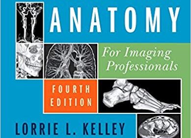
Sectional Anatomy for Imaging Professionals 4th Edition

[amazon_link asins=’0323414877′ template=’ProductAd’ store=’aishabano-20′ marketplace=’US’ link_id=’86bcea5b-eb62-11e8-8fd1-1fc59b7b0cd3′]

Sectional Anatomy for Imaging Professionals 4th Edition

DOWNLOAD THIS BOOK FREE HERE

https://upsto.re/DzKgY2U

An ideal resource for the classroom or clinical setting, Sectional Anatomy for Imaging Professionals, 4th Edition provides a comprehensive, and highly visual approach to the sectional anatomy of the entire body. Side-by-side presentations of actual diagnostic images from both MRI and CT modalities and corresponding anatomic line drawings illustrate the planes of anatomy most commonly demonstrated by diagnostic imaging. Easy-to-follow descriptions detail the location and function of the anatomy, while clearly labeled images help you confidently identify anatomic structures during clinical examinations. In all, it’s the one reference you need to consistently produce the best possible diagnostic images.


  • Side-by-side presentation of anatomy illustrations and corresponding CT and MRI images clarifies the location and structure of sectional anatomy.
  • More than 1,500 high-quality images and detailed line drawings demonstrate sectional anatomy for every body plane commonly imaged in the clinical setting.
  • Updated summary tables are used to simplify and organize key information in each chapter.
  • CT or MR images of special interest are featured on the opening page in each chapter to pique readers’ interest in the area about to be covered in the text.
  • Reference drawings and corresponding scanning planes appear on appropriate pages with the actual images, so they are easily referenced for correlation between the scanning planes and the resulting images.
  • Introductory chapter lays a foundation of the terminology that is related to sectional anatomy.


  • NEW! Updated content reflects the latest ARRT and ASRT curriculum guidelines.
  • NEW! Additional lymphatic system images give readers a better picture of this nuanced body system.
  • NEW! Additional pathology boxes help readers connect commonly encountered pathologies to related anatomy for greater diagnostic accuracy.
  • NEW! Updated line art familiarizes readers with the latest 3D and vascular imaging technology.
  • NEW! 2-color design makes difficult content easier to digest.

Differential Diagnosis in Neuroimaging: Brain and Meninges 1st Edition

[amazon_link asins=’1604067004′ template=’ProductAd’ store=’aishabano-20′ marketplace=’US’ link_id=’edb4cea2-e8ee-11e8-8b2b-f3f5e558b7d7′]

Differential Diagnosis in Neuroimaging: Brain and Meninges 1st Edition

DOWNLOAD THIS BOOK FREE HERE

https://upsto.re/DjHZxHg

This is a highly valuable book that contains a tremendous amount of information in words and pictures. It should be a must-have guide and reference manual for anyone involved in neurology and neurosurgery. — BIZ INDIA

…an outstanding way to learn imaging anatomy and corresponding diseases….This [series] is a highly desirable set of books and would be a valuable addition to any neuroradiologist’s personal collection, and certainly to any departmental or sectional library. — AJNR Blog

Authored by renowned neuroradiologist Steven P. Meyers, Differential Diagnosis in Neuroimaging: Brain and Meninges is a stellar guide for identifying and diagnosing brain pathologies based on location and neuroimaging results. The succinct text reflects more than 25 years of hands-on experience gleaned from advanced training and educating residents and fellows in radiology, neurosurgery, and neurology. The high-quality MRI, CT, PET, PET/CT, conventional angiography, and X-ray images have been collected over Dr. Meyers’s lengthy career, presenting an unsurpassed visual learning tool.

The distinctive ‘three-column table plus images’ format is easy to incorporate into clinical practice, setting this book apart from larger, disease-oriented radiologic tomes. The layout enables readers to quickly recognize and compare abnormalities based on high-resolution images.

Key Highlights

  • Tabular columns organized by anatomical abnormality include brain imaging findings and a summary of key clinical data that correlates to the images
  • Comprehensive imaging of the brain, ventricles, meninges, and neurovascular system in both children and adults, including congenital/developmental anomalies and acquired disease
  • More than 1,900 figures illustrate the radiological appearance of intracranial lesions, masses, neurodegenerative disorders, ischemia and infarction, and more

This visually rich resource is a must-have diagnostic tool for radiologists, neurosurgeons, and neurologists, and residents and fellows. The highly practical format makes it ideal for daily rounds, as well as a robust study guide for physicians preparing for board exams.

Radiology of Orthopedic Implants 1st ed

[amazon_link asins=’3319760076′ template=’ProductAd’ store=’aishabano-20′ marketplace=’US’ link_id=’d316b8e3-e77d-11e8-a98a-4fc57995624a’]

Radiology of Orthopedic Implants 1st ed

DOWNLOAD THIS BOOK FREE HERE

https://upsto.re/DKHDMaJ

There is an ever-expanding range of implants used in Orthopaedic Surgery. Nearly 200,000 joint replacement procedures are done in UK every year. The performance of these implants is assessed on radiographs. This is of interest to Orthopaedic surgeons and Radiologists alike.  Information on interpretation of these radiographs is not readily available in an easily readable format. This book will assist both trainees and practicing orthopedic surgeons and radiologists in assessing the radiologic appearance of implants and their potential for future performance.

Echocardiography in the CCU 1st ed

[amazon_link asins=’3319902776′ template=’ProductAd’ store=’aishabano-20′ marketplace=’US’ link_id=’7ea513fb-de14-11e8-b376-69161c9e23a9′]

Echocardiography in the CCU 1st ed

DOWNLOAD THIS BOOK FREE HERE

https://upsto.re/DTPN3dX

This book comprehensively reviews the use of echocardiography in the rapidly evolving field of critical care cardiology. Increasingly, cardiac care units (CCU) are focusing on the management of patients with multisystem diseases, advanced hemodynamics compromise, complex ventricular arrhythmias, and established or incipient multi-organ failure. This book covers ultrasound applications in such topics as hemodynamic assessment and the assessment of patients with intracardiac devices. Syndrome-based echocardiography in the CCU is also covered with an emphasis on using echocardiography in patients with acute dyspnea, acute chest pain and neurologic syndromes. The use of contrast echocardiography in the CCU is also covered.


Echocardiography in the CCU reveals the essential role of various echocardiographic modalities in modern acute cardiovascular care.  This is therefore a critical resource for all cardiology practitioners and trainees who use echocardiography in CCUs.

Perez & Brady’s Principles and Practice of Radiation Oncology Seventh Edition

[amazon_link asins=’1496386795′ template=’ProductAd’ store=’aishabano-20′ marketplace=’US’ link_id=’e32ef0a6-dc62-11e8-be84-7b67f7a031c7′]

Perez & Brady’s Principles and Practice of Radiation Oncology Seventh Edition

DOWNLOAD THIS BOOK FREE HERE

https://upsto.re/DwDmF3f

For more than 30 years, Perez and Brady’s Principles and Practice of Radiation Oncology has been the must-have standard reference for radiation oncologists and radiation oncology residents who need a comprehensive text covering both the biological and physical science aspects of this complex field as well as disease site-specific information on the integrated, multidisciplinary management of patients with cancer. The book has established itself as the discipline’s “text-of-record,” belonging on the shelf of all of those working in the field. The Seventh Edition continues this tradition of excellence with extensive updates throughout, many new chapters, and more than 1,400 full-color illustrations that highlight key concepts in tumor pathogenesis, diagnosis, and targeted radiation therapy.

  • Comprehensive coverage of the current state of knowledge of cancer biology; medical radiation physics; clinical radiation oncology; and radiation oncology economics, ethics, and public policy.
  • Regular, twice-yearly content updates available with the digital version of the text.
  • Ideal for radiation oncologists, radiation oncology residents, radiation biologists, medical radiation physicists, oncologists, physicists-in-training, radiation dosimetrists, and nurses and physician assistants working in radiation oncology departments.
  • New chapters include: Proton and Carbon Ion Beam Dosimetry and Treatment Planning; Stereotactic Radiation Therapy; Re-Irradiation; Error Avoidance; Undergraduate, Graduate, and Continuing Medical Education in Radiation Oncology; and a completely rewritten chapter on the economics of radiation oncology.
  • Designed to provide a better understanding of the natural history of cancer, the physical and technological methods of radiation application, the effects of irradiation on normal tissues, and the most judicious ways in which radiation therapy can be employed in the treatment of patients with cancer, either as a single modality or as part of a multi-modality treatment program.
  • All references printed in the book are now streamlined for quick access to further information.
  • Site-specific chapters include relevant background information on each tumor—including epidemiology, pathology, diagnostic work-up, prognostic factors, treatment techniques, applications of surgery, chemotherapy, immunotherapy and biological therapy, end results, and more.
  • Increased focus on new approaches and technologies with new and updated chapters.
  • Greater emphasis on palliative and supportive care reflects the role of radiation treatment in non-curative roles.
  • An international group of expert chapter authors keeps you well-informed.
  • A completely revised first chapter is designed to provide an overview of the discipline of radiation oncology. This will be particularly useful for residents beginning training and medical students beginning a radiation oncology clinical clerkship. The chapter is supplemented with multiple sidebars to augment the reader’s understanding of the field.

Enhance Your eBook Reading Experience

  • Read directly on your preferred device(s), such as computer, tablet, or smartphone.
  • Easily convert to audiobook, powering your content with natural language text-to-speech.

Diffusion Weighted Imaging of the Gastrointestinal Tract: Techniques and Clinical Applications 1st ed. 2019 Edition

[amazon_link asins=’331992818X’ template=’ProductAd’ store=’aishabano-20′ marketplace=’US’ link_id=’30c5489d-d6cf-11e8-b79a-75e19bd38594′]

Diffusion Weighted Imaging of the Gastrointestinal Tract: Techniques and Clinical Applications 1st ed. 2019 Edition

DOWNLOAD THIS BOOK FREE HERE

https://upsto.re/D698rTb

This book explains how diffusion weighted imaging has been incorporated in routine MRI examinations of the abdomen and pelvis: though its clinical role is still evolving, it is already considered an important tool for the assessment of rectal cancer treatment response, as was confirmed in recent ESGAR consensus statements. The standardization and clinical validation of quantitative DWI related biomarkers are still in progress, although certain efforts have been undertaken to establish imaging guidelines for different clinical indications/body parts.

The book reviews the technical aspects and clinical applications of DWI in imaging of the GI tract, and provides specific technical details (imaging protocols, artefacts, optimization techniques) for each GI tract division. This volume is mainly intended for radiologists who are interested in abdominal radiology, as well as radiology residents. Given that magnetic resonance physics is complex and can be cumbersome to learn, the authors have made it as simple and practical as possible.

Textbook of Uroradiology Fifth Edition

[amazon_link asins=’1451109164′ template=’ProductAd’ store=’aishabano-20′ marketplace=’US’ link_id=’d9983221-d6cd-11e8-8f58-d9f77b45e132′]

Textbook of Uroradiology Fifth Edition

DOWNLOAD THIS BOOK FREE HERE

https://upsto.re/D6a3zmW

OR

https://upsto.re/D6eg3Po

This 5th Edition of Textbook of Uroradiology focuses on subject matter that will provide critical learning to radiology and urology residents preparing for their board examinations as well as practicing radiologists.
Chapter-opener outlines and text boxes highlighting key points and differential diagnoses make this introductory textbook very user-friendly. The “Suggested Readings” at the end of each chapter are a valuable reference tool for those who desire additional information about a particular topic.
SPECIAL FEATURES
• Integrates all aspects of adult uroradiology including additional discussion on incidental findings and how to handle them
• More than 1,000 illustrations
• More Hybrid imaging including PET/CT and SPECT/CT
• More MR imaging including MR urography and MR angiography
• More material on CT dose reduction and 3D CT imaging

How to Perform Ultrasonography in Endometriosis 1st ed

[amazon_link asins=’3319711377′ template=’ProductAd’ store=’aishabano-20′ marketplace=’US’ link_id=’251b9577-d6cb-11e8-8d12-cfb17d198497′]

How to Perform Ultrasonography in Endometriosis 1st ed

DOWNLOAD THIS BOOK FREE HERE

https://upsto.re/DZvaQZT

This structured dynamic book outlines, step by step, an evidence-based systematic approach to the sonographic evaluation of the pelvis in women with suspected endometriosis. This “how to” guide is intended for those with basic ultrasonography skills who want to further develop their capabilities in performing the relevant sonographic techniques to identify endometriosis. Detailed schematics, and corresponding high-resolution ultrasound images and intuitive videos support readers in expanding their technical skills and bridging the gaps in their knowledge of endometriosis ultrasound. The International Deep Endometriosis Analysis (IDEA) group consensus statement was the culmination of the work of 29 authors from 5 continents. With the publication of How to Perform Ultrasonography in Endometriosis the authors intend to provide the basis for quality improvement and benchmarking of ultrasound in the world of endometriosis.

This book not only offers sonologists, radiologists and sonographers valuable insights into the field of endometriosis ultrasound, but also enables them to develop their practical skills in assessing women with chronic pelvic pain.

AIIMS-MAMC-PGI Imaging Series. Diagnostic Radiology. Gastrointestinal and Hepatobiliary Imaging, 3/E 3/E Edition

[amazon_link asins=’8184484348′ template=’ProductAd’ store=’aishabano-20′ marketplace=’US’ link_id=’f41492bf-d3cf-11e8-b7f3-9be655c3e5d5′]

DOWNLOAD THIS BOOK FREE HERE

https://upsto.re/DRNfBfC

PART ONE – GASTROINTESTINAL IMAGING – IMAGING TECHNIQUES; 1. Current Status of Conventional Techniques and Advances in GIT Imaging. THE ACUTE ABDOMEN; 2. Non-traumatic Acute Abdomen; 3. Imaging in Abdominal Trauma; INFECTIONS, INFLAMMATION AND NEOPLASMS; 4. Imaging of the Esophagus; 5. Benign Lesions of Stomach and Small Intestine; 6. Malignant Lesions of the Stomach and Small Intestine; 7. Abdominal Tuberculosis; 8. Non-Tubercular Inflammatory Bowel Diseases; 9. Colorectal Malignancies; 10. Lymphoma of Gastrointestinal Tract; 11. Imaging of Appendix; PART TWO – HEPATOBILIARY AND PANCREATIC IMAGING – LIVER AND BILIARY TRACT; 12. Liver Anatomy and Techniques of Imaging; 13. Benign Focal Lesions of Liver; 14. Malignant Focal Lesions of Liver; 15. Diffuse Liver Diseases; 16. Imaging of Obstructive Biliopathy; 17. Clinical Aspects of Liver Cirrhosis: A Perspective for the Radiologist; PANCREAS; 18. Imaging and Interventions in Pancreatitis; 19. Tumors of Pancreas; HEPATIC VASCULAR DISEASES; 20. Imaging in Portal Hypertension; 21. Hepatic Venous Outflow Tract Obstruction; INTERVENTIONS; 22. Gastrointestinal Haemorrhage; 23. Interventions in Obstructive Biliopathy; 24. Interventional Treatment of Liver Tumors; 25. Percutaneous Non-vascular GIT interventions; 26. Interventional Radiology in Portal Hypertension

1 22 23 24 25 26 61