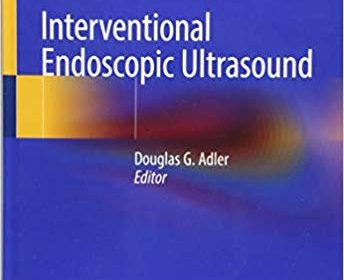
Interventional Endoscopic Ultrasound 1st ed

[amazon_link asins=’3319973754′ template=’ProductAd’ store=’aishabano-20′ marketplace=’US’ link_id=’b2d9f9e2-2c4b-4de9-914a-c4201f2698db’]

Interventional Endoscopic Ultrasound 1st ed

DOWNLOAD THIS BOOK FREE HERE

https://upsto.re/DoRu2Ho

This volume provides a comprehensive, state-of-the-art overview on interventional endoscopic ultrasound (EUS). Each chapter covers a single procedure or related group of procedures, features a detailed literature evaluation and tips on these cutting edge endoscopic techniques, and is accompanied by a comprehensive endoscopic video and illustrative endoscopic and ultrasonographic images. The text also reviews adverse events related to interventional EUS, both how to avoid them and how to manage them when they arise.
Written by to endoscopic experts in the field, Interventional Endoscopic Ultrasound is a valuable resource for practicing gastroenterologists who perform advanced endoscopy.

101 MRI Brain Solutions 1st Edition

[amazon_link asins=’9351525538′ template=’ProductAd’ store=’aishabano-20′ marketplace=’US’ link_id=’4404115b-53e5-4955-a0cc-6999531ed08d’]

101 MRI Brain Solutions 1st Edition

DOWNLOAD THIS BOOK FREE HERE

https://upsto.re/DeUwkWo

101 MRI Brain Solutions is a compilation of 101 cases related to congenital malformations and disorders affecting the brain. This comprehensive book details the use of MRI for diagnosis of infections including tuberculous meningitis, cerebral tuberculoma, multiple cerebral tuberculomas, viral encephalitis, Japanese encephalitis, neurocysticercosis, brain abscess and abscesses and ventriculitis. This book is divided into 12 chapters, with the first two discussing the basics of MRI and the anatomy of the brain. The following 9 chapters present a compilation of cases encountered in routine practice as well as more difficult cases. The last two chapters of this book are dedicated to the glossary of MRI Terms and MRI Acronyms. The use of MRI in the diagnosis of metachromatic leukodystrophy, Alexander disease, Canavan’s disease, adrenoleukodystrophy and multiple sclerosis, is highlighted with the help of clinical cases. 101 MRI Brain Solutions is an important resource for a broad audience, from post graduate students to practising radiologists and MR technical staff. Key Features Relevant to a wide audience Includes glossary of MRI terms and MRI acronyms Chapter on MR anatomy of the brain 166 full colour images and illustrations

Imaging for Otolaryngologists 1st Edition

[amazon_link asins=’3131463317′ template=’ProductAd’ store=’aishabano-20′ marketplace=’US’ link_id=’93406204-2a9a-42e8-91f5-a07917df11bf’]

Imaging for Otolaryngologists 1st Edition

DOWNLOAD THIS BOOK FREE HERE

https://upsto.re/DYHxweG

Imaging for Otolaryngologists distils the essentials of otolaryngologic imaging into a concise reference that concentrates on key topics that are of immediate interest to otolaryngologists practicing in a modern clinical environment.


Prepared by a renowned otolaryngologist, and reviewed and supplemented by expert radiologists, the book provides a well-rounded perspective. The central focus is on image interpretation, including the disease-specific characteristics, the features necessary for successful diagnosis, and the implications for surgery. Each of the 465 high-quality images is clearly labeled, and where appropriate comparisons are made between CT scans and MR images to show complementary functions and limitations.


Imaging for Otolaryngologists helps readers:


  • Evaluate the cross-sectional anatomy in rhinology, otology, and laryngology on plain films, CT scans, and MR images
  • Appreciate the contribution and limitations of plain films, CT, and MRI in the management of otolaryngologic diseases
  • Select the best imaging modality for chronic, acute, and emergency otolaryngologic conditions
  • Understand which radiological appearances to look for in the diagnosis of common and less common otolaryngologic diseases

Vascular Diagnosis with Ultrasound: Clinical Reference with Case Studies Volume 1: Cerebral and Peripheral Vessels (v. 1) 2nd revised edition

[amazon_link asins=’3131038322′ template=’ProductAd’ store=’aishabano-20′ marketplace=’US’ link_id=’0dbc302b-8c61-42d6-a577-2f742f6e3f71′]

Vascular Diagnosis with Ultrasound: Clinical Reference with Case Studies Volume 1: Cerebral and Peripheral Vessels (v. 1) 2nd revised edition

DOWNLOAD THIS BOOK FREE HERE

https://upsto.re/DL2sM6B

The critically acclaimed Vascular Diagnosis of Ultrasound returns in a new two-volume second edition, offering the most comprehensive information available on the broad spectrum of vascular ultrasound applications. Volume 1:Cerebral and Peripheral Vessels retains the accessible design and structure of the first edition to discuss the available ultrasound technologies, including continuous and pulsed-wave Doppler mode, b-mode, and conventional and color-coded duplex analysis in frequency and amplitude power modes. This text covers anatomy, physiology, normal and abnormal findings, test accuracy and sensitivity, providing the reader with the information essential to managing common clinical situations.


Highlights:


  • Provides comprehensive coverage of vascular
    ultrasonography in the arteries and veins of the cerebral circulation and the
    peripheral upper and lower limb circulation
  • Compares other diagnostic methods used in each region,
    such as conventional and noninvasive MR angiography
  • Assesses recent developments in ultrasound technology,
    including tissue perfusion studies, 3D and 4D imaging, contrast enhancement and
    microbubble applications, and their diagnostic, technological, and therapeutic
    implications
  • Cchallenging case studies for both the
    novice and the expert to review With contributions from experts in the field and
    more than 500 line drawings and images, this text is an indispensable reference for radiologists, vascular surgeons, and residents and students in these specialties.

Pocket Atlas of Echocardiography 1st Edition

[amazon_link asins=’3131412410′ template=’ProductAd’ store=’aishabano-20′ marketplace=’US’ link_id=’2ec1fc63-6493-4822-80f7-aa23e88ba836′]

Pocket Atlas of Echocardiography 1st Edition

DOWNLOAD THIS BOOK FREE HERE

https://upsto.re/DL2A4nL

In diagnostic cardiology, the usefulness and effectiveness of
state-of-the-art echocardiography is unsurpassed. This handy atlas includes all
of the information you need to navigate the numerous imaging planes that
transect the heart with ease and confidence.

Comprehensive coverage:


  • More than 400 illustrations, including, sharp, clear
    echocardiograms, full-color schematic diagrams, and 3-D images.
  • Detailed descriptions of all of the acoustic windows
    and imaging planes for every echocardiogram.
  • All major diseases depicted in B-mode, M-mode,
    Doppler and color Doppler.
  • A practical overview of the patient examination,
    including imaging and patient positioning.
  • All cardiac diseases are shown – valvular heart
    disease, coronary heart disease, cardiomyopathies, prosthetic valves,
    carditis, septal defects, hypertensive heart diseases, intracardiac masses.
  • Hundreds of vivid mnemonic devices and useful tips to
    help you locate, name, and remember all anatomical structures and features.

Comprehensive Textbook of Echocardiography 1st Edition 2 VOLUME SET

Comprehensive Textbook of Echocardiography 1st Edition 2 VOLUME SET

DOWNLOAD THIS BOOK FREE HERE

VOL I: https://upsto.re/DSR3nfL

VOL II: https://upsto.re/DSmkrQv

A practical guide to echocardiography for trainees, this text covers both the history and basics, as well as outlining how to perform different types of echocardiograph for various heart disease groups.

About the Author

Chest Radiology: Patterns and Differential Diagnoses 7th Edition

Chest Radiology: Patterns and Differential Diagnoses 7th Edition

DOWNLOAD THIS BOOK FREE HERE

https://upsto.re/DbBRy6z

Sharpen your skills in chest x-ray interpretation using this trusted clinical resource! Chest Radiology: Patterns and Differential Diagnoses, 7th Edition, by Dr. James Reed, walks you through a logical, sequential thought process for the differential diagnoses of 23 radiologic patterns of common chest diseases, using 150 superbly illustrated patient cases. You’ll gain a solid and thorough understanding of how to read and interpret chest x-rays with expert guidance on common disease patterns, differential diagnoses, narrowing down the diagnoses, and further studies (from additional radiographic exams to CT or to biopsy). Each chapter follows a consistent format: Presenting Case, Questions, Discussion, Top 5 Diagnoses, Summary, and Answer Guide.


  • Heavily illustrated with chest radiographs and additional CT, HRCT, and MR correlative images; plus bulleted summary boxes of comprehensive diagnoses lists and interpretation points.
  • An ideal resource for mastering this lower-cost modality before considering more complicated and costly procedures.
    • Illustrated case studies and quizzes feature new and improved questions that address the challenges seen in practice today.


    • Significant updates on carcinoid tumors and pulmonary nodules, including pathologic descriptions, detection, management, and new terminology; new lung cancer screening guidelines and benefits of CT vs. x-ray for screening; and CT description of nodules, detection of pulmonary nodules with chest x-rays, anatomic and perception problems with x-rays in identifying nodules.


    • New content on diffuse disease, including new pathologic terms and the latest impact of high resolution CT (HRCT).


    • Updates on radiation dosing and emphasis on lower dosing, and an expanded emphasis on how to interpret x-rays and CT scans.


  • Expert Consult™ eBook version included with purchase. This enhanced eBook experience allows you to search all of the text, figures, and references from the book on a variety of devices.

Chest Radiology: Patterns and Differential Diagnoses

[amazon_link asins=’0323498310′ template=’ProductAd’ store=’aishabano-20′ marketplace=’US’ link_id=’982e18e2-07ba-11e9-8e71-5bdbcffd66b6′]

Chest Radiology: Patterns and Differential Diagnoses

DOWNLOAD THIS BOOK FREE HERE

https://upstore.net/DbBRy6z

Sharpen your skills in chest x-ray interpretation using this trusted clinical resource! Chest Radiology: Patterns and Differential Diagnoses, 7th Edition, by Dr. James Reed, walks you through a logical, sequential thought process for the differential diagnoses of 23 radiologic patterns of common chest diseases, using 150 superbly illustrated patient cases. You’ll gain a solid and thorough understanding of how to read and interpret chest x-rays with expert guidance on common disease patterns, differential diagnoses, narrowing down the diagnoses, and further studies (from additional radiographic exams to CT or to biopsy). Each chapter follows a consistent format: Presenting Case, Questions, Discussion, Top 5 Diagnoses, Summary, and Answer Guide.

  • Heavily illustrated with chest radiographs and additional CT, HRCT, and MR correlative images; plus bulleted summary boxes of comprehensive diagnoses lists and interpretation points.
  • An ideal resource for mastering this lower-cost modality before considering more complicated and costly procedures.
    • Illustrated case studies and quizzes feature new and improved questions that address the challenges seen in practice today.

    • Significant updates on carcinoid tumors and pulmonary nodules, including pathologic descriptions, detection, management, and new terminology; new lung cancer screening guidelines and benefits of CT vs. x-ray for screening; and CT description of nodules, detection of pulmonary nodules with chest x-rays, anatomic and perception problems with x-rays in identifying nodules.

    • New content on diffuse disease, including new pathologic terms and the latest impact of high resolution CT (HRCT).

    • Updates on radiation dosing and emphasis on lower dosing, and an expanded emphasis on how to interpret x-rays and CT scans.

  • Expert Consult™ eBook version included with purchase. This enhanced eBook experience allows you to search all of the text, figures, and references from the book on a variety of devices.


Introduction to Medical Imaging: Physics, Engineering and Clinical Applications (Cambridge Texts in Biomedical Engineering)

[amazon_link asins=’0521190657′ template=’ProductAd’ store=’aishabano-20′ marketplace=’US’ link_id=’ecce45af-fd8c-11e8-8783-1f61fbae02df’]

Introduction to Medical Imaging: Physics, Engineering and Clinical Applications (Cambridge Texts in Biomedical Engineering)

DOWNLOAD THIS BOOK FREE HERE

https://upstore.net/DfCKXgj

Covering the basics of X-rays, CT, PET, nuclear medicine, ultrasound, and MRI, this textbook provides senior undergraduate and beginning graduate students with a broad introduction to medical imaging. Over 130 end-of-chapter exercises are included, in addition to solved example problems, which enable students to master the theory as well as providing them with the tools needed to solve more difficult problems. The basic theory, instrumentation and state-of-the-art techniques and applications are covered, bringing students immediately up-to-date with recent developments, such as combined computed tomography/positron emission tomography, multi-slice CT, four-dimensional ultrasound, and parallel imaging MR technology. Clinical examples provide practical applications of physics and engineering knowledge to medicine. Finally, helpful references to specialised texts, recent review articles, and relevant scientific journals are provided at the end of each chapter, making this an ideal textbook for a one-semester course in medical imaging.

Color Atlas of Ultrasound Anatomy

[amazon_link asins=’3131390522′ template=’ProductAd’ store=’aishabano-20′ marketplace=’US’ link_id=’89812f44-fb8f-11e8-bf69-e77a2a205d65′]

Color Atlas of Ultrasound Anatomy

DOWNLOAD THIS BOOK FREE HERE

https://upstore.net/DrEWTd6

Color Atlas of Ultrasound Anatomy, Second Edition presents a systematic, step-by-step introduction to normal sectional anatomy of the abdominal and pelvic organs and thyroid gland, essential for recognizing the anatomic landmarks and variations seen on ultrasound. Its convenient, double-page format, with more than 250 image quartets showing ultrasound images on the left and explanatory drawings on the right, is ideal for rapid comprehension. In addition, each image is accompanied by a line drawing indicating the position of the transducer on the body and a 3-D diagram demonstrating the location of the scanning plane in each organ.

Special features:

More than 60 new ultrasound images in the second edition that were obtained with state-of-the-art equipment for the highest quality resolution A helpful foundation on standard sectional planes for abdominal scanning, with full-color photographs demonstrating probe placement on the body and diagrams of organs shown Front and back cover flaps displaying normal sonographic dimensions of organs for easy reference

Covering all relevant anatomic markers, measurable parameters, and normal values, and including both transverse and longitudinal scans, this pocket-sized reference is an essential learning tool for medical students, radiology residents, ultrasound technicians, and medical sonographers.

1 21 22 23 24 25 61