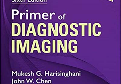
Primer of Diagnostic Imaging 6th Edition

Primer of Diagnostic Imaging 6th Edition

Widely known as THE survival guide for radiology residents, fellows, and junior faculty, the “purple book” provides comprehensive, up-to-date coverage of diagnostic imaging in an easy-to-read, bulleted format. Focusing on the core information you need for learning and practice, this portable resource combines the full range of diagnostic imaging applications with the latest imaging modalities, making it the perfect clinical companion and review tool.
  • Features more than 1,200 detailed illustrations now in full color, plus images that clearly depict the latest applications of CT, MRI, PET/CT, and other diagnostic imaging modalities.
  • Provides new coverage of non-interpretive skills such as quality and safe dosing.
  • Balances new information and anatomic drawings with timeless, relevant material to fully prepare you for the boards and for daily practice.
  • Explains the nuances of key diagnostic details for all body systems, including signs and symptoms, anatomic landmarks, and common radiologic-pathologic alterations, for the full range of radiologic modalities and specialties.
  • Uses a bulleted format and provides mnemonics, descriptive terminology, and space for note taking that make it easy to learn and remember key facts, techniques, and images.
  • Allows you to work through diagnoses with hundreds of differentials for board certification preparation.
  • Clarifies the impact of the latest disease entities on the interpretation of radiologic findings.
  • Expert Consult™ eBook version included with purchase. This enhanced eBook experience allows you to search all of the text, figures, and references from the book on a variety of devices.

DOWNLOAD THIS MEDICAL BOOK

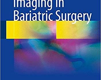
Imaging in Bariatric Surgery 1st ed

Imaging in Bariatric Surgery 1st ed

This book offers detailed guidance on the use of imaging in the context of bariatric surgery. After a summary of the types of surgical intervention, the role of imaging prior to and after surgery is explained, covering both the normal patient and the patient with complications. The most common pathologic features that may be encountered in daily practice are identified and illustrated, and in addition the treatment of complications by means of interventional radiology and endoscopy is described. The authors are acknowledged international experts in the field, and the text is supported by surgical graphs and flow charts as well as numerous images. Overweight and obesity are very common problems estimated to affect nearly 30% of the world’s population; nowadays, bariatric surgery is a safe and effective treatment option for people with severe obesity. The increasing incidence of bariatric surgery procedures makes it imperative that practitioners have a sound knowledge of the imaging appearances of postoperative anatomy and potential complications, and the book has been specifically designed to address the lack of knowledge in this area.

DOWNLOAD THIS MEDICAL BOOK


Textbook of Clinical Echocardiography 6th Edition

Textbook of Clinical Echocardiography 6th Edition

Today’s echocardiography continues to be a low-cost, minimal-risk procedure with the potential to yield a vast amount of detailed, precise anatomic and physiologic information. Dr. Catherine Otto’s Textbook of Clinical Echocardiography, 6th Edition, clearly outlines how to master the core principles of echocardiographic imaging in order to make an initial diagnosis and integrate this data in clinical decision making for patients with a wide range of cardiovascular diseases. Ideal for cardiology fellows, medicine residents, and cardiac sonography students, this bestselling text teaches all the essential elements of ultrasound physics, tomographic and 3D anatomy, image acquisition, advanced imaging modalities, and application in specific disease categories– all with a practical, problem-based approach.

    • Concentrates on the foundational concepts you need to know to perform and interpret echocardiographic studies and to pass your board exams.

  • Covers all advanced echo techniques, including contrast echo, 3D echo, and myocardial mechanics, as well as intraoperative and intra-procedural transesophageal echocardiography (TEE).
  • Discusses what alternative diagnostic approaches to initiate when echocardiography does not provide a definitive answer.
    • Includes new Echo Math boxes in each chapter for quick review and greater comprehension.
    • Provides new ASE recommendations for chamber quantitation, including updated tables of normal measurements.
    • Matches full-color anatomic drawings of heart structures with the 2D and 3D echocardiographic views.
    • Pairs state-of-the-art echo images with more than 360 videos that illustrate the full range of cardiac disease diagnosed with this powerful imaging approach.

  • Expert Consult™ eBook version included with purchase. This enhanced eBook experience allows you click on each figure to see the corresponding video, as well as search all of the text, figures, and references from the book on a variety of devices.

DOWNLOAD THIS MEDICAL BOOK HERE

https://upsto.re/Cibdhgy

Basic Clinical Radiobiology 5th Edition

Basic Clinical Radiobiology is a concise but comprehensive textbook setting out the essentials of the science and clinical application of radiobiology for those seeking accreditation in radiation oncology, clinical radiation physics, and radiation technology.

Fully revised and updated to keep abreast of current developments in radiation biology and radiation oncology, this fifth edition continues to present in an interesting way the biological basis of radiation therapy, discussing the basic principles and significant developments that underlie the latest attempts to improve the radiotherapeutic management of cancer.

This new edition is highly illustrated with attractive 2-colour presentation and now includes new chapters on stem cells, tissue response and the convergence of radiotherapy, radiobiology, and physics. It will be invaluable for FRCR (clinical oncology) and equivalent candidates, SpRs (and equivalent) in radiation oncology, practicing radiation oncologists and radiotherapists, as well as radiobiologists and radiotherapy physicists.

DOWNLOAD THIS MEDICAL BOOK HERE

https://upsto.re/CPEKxBL

ALTERNATE LINK

https://douploads.com/iams14yw1fa1

Caffey’s Pediatric Diagnostic Imaging, 2-Volume Set

Caffey’s Pediatric Diagnostic Imaging, 2-Volume Set

For more than 70 years, Caffey’s Pediatric Diagnostic Imaging has been the comprehensive, go-to reference that radiologists have relied upon for dependable coverage of all aspects of pediatric imaging. In the 13th Edition, Dr. Brian Coley leads a team of experts to bring you up to date with today’s practice standards in radiation effects and safety, as well as in head and neck, neurologic, thoracic, cardiac, gastrointestinal, genitourinary, and musculoskeletal pediatric imaging. This two-volume bestselling reference is a must-have resource for pediatric radiologists, general radiologists, pediatric subspecialists, pediatricians, hospitals, and more – anywhere clinicians need to ensure safe, effective, and up-to-date imaging of children. View more >

Key Features
  • Provides access to 50 online videos, including hypertrophic pyloric stenosis, disorders of swallowing, fetal swallowing, fetal bowel obstruction, upper GI and ultrasound evaluation of malrotation and volvulus, congenital heart disease MRI evaluation, and many more.
  • Includes separate chapters on radiation effects and safety, pre-natal imaging, neoplasms, trauma, techniques, embryology, genetic anomalies, and common acquired conditions.

DOWNLOAD THIS MEDICAL BOOK HERE

https://upsto.re/CgyZvYR

Osborn’s Brain 2nd Edition

[amazon_link asins=’0323477763′ template=’ProductAd’ store=’aishabano-20′ marketplace=’US’ link_id=’4fa391a9-316f-4c59-94d6-e7aeffa2a3e1′]

Comprehensive, visually appealing, and easy to understand, Osborn’s Brain, second edition, by the highly esteemed Dr. Anne G. Osborn, provides a solid framework for understanding the complex subject of brain imaging when studied cover to cover. Almost completely rewritten and featuring 75% new illustrations, it combines essential anatomy with gross pathology and imaging, clearly demonstrating why and how diseases appear the way they do. The most immediate emergent diagnostic topics are followed by nonemergent pathologies, integrating the most relevant information from Dr. Osborn’s entire career of accumulated knowledge, experience, and interest in neuropathology, neurosurgery, and clinical neurosciences.

    • Covers the “must-know” aspects of brain imaging together with spectacular pathology examples, relevant anatomy, and up-to-date techniques in neuroradiology―perfect for radiologists, neuroradiologists, neurosurgeons, and neurologists at all levels
    • Begins with emergent topics such as trauma, nontraumatic hemorrhage, stroke, and vascular lesions, followed by infections, demyelinating and inflammatory diseases, neoplasms, toxic-metabolic-degenerative disorders, and congenital brain malformations
    • Features more than 4,000 stunning, high-resolution radiologic images and medical illustrations, all of which are annotated to describe the most clinically significant features
    • Includes Dr. Osborn’s trademark summary boxes scattered throughout for quick review of essential facts, as well as the most recent and up-to-date references available


  • Helps readers think clearly about diagnoses, types of diagnoses, and the various pathologies that can affect the brain
    • Includes new WHO classifications of brain tumors, new entities including IgG4-related disease and CLIPPERS, new and emerging infectious diseases, and updated insights into brain trauma and brain degeneration


  • Expert Consult™ eBook version included with purchase. This enhanced eBook experience allows you to search all of the text, figures, Q&As, and references from the book on a variety of devices.

DOWNLOAD THIS MEDICAL BOOK HERE

https://upsto.re/CXHh9M6

ALTERNATE LINK


Diagnostic Ultrasound, 2-Volume Set 5th Edition

[amazon_link asins=’0323401716′ template=’ProductAd’ store=’aishabano-20′ marketplace=’US’ link_id=’a31e0368-8c87-4348-8c74-829c659085fd’]

Now fully updated with more than 2,000 new images, 200 new videos, and new content throughout,Diagnostic Ultrasound, 5th Edition, by Drs. Carol M. Rumack and Deborah Levine, remains the most comprehensive and authoritative ultrasound resource available. Spanning a wide range of medical specialties and practice settings, it provides complete, detailed information on the latest techniques for ultrasound imaging of the whole body; image-guided procedures; fetal, obstetric, and pediatric imaging; and much more. Up-to-date guidance from experts in the field keep you abreast of expanding applications of this versatile imaging modality and help you understand the “how” and “why” of ultrasound use and interpretation.

  • Covers all aspects of diagnostic ultrasound with sections for Physics; Abdominal, Pelvic, Small Parts, Vascular, Obstetric, and Pediatric Sonography.
  • Uses a straightforward writing style and extensive image panels with correlative findings.
    • Features 5,000 images – more than 2,000 brand-new – including new 2D and 3D imaging as well as the use of contrast agents and elastography.
    • Includes a new virtual chapter on artifacts with individually labelled images from throughout the book, displaying artifacts with descriptive legends by category and how they can be used in diagnosis or corrected for better quality imaging.
    • Features more images and new uses for contrast agents in the liver, breast, and in pediatric applications. Includes current information on imaging more diagnostic dilemmas, such as Zika virus in the fetus and newborn.
    • Includes 400 video clips showing real-time scanning of anatomy and pathology.


  • Expert Consult™ eBook version included with purchase. This enhanced eBook experience allows you to search all of the text, figures, Q&As, and references from the book on a variety of devices.

DOWNLOAD THIS MEDICAL BOOK HERE

https://upsto.re/CgTNc5R

ALTERNATE LINK

https://douploads.com/z5hengj831t2

Chest Radiology: Patterns and Differential Diagnoses 7th Edition

[amazon_link asins=’0323498310′ template=’ProductAd’ store=’aishabano-20′ marketplace=’US’ link_id=’c46e34b3-7663-43b9-b22c-a3b620c505a2′]

Sharpen your skills in chest x-ray interpretation using this trusted clinical resource! Chest Radiology: Patterns and Differential Diagnoses, 7th Edition, by Dr. James Reed, walks you through a logical, sequential thought process for the differential diagnoses of 23 radiologic patterns of common chest diseases, using 150 superbly illustrated patient cases. You’ll gain a solid and thorough understanding of how to read and interpret chest x-rays with expert guidance on common disease patterns, differential diagnoses, narrowing down the diagnoses, and further studies (from additional radiographic exams to CT or to biopsy). Each chapter follows a consistent format: Presenting Case, Questions, Discussion, Top 5 Diagnoses, Summary, and Answer Guide.


  • Heavily illustrated with chest radiographs and additional CT, HRCT, and MR correlative images; plus bulleted summary boxes of comprehensive diagnoses lists and interpretation points.
  • An ideal resource for mastering this lower-cost modality before considering more complicated and costly procedures.
    • Illustrated case studies and quizzes feature new and improved questions that address the challenges seen in practice today.


    • Significant updates on carcinoid tumors and pulmonary nodules, including pathologic descriptions, detection, management, and new terminology; new lung cancer screening guidelines and benefits of CT vs. x-ray for screening; and CT description of nodules, detection of pulmonary nodules with chest x-rays, anatomic and perception problems with x-rays in identifying nodules.


    • New content on diffuse disease, including new pathologic terms and the latest impact of high resolution CT (HRCT).


    • Updates on radiation dosing and emphasis on lower dosing, and an expanded emphasis on how to interpret x-rays and CT scans.


  • Expert Consult™ eBook version included with purchase. This enhanced eBook experience allows you to search all of the text, figures, and references from the book on a variety of devices.

DOWNLOAD THIS MEDICAL BOOK HERE

https://upsto.re/CXyfuga

ALTERNATE LINK

https://douploads.com/oqma14skarz6

Obstetric Imaging: Fetal Diagnosis and Care (Expert Radiology) 2nd Edition

[amazon_link asins=’0323445489′ template=’ProductAd’ store=’aishabano-20′ marketplace=’US’ link_id=’4cc1a5e5-29fc-4ab4-a8ec-c099d99e3d83′]

Richly illustrated and comprehensive in scope, Obstetric Imaging, 2nd Edition, provides up-to-date, authoritative guidelines for more than 200 obstetric conditions and procedures, keeping you at the forefront of this fast-changing field. This highly regarded reference covers the extensive and ongoing advances in maternal and fetal imaging in a concise, newly streamlined format for quicker access to common and uncommon findings. Detailed, expert guidance, accompanied by superb, high-quality images, helps you make the most of new technologies and advances in obstetric imaging.

  • Features more than 1,350 high-quality images, including 400 in color, and over 85 videos online.
  • Helps you select the best imaging approaches and effectively interpret your findings with a highly templated, bulleted, at-a-glance organization.
  • Reflects all the latest developments in the field, including genetics, open fetal surgery, fetal echocardiography, Zika virus, and 3D imaging, so you can provide the safest and most responsive care to both mother and fetus.
  • Includes new chapters on Limbs and Bones Overview; Open Fetal Surgery; Biophysical Profile; Ultrasound Physics; Elastography; Doppler; MRI; Echogenic Bowel; Pregnancy of Unknown Location (PUL), Failed Pregnancy and Ectopic Pregnancy, Cesarean Scar Pregnancy; Cytomegalovirus (CMG), Rubella, Toxoplasmosis, Herpes, Varicella; and Congenital Syphilis; plus a new chapter on Zika Virus written by imaging experts from the “hot zone.”
    • Keeps you up to date with the latest developments in multimodality imaging and optimizing diagnostic accuracy from ultrasound, 3D ultrasound, Doppler, MRI, elastography, image-guided interventions, and much more.


  • Expert Consult™ eBook version included with purchase. This enhanced eBook experience allows you to search all of the text, figures, Q&As, and references from the book on a variety of devices.

DOWNLOAD THIS MEDICAL BOOK HERE

https://upsto.re/CXnHCuV

Medical Imaging for Health Professionals: Technologies and Clinical Applications 1st Edition

[amazon_link asins=’1119120284′ template=’ProductAd’ store=’aishabano-20′ marketplace=’US’ link_id=’845ba0ff-7d50-4ce6-8700-345fff1d0e52′]

Medical Imaging for Health Professionals: Technologies and Clinical Applications 1st Edition

Describes the most common imaging technologies and their diagnostic applications so that pharmacists and other health professionals, as well as imaging researchers, can understand and interpret medical imaging science

This book guides pharmacists and other health professionals and researchers to understand and interpret medical imaging. Divided into two sections, it covers both fundamental principles and clinical applications. It describes the most common imaging technologies and their use to diagnose diseases. In addition, the authors introduce the emerging role of molecular imaging including PET in the diagnosis of cancer and to assess the effectiveness of cancer treatments. The book features many illustrations and discusses many patient case examples.

Medical Imaging for Health Professionals: Technologies and Clinical Applications offers in-depth chapters explaining the basic principles of: X-Ray, CT, and Mammography Technology; Nuclear Medicine Imaging Technology; Radionuclide Production and Radiopharmaceuticals; Magnetic Resonance Imaging (MRI) Technology; and Ultrasound Imaging Technology. It also provides chapters written by expert radiologists in well-explained terminology discussing clinical applications including: Cardiac Imaging; Lung Imaging; Breast Imaging; Endocrine Gland Imaging; Abdominal Imaging; Genitourinary Tract Imaging; Imaging of the Head, Neck, Spine and Brain; Musculoskeletal Imaging; and Molecular Imaging with Positron Emission Tomography (PET).

  • Teaches pharmacists, health professionals, and researchers the basics of medical imaging technology
  • Introduces all of the customary imaging tools—X-ray, CT, ultrasound, MRI, SPECT, and PET—and describes their diagnostic applications
  • Explains how molecular imaging aids in cancer diagnosis and in assessing the effectiveness of cancer treatments
  • Includes many case examples of imaging applications for diagnosing common diseases

Medical Imaging for Health Professionals: Technologies and Clinical Applications is an important resource for pharmacists, nurses, physiotherapists, respiratory therapists, occupational therapists, radiological or nuclear medicine technologists, health physicists, radiotherapists, as well as researchers in the imaging field.

DOWNLOAD THIS MEDICAL BOOK HERE

https://upsto.re/CqCsuQM

1 19 20 21 22 23 61