Cardiovascular Intervention: A Companion to Braunwald’s Heart Disease 1st Edition
Cardiovascular Intervention: A Companion to Braunwald’s Heart Disease 1st Edition

Introducing Cardiovascular Interventiona comprehensive companion volume to Braunwald’s Heart Disease. This medical reference book contains focused chapters on how to utilize cutting-edge interventional technologies, with an emphasis on the latest protocols and standards of care. Cardiovascular Intervention also includes an eBook updated with late-breaking clinical trials, “Hot off the Press” commentary, and Focused Reviews that are relevant to interventional cardiology.


  • View immersive videos from an online library of procedural clips located on Expert Consult, and stay up to date in the field with interventional topics regularly added online.
  • Remain abreast of the newest interventional techniques, including next-generation stents, invasive lesion assessment, and methods to tackle complex anatomy.
  • Provide optimal patient care with help from easy-to-access information on the latest diagnostic and treatment advances, discussions on percutaneous approaches to structural heart disease, and new developments in treating heart valve disease.
  • Expert Consult eBook version included with purchase. This enhanced eBook experience allows you to search all of the text, figures, references, and videos from the book on a variety of devices.

DOWNLOAD THIS MEDICAL BOOK

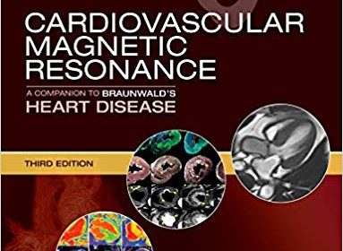
Cardiovascular Magnetic Resonance: A Companion to Braunwald’s Heart Disease 3rd Edition

Cardiovascular Magnetic Resonance: A Companion to Braunwald’s Heart Disease 3rd Edition

Written by an expert team of cardiologists, radiologists, and basic scientists, this third edition of Cardiovascular Magnetic Resonance continues to bridge the divide among specialty areas in with cohesive presentation of this complex and fast-changing field. Offering comprehensive coverage of CMR and the latest cardiology applications, this practical reference enhances the understanding of cardiac physiology and the interpretation and diagnosis of cardiovascular disease. This is an ideal resource for cardiologists, cardiovascular and general radiologists, and anyone who needs up-to-date information on CMR’s uses, benefits, and limitations in cardiovascular care.

  • Provides state-of-the-art coverage of CMR technologies and guidelines, including basic principles, imaging techniques, ischemic heart disease, right ventricular and congenital heart disease, vascular and pericardium conditions, and functional cardiovascular disease.
  • Includes new chapters on non-cardiac pathologypacemaker safety, economics of CMR, and guidelines as well as new coverage of myocarditis and its diagnosis and assessment of prognosis by cardiovascular magnetic resonance, and the use of PET/CMR imaging of the heart, especially in sarcoidosis.
  • Features more than 1,100 high-quality images representing today’s CMR imaging.
  • Covers T1, T2 and ECV mapping, as well as T2* imaging in iron overload, which has been shown to save lives in patients with thalassaemia major.
  • Discusses the cost-effectiveness of CMR.
  • Expert Consult™ eBook version included with purchase. This enhanced eBook experience allows you to search all of the text, figures, and references from the book on a variety of devices.
 

DOWNLOAD THIS MEDICAL BOOK

Echocardiography Pocket Guide: The Transthoracic Examination (Echocardiography Pocket Guides) 1st Edition

Echocardiography Pocket Guide: The Transthoracic Examination (Echocardiography Pocket Guides) 1st Edition

Equip yourself with the most highly illustrated step-by-step guide to echocardiography! Developed for medical students, residents, cardiologists, and sonographers, Echocardiography Pocket Guide: The Transthoracic Examination presents a comprehensive, easy-to-understand, and practical guide to the performance and interpretation of the transthoracic examination. Such knowledge and skills are essential for cardiologists and non-cardiologists alike as echocardiography – the most widely used cardiac imaging technique – becomes more available as hand-carried and pocket-sized devices. Featuring nearly 1,000 anatomically accurate illustrations and images, this outstanding guide presents a step-by-step assessment of cardiac structure and function. It summarizes normal and abnormal findings of cardiac chamber and valvular structure and function. This unrivaled work, Echocardiography Pocket Guide: The Transthoracic Examination has the information you need to accurately perform and interpret the comprehensive transthoracic echocardiography examination.


DOWNLOAD THIS MEDICAL BOOK

Feigenbaum’s Echocardiography Eighth Edition

Feigenbaum’s Echocardiography Eighth Edition

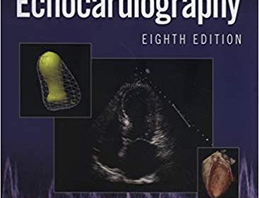
Continuing the long-standing Feigenbaum tradition as an authoritative, comprehensive echocardiography resource, the thoroughly revised Feigenbaum’s Echocardiography, Eighth Edition, helps echocardiographers, fellows, clinicians, and sonographers master the art and science of echocardiography and stay current with all that’s new in the field. Written by William F. Armstrong and Thomas Ryan, it guides you through pertinent physics, technology, clinical applications, and new developments in the field. As in the past, the book is written primarily for the practitioner who uses echocardiographic methods to care for and manage patients, with a focus on appropriate clinical applications.

  • Features enhanced online content including echo clips, case studies, and more.
  • Includes new content specifically for cardiac sonographers.
  • Reflects new training guidelines for cardiology fellows emphasizing the importance of echocardiography in the general practice of cardiology.
  • Describes the interrelationship of other imaging technologies with echocardiography and the role of echo as part of a multimodality approach to patients.
  • Contains more than 1,600 high-quality illustrations – 600 in full color.
  • Covers the use of 3D echocardiography and perfusion imaging; current AHA/ACC guidelines, appropriate use guidelines, and the mechanics and utility of strain and strain rate imaging.


DOWNLOAD THIS MEDICAL BOOK

External Beam Therapy (Radiotherapy in Practice) 3rd Edition

External Beam Therapy (Radiotherapy in Practice) 3rd Edition

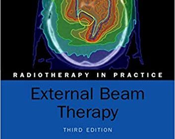
External beam therapy is the most common form of radiotherapy, delivering ionizing radiation such as high-energy x-rays, gamma rays, or electron beams directly into the location of the patient’s tumour. Now in its third edition, this book is an essential, practical guide to external beam radiotherapy planning and delivery, covering the rapid technological advances made in recent years.

The initial chapters give a detailed insight into the fundamentals of clinical radiotherapy. This is followed by systematic details for each tumour site commonly treated with radiotherapy, covering indications, treatment, and planning. The final chapter covers the all important aspect of quality assurance in radiotherapy delivery.

This third edition has been fully updated and revised to reflect new techniques, including details of intensity modulated radiotherapy (IMRT), image guided radiotherapy (IGRT), stereotactic body radiotherapy (SBRT), and proton therapy.

Written by experts in each field, External Beam Therapy is an invaluable companion to professionals and trainees in medical physics, therapeutic radiology, and clinical or radiation oncology.

ABOUT THE SERIES
Radiotherapy remains the major non-surgical treatment modality for the management of malignant disease. It is based on the application of the principles of applied physics, radiobiology, and tumour biology to clinical practice. Each volume in the series takes the reader through the basic principles of the use of ionizing radiation and then develops this by individual sites. This series of practical handbooks is aimed at physicians both training and practising in radiotherapy, as well as medical physics, dosimetrists, radiographers, and senior nurses.


DOWNLOAD THIS MEDICAL BOOK HERE

Cardiovascular Imaging: Arterial and Aortic Valve Inflammation and Calcification

Cardiovascular Imaging: Arterial and Aortic Valve Inflammation and Calcification

This book provides comprehensive reviews on our most recent understanding of the molecular and cellular mechanisms underlying atherosclerosis and calcific aortic valve disease (CAVD) as visualized in animal models and patients using optical molecular imaging, PET-CT, ultrasound and MRI. In addition to presenting up-to-date information on the multimodality imaging of specific pro-inflammatory or pro-calcification pathways in atherosclerosis and CAVD, the book addresses the intriguing issue of whether cardiovascular calcification is an inflammatory disease, as has been recently supported by several preclinical and clinical imaging studies. In order to familiarize researchers and clinicians from other specialties with the basic mechanisms involved, chapters on the fundamental pathobiology of atherosclerosis and CAVD are also included. The imaging chapters, written by some of the foremost investigators in the field, are so organized as to reveal the nature of the involved mechanisms as disease progresses.


DOWNLOAD THIS MEDICAL BOOK HERE


Contrast Echocardiography: Compendium for Clinical Practice 1st ed

This book provides a comprehensive overview of the practical aspects of contrast echocardiography. It also covers all the material in the guidelines published by the American Society of Echocardiography (ASE) in 2018 and the recommendations set out by the European Association of Cardiovascular Imaging (EACVI) in 2017. Contrast echocardiography at present is only used in 5-10% of cases, but this is expected to grow rapidly following the recommendations of the ASE and EACVI. The chapters cover the approved indications and provide practical advice on how to administer the contrast agents and how to optimize the recordings as well as how to deal with the pitfalls. The reader will find all the information on how to use contrast agents for assessment of shunts, LV volumes and function as well as myocardial diseases and masses.  Detailed protocols are included for stress echocardiography and myocardial perfusion imaging. Other topics covered include the use of contrast agents for coronary sonography and transesophageal echocardiography.


Contrast Echocardiography: Compendium for Clinical Practice comprehensively covers all aspects of the clinical use of contrast echocardiography and has been written by two cardiologists who share their experience from their high volume echo laboratories. One of the authors has been a member of both the ASE guidelines and EACVI recommendation writing groups.  It is therefore, a critical text for echocardiographers and sonographers who perform echocardiography.


DOWNLOAD THIS MEDICAL BOOK HERE

Cardiac Electrophysiology Without Fluoroscopy 1st ed

This book reflects how the concern regarding the effects of radiation exposure in patients and health personnel involved in cardiac electrophysiology (EP) has inspired new developments in cardiac electrophysiology procedures without the use of fluoroscopy. This innovative method has become a subspecialty within electrophysioloy with several EP laboratories around the world adopting an exclusive non-fluoroscopy approach. It features guidance on how to use three dimensional (3D) navigation systems, ablation energy sources and zero-fluorospic implantation of cardiac electronic devices. The potential complications and associated preventative methods with utilising RFCA are also described.

Cardiac Electrophysiology Without Fluoroscopy offers a thorough description of the technique correlated to the performance of EP procedure without the use of radiation, and provides a valuable resource for those seeking a practically applicable guide on how to perform cardiac EP without fluoroscopy, including practising and trainee electrophysiologists, cardiac imagers, general cardiologists and emergency medicine physicians.


DOWNLOAD THIS MEDICAL BOOK HERE

Advanced Biomedical Image Analysis 1st Edition

Advanced Biomedical Image Analysis 1st Edition

A comprehensive reference of cutting-edge advanced techniques for quantitative image processing and analysis

Medical diagnostics and intervention, and biomedical research rely progressively on imaging techniques, namely, the ability to capture, store, analyze, and display images at the organ, tissue, cellular, and molecular level. These tasks are supported by increasingly powerful computer methods to process and analyze images. This text serves as an authoritative resource and self-study guide explaining sophisticated techniques of quantitative image analysis, with a focus on biomedical applications. It offers both theory and practical examples for immediate application of the topics as well as for in-depth study.

Advanced Biomedical Image Analysis presents methods in the four major areas of image processing: image enhancement and restoration, image segmentation, image quantification and classification, and image visualization. In each instance, the theory, mathematical foundation, and basic description of an image processing operator is provided, as well as a discussion of performance features, advantages, and limitations. Key algorithms are provided in pseudo-code to help with implementation, and biomedical examples are included in each chapter.

Image registration, storage, transport, and compression are also covered, and there is a review of image analysis and visualization software. The accompanying live DVD contains a selection of image analysis software, and it provides most of the algorithms from the book so readers can immediately put their new knowledge to use.

Members of the academic community involved in image-related research as well as members of the professional R&D sector will rely on this volume.
It is also well suited as a textbook for graduate-level image processing classes in the computer science and engineering fields.


DOWNLOAD THIS MEDICAL BOOK HERE


ULTIMATE SHOPPING EXPERIENCE https://manzastore.com/

Radionuclide Parathyroid Imaging: Book and Atlas 1st ed

Radionuclide Parathyroid Imaging: Book and Atlas 1st ed

This atlas, compiled by experienced specialists in the field, is designed as a ready reference on the use of parathyroid scintigraphy in patients with hyperparathyroidism, both for the localisation of parathyroid pathology and as an aid to surgery. The introductory chapters review the basic core knowledge on the subject. Eighty case reviews are then presented, covering gamma camera planar imaging, SPECT, hybrid SPECT-CT, and also PET-CT. In total, 240 illustrations are included, comprising 160 grey-scale photos depicting nuclear medicine and CT images and 80 dual-modality fusion colour photos. This compilation of illustrative clinical cases will greatly assist clinicians and imaging specialists in image interpretation in different settings. The images replicate normal conventional formats used for routine reporting and hence facilitate fast and reliable diagnosis. Each of the case reviews includes documentation of the procedure, findings, and conclusions with relevant commentary. Surgeons, nuclear medicine physicians, and radiologists will find the Radionuclide Parathyroid Imaging: Book and Atlas to be a valuable practical tool and learning aid.


DOWNLOAD THIS MEDICAL BOOK HERE


ULTIMATE SHOPPING EXPERIENCE https://manzastore.com/


1 16 17 18 19 20 62