Image-Guided Interventions: Expert Radiology Series 3rd Edition

Image-Guided Interventions: Expert Radiology Series 3rd Edition
Image-Guided Interventions: Expert Radiology Series 3rd Edition

Completely revised to reflect recent, rapid changes in the field of interventional radiology (IR), Image-Guided Interventions, 3rd Edition, offers comprehensive, narrative coverage of vascular and nonvascular interventional imaging―ideal for IR subspecialists as well as residents and fellows in IR. This award-winning title provides clear guidance from global experts, helping you formulate effective treatment strategies, communicate with patients, avoid complications, and put today’s newest technology to work in your practice.

  • Offers step-by-step instructions on a comprehensive range of image-guided intervention techniques, including discussions of equipment, contrast agents, pharmacologic agents, antiplatelet agents, and classic signs, as well as detailed protocols, algorithms, and SIR guidelines.
  • Includes new chapters on Patient Preparation, Prostate Artery Embolization, Management of Acute Aortic Syndrome, Percutaneous Arterial Venous Fistula Creation, Lymphatic Interventions, Spinal and Paraspinal Nerve Blocks, and more.
  • Employs a newly streamlined format with shorter, more digestible chapters for quicker reference.
  • Integrates new patient care and communication tips throughout to address recent changes in practice.
  • Highlights indications and contraindications for interventional procedures, and provides tables listing the materials and instruments required for each.
  • Features more than 2,300 state-of-the-art images demonstrating IR procedures, full-color illustrations of anatomical structures and landmarks, and video demonstrations online.

DOWNLOAD THIS BOOK


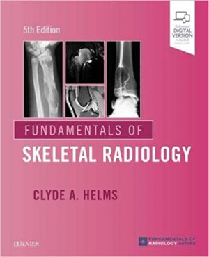
Fundamentals of Skeletal Radiology 5th Edition

Fundamentals of Skeletal Radiology 5th Edition

Fundamentals of Skeletal Radiology 5th Edition

Trusted by thousands of radiology residents, students, and clinicians, the “pink book” continues to be the perfect first book for essential, easily accessible information in skeletal imaging. Fundamentals of Skeletal Radiology, 5th Edition, provides an authoritative introduction to x-rays, MR, and other skeletal imaging modalities, offering a quick, effective review of musculoskeletal imaging in a concise, easy-to-read style.

  • Depicts musculoskeletal imaging concepts and techniques through hundreds of high-quality digital radiographs, MRIs, bone scans, and CT images.
  • Uses a succinct, highly accessible writing style for easy, straightforward understanding of complex material.
  • Updates include numerous new, high-quality MR images and extensive coverage of MRI of the spine and joints, including imaging protocols, common pathologies, and detailed specifics on reading and interpretation.
  • Presents full-color imaging examples to help you discern subtleties and nuances for efficient and accurate interpretation.
  • Discusses radiation dosage concerns, early detection, avoiding unnecessary exams, and common skeletal conditions, including a chapter on trauma.

DOWNLOAD THIS BOOK


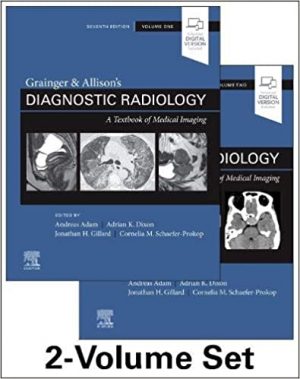
Grainger & Allison’s Diagnostic Radiology 2-Volume Set 7th Edition

Grainger & Allison’s Diagnostic Radiology 2-Volume Set 7th Edition

Grainger & Allison’s Diagnostic Radiology 2-Volume Set 7th Edition

Master the information you need to know for practice and prepare for certification or recertification with a succinct, comprehensive account of the entire spectrum of imaging modalities and their clinical applications. Throughout six outstanding editions, Grainger and Allison’s Diagnostic Radiology has stood alone as the single comprehensive reference on general diagnostic radiology. Now in two succinct volumes, the 7th Edition of this landmark text continues to provide complete coverage of all currently available imaging techniques and their clinical applications – the essential information you need to succeed in examinations and understand current best practices in radiological diagnosis.

  • Organizes content along an organ and systems basis, covering all diagnostic imaging techniques in an integrated, correlative fashion, with a focus on the topics that matter most to a trainee radiologist in the initial years of training.
  • Contains more than 4,000 high-quality illustrations that enhance and clarify the text.
  • Features an expanded section on cardiac imaging to reflect major developments in cardiac MRI, including 3D ultrasound, PET, and SPECT.
  • Integrates functional and molecular imaging throughout each section, and includes the latest image-guided biopsy and ablation techniques.
  • Provides an ideal resource for written, oral, and re-certifying board study as well as for a clinical practice refresher on topics that may have been forgotten.

DOWNLOAD THIS BOOK


Textbook of Three-Dimensional Echocardiography 2nd ed

Textbook of Three-Dimensional Echocardiography 2nd ed

Textbook of Three-Dimensional Echocardiography 2nd ed

Textbook of Three-Dimensional Echocardiography 2nd ed

This thoroughly revised textbook provides a practically applicable guide to three-dimensional echocardiography (3DE). Background is provided on the evolution of the technology and physics that support the implementation of both transthoracic and transesophageal approaches to 3DE. The incremental value of 3DE to assess cardiac chambers is also described. Moreover, a range of cardiac valvular diseases including the mitral, aortic, and tricuspid valve have been portrayed and illustrated in depth.These include congenital abnormalities, regurgitation and stenosis. Emphasis is also placed on technical aspects of the technique and where it can provide added value, including post-surgery assessments and evaluation of cardiac masses.

FOR MORE BOOKS VISIT EDOWNLOADS.ME

Textbook of Three-Dimensional Echocardiography enables readers to develop a deep understanding of how to use this imaging modality. It provides a valuable resource for the echocardiography trainee looking to develop their knowledge and for the experienced practitioner seeking a comprehensive up-to-date reference.

DOWNLOAD THIS BOOK


Errors in Emergency and Trauma Radiology

Errors in Emergency and Trauma Radiology

Errors in Emergency and Trauma Radiology

Errors in Emergency and Trauma Radiology

This book describes and illustrates the gamut of errors that may arise during the performance and interpretation of imaging of both nontraumatic and traumatic emergencies, using a head-to-toe approach. The coverage encompasses mistakes related to suboptimal imaging protocols, failure to review a portion of the examination, satisfaction of search error, and misinterpretation of imaging findings.

FOR MORE BOOKS VISIT EDOWNLOADS.ME

The book opens with an overview of an evidence-based approach to errors in imaging interpretation in patients in the emergency setting. Subsequent chapters describe errors in radiographic, US, multidetector CT, dual-energy CT, and MR imaging of common as well as less common acute conditions, including disorders in the pediatric population, and the unique mistakes in the imaging evaluation of pregnant patients. The book is written by a group of leading North American and European Emergency and Trauma Radiology experts. It will be of value to emergency and general radiologists, to emergency department physicians and related personnel, to general and trauma surgeons, and to trainees in all of these specialties.

DOWNLOAD THIS BOOK


Diagnostics in Ocular Imaging

Diagnostics in Ocular Imaging: Cornea, Retina, Glaucoma and Orbit

Diagnostics in Ocular Imaging

This book presents a new avenue in the field of ophthalmology and sheds light on the field of eye imaging. With the increasing availability of electronic devices and their important role in both personal and professional aspects of human life, there is a growing need for perfect vision. Ophthalmic imaging is a major tool for screening and documenting eye diseases in both medical and surgical fields of ophthalmology and is also of use for ophthalmologists around the globe.

FOR MORE MEDICAL BOOKS VISIT EDOWNLOADS.ME

The number of eye-imaging devices has increased dramatically, however undiagnosed or poorly managed eye diseases remain a significant cause of ocular and visual problems worldwide. This essential guide addresses the need for a book that is dedicated to ophthalmic imaging, covering the cornea, glaucoma, retina and orbital imaging with updates on medical and surgical aspects of the topic.

DOWNLOAD THIS BOOK


Pediatric Body MRI: A Comprehensive, Multidisciplinary Guide

Pediatric Body MRI: A Comprehensive, Multidisciplinary Guide

Pediatric Body MRI: A Comprehensive, Multidisciplinary Guide

This book is a unique, authoritative and clinically oriented text on pediatric body MRI. It is your one-step reference for current information on pediatric body MRI addressing all aspects of congenital and acquired disorders. The easy-to-navigate text is divided into 17 chapters. Each chapter is organized to comprehensively cover the latest MRI techniques, fundamental embryology and anatomy, normal development and anatomic variants, key clinical presentation, characteristic imaging findings with MRI focus, differential diagnosis and pitfalls, as well as up-to-date management and treatment.

FOR MORE MEDICAL BOOKS AND TUTORIALS VISIT EDOWNLOADS.ME

Written by internationally known pediatric radiology experts and editorial team lead by acclaimed author, Edward Y. Lee, MD, MPH, this book is an ideal guide for practicing radiologists, radiology trainees, MRI technologists as well as clinicians in other specialties who are interested in pediatric body MRI.

DOWNLOAD THIS BOOK


Pediatric Musculoskeletal Ultrasonography

Pediatric Musculoskeletal Ultrasonography

Pediatric Musculoskeletal Ultrasonography

This book provides a comprehensive compilation of musculoskeletal ultrasonography (MSUS) fundamentals in pediatric rheumatology with emphasis on imaging techniques, normal anatomy, approaches towards standardization, and the spectrum of pathologic findings seen in the pediatric population. It examines the techniques and pitfalls of MSUS in pediatrics and compares sonoanatomy in pediatric patients versus adults. Chapters cover a range of anatomical sites, including shoulder and arm, elbow and forearm, wrists and hands, hip and thigh, knee and leg, and ankle and feet.

FOR MORE MEDICAL BOOKS VISIT EDOWNLOADS.ME

The text also discusses the use of ultrasonography in juvenile inflammatory arthritic conditions and sports-related injuries. Finally, the book concludes with a summary of the recent advances in pediatric musculoskeletal ultrasonography. Featuring contributions from a large international group of leaders in the field, Pediatric Musculoskeletal Ultrasonography is an authoritative reference for pediatric and adult rheumatologists, sonographers, radiologists, physiotherapists, and orthopedic specialists.

DOWNLOAD THIS BOOK


Atlas of Pediatric Echocardiography

Atlas of Pediatric Echocardiography

Written by Dr. Filip Kucera, a pediatric cardiology consultant at Great Ormond Street Hospital, Atlas of Pediatric Echocardiography provides “next level” guidance for clinicians who want to improve their echocardiographic skills. It covers a wide range of levels, from a beginner to an advanced level. This highly illustrated atlas is an excellent resource not only for pediatric cardiologists and trainees in pediatric cardiology, but also neonatologists, pediatric intensivists, and pediatricians with an interest in echocardiography.

•Contains over 800 high-quality echocardiograms, depicting normal views followed by congenital and acquired cardiovascular defects and other conditions.

•Provides clear explanations for all pathology images for a clear understanding of the diagnosis.

•Includes a guide to normal echocardiographic examination.

•Covers segmental approach to congenital heart disease; atrial, ventricular and atrio-ventricular septal defects; diseases of the mitral, tricuspid, left and right ventricular outflow tract; double outlet right ventricle; tetralogy of Fallot; transposition of the great arteries; truncus arteriosus; functionally single ventricle; PDA; coarctation and interruption of the aorta; vascular rings; pulmonary and systemic venous anomalies; congenital coronary artery abnormalities; myocarditis; cardiomyopathies; Kawasaki disease; rheumatic heart disease; infective endocarditis; pericardial disease; cardiac tumors; pulmonary hypertension; genetic syndromes; mechanical circulatory support; heart transplantation and more.

DOWNLOAD THIS BOOK


Incidental Findings in Neuroimaging and Their Management

Incidental Findings in Neuroimaging and Their Management

Incidental Findings in Neuroimaging and Their Management

Incidental Findings in Neuroimaging and Their Management: A Guide for Radiologists, Neurosurgeons, and Neurologists presents a streamlined, case-based approach to 50 commonly seen incidental findings in neuroimaging. Edited by Kaye Westmark, Dong Kim, and Roy Riascos, this unique book provides the necessary knowledge to manage significant unexpected findings—from identification and analysis to efficacious interventions. With collaborative contributions from neuroradiologists, neurosurgeons, neurologists, otolaryngologists, body and musculoskeletal imaging experts, endocrinologists and hematologists/oncologists, this resource encompasses a wide spectrum of incidental findings.

FOR MORE BOOKS AND TUTORIALS VISIT EDOWNLOADS.ME

Organized by six sections, the book starts with normal variants that are extremely important to recognize in order to avoid unwarranted additional testing and unnecessary stress for the patient. Subsequent sections detail abnormalities that require extensive clinical evaluation in order to determine ideal management. These include incidental findings for extracranial, extra-spinal, intracranial, and intraspinal imaging. The final section outlines CT and MR imaging artifacts that are particularly concerning because they may mimic more dangerous pathologies while degrading imaging quality and obscuring real findings.

Key Features

  • Key findings and differential diagnosis are listed for each entity
  • Diagnostic decision trees present algorithms in an easy-to-understand manner
  • Artifact analyses explain the technical reason for each artifact and what can be done to mitigate effects
  • Clinical Q&As connect the radiologic diagnosis with actual case management decisions and provide in-depth background information that is applicable to management in various scenarios

This essential guide will help trainee and practicing neuroradiologists, neurosurgeons, and neurologists interpret incidental spine and brain imaging findings and make clinically informed, complex treatment decisions.

DOWNLOAD THIS BOOK


1 8 9 10 11 12 62