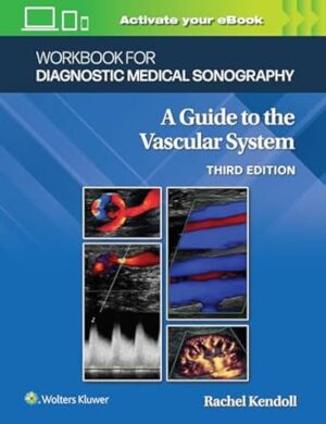
Workbook for Diagnostic Medical Sonography: The Vascular Systems (Lippincott Connect) Third Edition

Workbook for Diagnostic Medical Sonography: The Vascular Systems (Lippincott Connect) Third Edition

Designed to accompany the 3rd Edition of Anne Marie Kupinski’s text, Workbook for Diagnostic Medical Sonography: A Guide to the Vascular System, 3rd Edition, by Rachel Kendoll, offers a full complement of self-study aids for active learning that enable you to assess and build your knowledge as you advance through the text. Most importantly, it helps you get the most out of your study time, with a variety of custom-designed exercises to help you master each objective.

  • Features a new, full-color format throughout 
  • Contains glossary terms reviews, illustrated anatomy and physiology reviews with image labeling, and chapter reviews with multiple-choice, fill-in-the-blank, and short answer questions 
  • Features up-to-date sonograms and relevant content throughout, including new coverage of ergonomics 


Workbook for Diagnostic Medical Sonography: A Guide to the Vascular System, 3rd Edition, is now available with Lippincott® Connect! Lippincott® Connect enables you to personalize your teaching and enhance your students’ course experience in a customizable, all-in-one learning solution combining assessment, multimedia content, and an interactive eBook. Instructors can create assignments, assess students’ knowledge, and track their progress. Students maximize efficiency through valuable feedback and remediation. Key performance insights are reported in a user-friendly dashboard that allows instructors and students to tailor their teaching and learning experiences.  If you would like to use  Workbook for Diagnostic Medical Sonography: A Guide to the Vascular System, 3rd Edition, in your course, please contact medicaleducationinquiries@wolterskluwer.com. 
 

Enrich Your eBook Reading Experience 

  • Read directly on your preferred device(s), such as computer, tablet, or smartphone. 
  • Easily convert to audiobook, powering your content with natural language text-to-speech. 

DOWNLOAD THIS MEDICAL BOOK

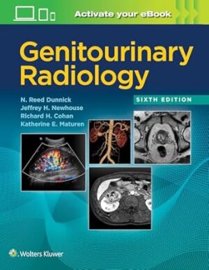
Genitourinary Radiology 6th Edition

Genitourinary Radiology 6th Edition

Selected as a Doody’s Core Title for 2022 and 2023!

Previously known as the 
Textbook of Uroradiology , the newly retitled Genitourinary Radiology continues to bring you top-flight expertise in interpreting imaging studies of the genitourinary tract. A team of leading authorities walk you through the full range of relevant modalities and findings for each anatomical region, providing a multitude of high-quality representative images that capture the characteristic appearance of the conditions you’re likely to encounter. The result remains an indispensible resource for diagnosing genitourinary diseases and disorders.
Features

  • Identify abnormalities using the latest imaging technology thanks to comprehensive updates throughout, including PET/CT and SPECT/CT, MR urography and MR angiography, CT dose reduction and 3D CT imaging, and more.
  • Recognize what to look for by reviewing more than 1,000 images and accompanying discussions that pinpoint the most important diagnostic features for virtually every genitourinary disorder.
  • Quickly find the guidance you need through a practical, intuitive organization that concisely summarizes the disorders found in each anatomical area and then walks you through how these disorders present via the most relevant imaging modalities.


Your book purchase includes a complimentary download of the enhanced eBook for iOS, Android, PC & Mac.

Take advantage of these practical features that will improve your eBook experience:

  • The ability to download the eBook on multiple devices at one time — providing a seamless reading experience online or offline
  • Powerful search tools and smart navigation cross-links that allow you to search within this book, or across your entire library of VitalSource eBooks
  • Multiple viewing options that enable you to scale images and text to any size without losing page clarity, as well as responsive design
  • The ability to highlight text and add notes with one click

DOWNLOAD THIS MEDICAL BOOK 

An Atlas of Neonatal Brain Sonography, 2nd Edition (182) 2nd Edition

An Atlas of Neonatal Brain Sonography, 2nd Edition (182) 2nd Edition

This Atlas covers the entire spectrum of brain disease as studied with ultrasound, illustrated throughout with superb-quality images. It is aimed at neonatologists and radiologists confronted with everyday clinical questions on the neonatal ward. Most newborn brain disorders can be identified with ultrasound; this book will therefore be particularly useful in settings with limited MRI facilities. Prenatal ultrasound specialists will also find it valuable as a postnatal reference in their field of interest. Suggestions for differential diagnosis accompany all the sonographic findings, guiding the clinician in proceeding from an abnormal image to a diagnosis. This second edition of the Atlas has been brought up to date to include the many advances in technique and interpretation that have been made in the past decade. The images have been replaced with new ones of higher quality, and all the line artwork has been standardised and improved.

DOWNLOAD THIS MEDICAL BOOK 


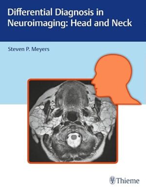
Differential Diagnosis in Neuroimaging: Head and Neck 1st Edition

Differential Diagnosis in Neuroimaging: Head and Neck 1st Edition

Authored by renowned neuro-radiologist Steven P. Meyers, Differential Diagnosis in Neuroimaging: Head and Neck is a stellar guide for identifying and diagnosing head and neck disease based on location and neuroimaging results. The succinct text reflects more than 25 years of hands-on experience gleaned from advanced training and educating residents and fellows in radiology, neurosurgery, and otolaryngology. The high-quality MRI and CT scans have been collected over Dr. Meyers’s lengthy career, presenting an unsurpassed visual learning tool.

The distinctive ‘three-column table plus images’ format is easy to incorporate into clinical practice, setting this book apart from larger, disease-oriented radiologic tomes. This layout enables readers to quickly recognize and compare abnormalities based on more than 1,500 high-resolution images. Chapters cover skull imaging, temporal bone imaging, orbital imaging, paranasal imaging, suprahyoid neck imaging, and infrahyoid neck imaging, for a full spectrum of head and neck pathologies.

Key Highlights

  • Tabular columns organized by anatomical abnormality include neck, facial, and skull based imaging findings and a summary of key clinical data that correlate to the images
  • Congenital/developmental and acquired abnormalities including solitary or multiple orbital lesions; and solitary, multifocal, or diffuse sinonasal disease
  • Abnormalities of the skull, craniovertebral junction, tempormandibular joint, infrahyoid neck, anterior and posterior cervical space, perivertebral space, and brachial plexus

This visually rich resource is a must-have diagnostic tool for residents, fellows, and practitioners in radiology, otolaryngology-head and neck surgery, and neurosurgery. The highly practical format makes it ideal for daily rounds, as well as a robust study guide for physicians preparing for board exams.

DOWNLOAD THIS MEDICAL BOOK 

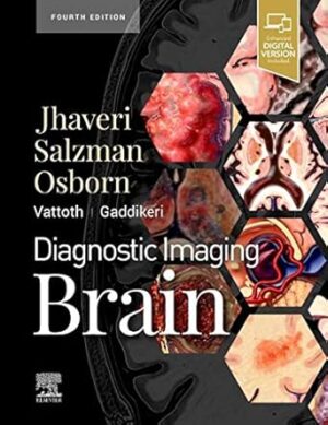
Diagnostic Imaging: Brain 4th Edition

Diagnostic Imaging: Brain 4th Edition

Covering the entire spectrum of this fast-changing field, Diagnostic Imaging: Brain, fourth edition,is an invaluable resource for neuroradiologists, general radiologists, and trainees―anyone who requires an easily accessible, highly visual reference on today’s neuroimaging of both common and rare conditions. World-renowned authorities provide updated information on more than 300 diagnoses, all lavishly illustrated, delineated, and referenced, making this edition a useful learning tool as well as a handy reference for daily practice.

  • Provides authoritative, comprehensive guidance on both pathology-based and anatomy-based diagnoses to help you diagnose the full range of brain and CNS conditions

  • Features thousands of extensively annotated images, including a large number of full-color illustrations―greatly expanded since the previous edition

  • Details 31 new diagnoses, covering key topics such as critical illness-associated microbleeds, autoimmune encephalitis, multinodular and vacuolating tumor of cerebrum, calcifying pseudoneoplasm of neuraxis (CAPNON), uremic encephalopathy, gadolinium deposition and associated controversies, ataxia-telangiectasia, and Zika virus infection

  • Reflects updates from the most recent WHO Classification of Tumors of the CNS, which presents major restructuring of brain tumor categories and incorporates new entities that are defined by both histology and molecular features

  • Includes updates to the 2016 WHO Classification of Tumors of the CNS by cIMPACT-NOW based on recent and ongoing advances in molecular pathogenesis

    • Covers recent neuroimaging advances, such as 7T MRI scanners and dual-energy/dual-source CT imaging

    • Uses bulleted, succinct text and highly templated chapters for quick comprehension of essential information at the point of care
    • Enhanced eBook version included with purchase. Your enhanced eBook allows you to access all of the text, figures, and references from the book on a variety of devices


DOWNLOAD THIS MEDICAL BOOK 

Detection Systems in Lung Cancer and Imaging (Volume 1)

Detection Systems in Lung Cancer and Imaging (Volume 1)

This book focuses on major trends and challenges in the detection of lung cancer, presenting work aimed at identifying new techniques and their use in biomedical analysis. Volume 1 examines the main applications of computer-aided diagnosis to lung cancer. It is ideal for academics, industry professionals and advanced students.

DOWNLOAD THIS MEDICAL BOOK 

Netter’s Concise Radiologic Anatomy: With STUDENT CONSULT Online Access (Netter Basic Science) 2nd Edition

Netter’s Concise Radiologic Anatomy: With STUDENT CONSULT Online Access (Netter Basic Science) 2nd Edition

Designed to make learning more interesting and clinically meaningful, Netter’s Concise Radiologic Anatomy, 2nd Edition matches radiologic images―from MR and ultrasound to CT and advanced imaging reconstructions―to the exquisite artwork of master medical illustrator Frank H. Netter, MD. As a companion to the bestselling Netter’s Atlas of Human Anatomy, this updated medical textbook begins with the anatomy and matches radiologic images to the anatomic images; the result is a concise, visual guide that shows how advanced diagnostic imaging is an amazing “dissection tool” for viewing human anatomy in the living patient!

“Overall I feel that this textbook is a good overview of radiology, and perfectly adequate for medical students as we are not expected to have a very in depth knowledge of radiology past chest and abdominal x rays. It may well prove valuable for the Netter-collectors out there, and provides an excellent quick guide to radiology.” Reviewed by: TheMedicalStudent.co.uk Date: July 2014

  • Quickly review key information with a concise, user-friendly format that is organized and color-coded to be in-line with Netter’s Atlas of Human Anatomy, 6th Edition.
  • View direct, at-a-glance comparisons between idealized anatomic illustrations and real-life medicine with side-by-side radiology examples of normal anatomy and common variants with corresponding anatomy illustrations.
  • Improve upon your knowledge with a brief background in basic radiology, including reconstructions and a list of common abbreviations for the images presented.
  • Broaden your visual comprehension with the help of 30 brand-new ultrasound images.
  • Access the complete contents online at Student Consult.

DOWNLOAD THIS MEDICAL BOOK 

MRI Made Easy (for beginners) 2nd Edition

MRI Made Easy (for beginners) 2nd Edition

This book explains all the basic entities related to magnetic resonance imaging (MRI) for the beginners. It begins by explaining basics of magnetism, magnetic field, and basic principles behind MRI. Scanning parameters, accessory techniques, MR interpretation, clinical applications of MRI, sequences and artifacts are discussed briefly with representative pictures. Different kinds of MR instrumentation, scanning options are also explained based on which we can chose the instrument to identify a particular ailment. While using MR imaging safety options should also be considered as it may damage skin, and tissue heating. Principles and advantages of neuroimaging is discussed, which helps in interpreting MR imaging apart from anatomical and pathological knowledge of the brain. Bodyimaging is the new aspect of MRI, which helps to assess staging and treatment planning in musculoskeletal tumors. Other instrumentation discussed here includes MR angiography, MR diffusion and its clinical applications, MR spectroscopy, MR Cholangiopancreatography. Advancement and higher MR technologies like 3-Tesla MRI which is superior in epilepsy, inner ear and functional imaging are also discussed briefly.

DOWNLOAD THIS MEDICAL BOOK 

Grainger & Allison’s Diagnostic Radiology: 2-Volume Set 7th Edition

Grainger & Allison’s Diagnostic Radiology: 2-Volume Set 7th Edition

Master the information you need to know for practice and prepare for certification or recertification with a succinct, comprehensive account of the entire spectrum of imaging modalities and their clinical applications. Throughout six outstanding editions, Grainger and Allison’s Diagnostic Radiology has stood alone as the single comprehensive reference on general diagnostic radiology. Now in two succinct volumes, the 7th Edition of this landmark text continues to provide complete coverage of all currently available imaging techniques and their clinical applications – the essential information you need to succeed in examinations and understand current best practices in radiological diagnosis.

  • Organizes content along an organ and systems basis, covering all diagnostic imaging techniques in an integrated, correlative fashion, with a focus on the topics that matter most to a trainee radiologist in the initial years of training.

  • Contains more than 4,000 high-quality illustrations that enhance and clarify the text.

  • Features an expanded section on cardiac imaging to reflect major developments in cardiac MRI, including 3D ultrasound, PET, and SPECT.

  • Integrates functional and molecular imaging throughout each section, and includes the latest image-guided biopsy and ablation techniques.

  • Provides an ideal resource for written, oral, and re-certifying board study as well as for a clinical practice refresher on topics that may have been forgotten.
  • Enhanced eBook version included with purchase. Your enhanced eBook allows you to access all of the text, figures, and references from the book on a variety of devices.

DOWNLOAD THIS MEDICAL BOOK 

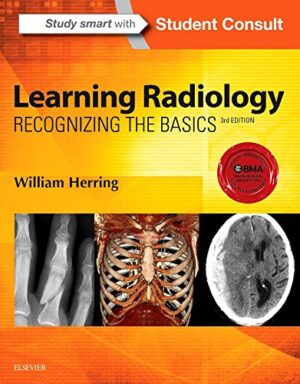
Learning Radiology: Recognizing the Basics 3rd Edition

Learning Radiology: Recognizing the Basics 3rd Edition

must-have for anyone who will be required to read and interpret common radiologic images, Learning Radiology: Recognizing the Basics is an image-filledpractical, and easy-to-read introduction to key imaging modalities. Skilled radiology teacher William Herring, MD, masterfully covers exactly what you need to know to effectively interpret medical images of all modalities. Learn the latest on ultrasound, MRI, CT, patient safety, dose reduction, radiation protection, and more, in a time-friendly format with brief, bulleted text and abundant high-quality images. Then ensure your mastery of the material with additional online content, bonus images, and self-assessment exercises at Student Consult.

  • Identify a wide range of common and uncommon conditions based upon their imaging findings.

  • Arrive at diagnoses by following a pattern recognition approach, and logically overcome difficult diagnostic challenges with the aid of decision trees.

    • Quickly grasp the fundamentals you need to know through more than 700 images and an easy-to-use format and pedagogy, including: bolding of key points and icons designating special content; Diagnostic Pitfalls; Really, Really Important Points; Weblinks; and Take-Home Points.

    • Gauge your mastery of the material and build confidence with extra images, bonus content, interactive self-assessment exercises, and USMLE-style Q&A that provide effective chapter review and quick practice for your exams.

    • Apply the latest recommendations on patient safety, dose reduction and radiation protection

    • Benefit from the extensive knowledge and experience of esteemed author Dr. William Herring―a skilled radiology teacher and the host of his own specialty website, www.learningradiology.com.
    • Stay current in the latest advancements and developments with meticulous updates throughout including a new chapter on Pediatric Radiology as well as more than 60 new and updated photos, many highlighting newer imaging modalities.

    • Maximize your learning experience with interactive Student Consult extras videos/images of 3D images, functional imaging examinations, dynamic studies, and additional assessments.

      • Student Consult eBook version included with purchase. This enhanced eBook experience allows you to search all of the text, figures, references, and videos from the book on a variety of devices.


DOWNLOAD THIS MEDICAL BOOK 


1 2 3 62