Pediatric Emergency Radiology (WHAT DO I DO NOW EMERGENCY MEDICINE)

Pediatric Emergency Radiology (WHAT DO I DO NOW EMERGENCY MEDICINE) 
Part of the “What Do I Do Now?: Emergency Medicine” series, Pediatric Emergency Radiology uses a case-based approach to cover common and important topics in radiology imaging for pediatric emergency care. Each chapter provides a discussion of the diagnosis, key points to remember, and selected references for further reading. Areas of controversy are clearly delineated with a discussion regarding evidence-based options and a balanced view of treatment and disposition decisions. The book addresses a wide range of topics including neonatal respiratory distress, foreign body ingestion, Bronchiolitis, and related radiology issues faced by emergency medicine providers and pediatricians.

Pediatric Emergency Radiology is an engaging collection of thought-provoking cases which clinicians can utilize for effective imaging of pediatric patients. The volume is also a self-assessment tool that tests the reader’s ability to answer the question, “What do I do now?”

DOWNLOAD THIS MEDICAL BOOK

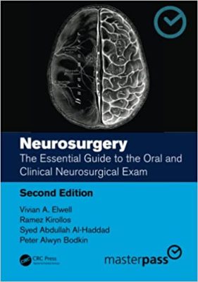
Neurosurgery (MasterPass) 2nd Edition

Neurosurgery (MasterPass) 2nd Edition

This new edition of Neurosurgery: The Essential Guide to the Oral and Clinical Neurosurgical Exam provides a concise and practical guidebook of the core knowledge and principles for the International and Intercollegiate FRCS Specialty Examination in Neurosurgery. It is a vital resource for the American Board of Neurological Surgery (ABNS) and other neurosurgical examinations around the world.

Written by neurosurgeons at the top of their field and based on new guidelines, this book takes students through how to succeed in the FRCS neurosurgery exams and provides an overview of crucial short and intermediate cases designed to mirror the exam’s testing of a candidate’s clinical knowledge, diagnostic acumen, investigation and interpretation, treatment options and taking consent.

Including 72 vital online revision flash cards, covering critical and diverse examination cases from trauma to paediatric spine exams, this edition also contains crucial guidance to Vivas on the following:


  • Operative surgery and surgical anatomy


  • Investigation of the neurosurgical patient


  • The non-operative clinical practice of neurosurgery

This book is a must-read for candidates preparing for the final Intercollegiate Specialty Examination in Neurosurgery (UK), International FRCS Specialty Examination in Neurosurgery (UK) International FRCS Specialty Examination in Neurosurgery as well as the American, Canadian, European and Australasian exams. In addition to helping candidates pass their final exams, the book provides wonderful insight into Neurosurgery for Medical Students, Surgical Residents and Neurosurgical Consultants.

DOWNLOAD THIS MEDICAL BOOK

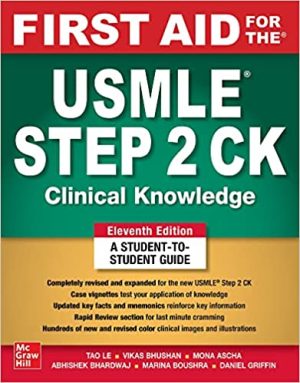
First Aid for the USMLE Step 2 CK, Eleventh Edition 11th Edition

First Aid for the USMLE Step 2 CK, Eleventh Edition 11th Edition

The most comprehensive and up-to-date high-yield review available for the USMLE® Step 2 CK―completely revised and better than ever!

The expert author team that guided students to success on the USMLE® Step 1 presents the latest edition of this skill-sharpening review for the USMLE® Step 2 CK. With an easy-to-follow bulleted presentation of must-know diseases and disorders, this one-of-a-kind study companion offers the most current overview of all core areas on the boards. Included is a host of learning tools, from key facts and mnemonics to full-color illustrations and proven test-taking strategies―everything students need to pass the exam with flying colors.

  • Co-written by students who excelled on the recent exam and reviewed by top faculty
  • Concise summaries of more than 1,000 commonly tested clinical topics for fast, high-yield study
  • Key Facts and mnemonics reinforce must-know concepts
  • Expert coverage of best initial steps in diagnosis and management
  • Updated Rapid Review section facilitates last-minute cramming
  • Hundreds of full-color photographs and illustrations
  • Revised study and test-taking strategies
  • A completely updated listing of top-rated review sources

DOWNLOAD THIS MEDICAL BOOK

Huppert’s Notes: Pathophysiology and Clinical Pearls for Internal Medicine 1st Edition

Huppert’s Notes: Pathophysiology and Clinical Pearls for Internal Medicine 1st Edition

Bridge the gap between pathophysiology and clinical medicine in a succinct outline of core internal medicine topics!

Originally created and road-tested by a resident and then updated by a team of resident authors, Huppert’s Notes succinctly organizes the foundational science covered early in medical school and the clinical approaches encountered in clerkships and beyond. This marriage of pathophysiology and clinical medicine provides a framework for how to approach internal medicine concepts mechanistically, rather than through memorization.

You’ll find concise descriptions of common medical conditions with diagnostic and management pearls, as well as high-yield diagrams and tables to emphasize key concepts. Covering all internal medicine subspecialties, each Huppert’s Notes chapter is organized in an intuitive and consistent outline format for rapid access:
 

  • Anatomy & Physiology
  • Diagnostics
  • Approaches & Chief Complaints
  • Diseases & Pathophysiology
  • Key Medications & Interventions
  • Key Clinical Trials & Publications
  • Space for your personal notes

DOWNLOAD THIS MEDICAL BOOK

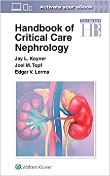
Handbook of Critical Care Nephrology First Edition

Handbook of Critical Care Nephrology First Edition

Designed specifically for nephrologists and trainees practicing in the ICU, Handbook of Critical Care Nephrology is a portable critical care reference with a unique and practical nephrology focus. Full-color illustrations, numerous algorithms, and intuitively arranged contents make this manual a must-have resource for nephrology in today’s ICU.
  • Covers all essential topics related to nephrology in the ICU, including ICU monitoring, drugs and blood products, imaging, acute kidney injury, electrolytes and acid-base disorders, poisonings and intoxications, extracorporeal therapies, organ transplantation, and ethics and palliative care.
  • Provides valuable information on specific conditions including sepsis, obstetrics, abdominal compartment syndrome, AKI in burns and trauma, gastrointestinal bleeding, neurological emergencies, environmental topics such as hemorrhagic fever and terrorism, hematologic/oncologic emergencies, and more.
  • Includes easy-to-understand visual abstracts of key studies.

Enrich Your Ebook Reading Experience 

  • Read directly on your preferred device(s), such as computer, tablet, or smartphone.
  • Easily convert to audiobook, powering your content with natural language text-to-speech.

DOWNLOAD THIS MEDICAL BOOK

Integrated Non-Invasive Cardiovascular Imaging: A Guide for the Practitioner (Non-serial Publications)

Integrated Non-Invasive Cardiovascular Imaging: A Guide for the Practitioner (Non-serial Publications) 

Integrated cardiovascular imaging is the optimal use of multiple imaging modalities to obtain complementary information about cardiac diseases, to aid in diagnosis, determine aetiology and prognosis, with the ultimate objective of effectively guiding clinical decision making. Over the past few decades, advances in technology have contributed to the development of new imaging modalities and refinement of existing ones, leading to major improvements in the accuracy of diagnosing cardiovascular disease. While modality-centric expertise has been the primary driver of improvements in each modality, this has also contributed to imagers working in silos resulting with limited inter-modality coordination and collation of information relevant for patient care. This publication provides comprehensive guidance on the rationale and implementation of integrated cardiovascular imaging for practitioners.

Atlas of the Human Hypothalamus: Anatomy, Blood Supply, Myelo-, and Cytoarchitecture 1st Edition

Atlas of the Human Hypothalamus: Anatomy, Blood Supply, Myelo-, and Cytoarchitecture 1st Edition

Lower Urinary Tract Symptoms and Benign Prostatic Hyperplasia: From Research to Bedside 1st Edition

Lower Urinary Tract Symptoms and Benign Prostatic Hyperplasia: From Research to Bedside 1st Edition

Lower Urinary Tract Symptoms and Benign Prostatic Hyperplasia: From Research to Bedside offers full acknowledgment of the basic research of lower urinary tract symptoms (LUTS) and benign prostatic hyperplasia (BPH), also connecting the clinical and practice management of the disease. It provides a full comprehension of LUTS and BPH from several aspects, allowing for a schematic interpretation of the disease and subsequent medical management. Going beyond the guidelines in the field, this title enhances the knowledge of BPH onset, allowing for the advancement of research, beneficial clinical implication and treatment.

Perfect for researchers, urologists, pathologists and endocrinologists, this must-have reference provides what is needed to understand LUTS and BPH in one easy to reference place.

DOWNLOAD THIS MEDICAL BOOK

Imaging of the Cardiovascular System, Thorax, and Abdomen (Magnetic Resonance Imaging Handbook) 1st Edition

Imaging of the Cardiovascular System, Thorax, and Abdomen (Magnetic Resonance Imaging Handbook) 1st Edition

Magnetic resonance imaging (MRI) is a technique used in biomedical imaging and radiology to visualize internal structures of the body. Because MRI provides excellent contrast between different soft tissues, the technique is especially useful for diagnosticimaging of the brain, muscles, and heart.

In the past 20 years, MRI technology has improved significantly with the introduction of systems up to 7 Tesla (7 T) and with the development of numerous post-processing algorithms such as diffusion tensor imaging (DTI), functional MRI (fMRI), and spectroscopic imaging. From these developments, the diagnostic potentialities of MRI have improved impressively with an exceptional spatial resolution and the possibility of analyzing the morphology and function of several kinds of pathology.

Given these exciting developments, the Magnetic Resonance Imaging Handbook: Imaging of the Cardiovascular System, Thorax, and Abdomen is a timely addition to the growing body of literature in the field. Offering comprehensive coverage of cutting-edge imaging modalities, this book:


  • Discusses MRI of the heart, blood vessels, lungs, breasts, diaphragm, liver, gallbladder, spleen, pancreas, adrenal glands, and gastrointestinal tract
  • Explains how MRI can be used in vascular, posttraumatic, postsurgical, and computer-aided diagnostic (CAD) applications
  • Highlights each organ’s anatomy and pathological processes with high-quality images
  • Examines the protocols and potentialities of advanced MRI scanners such as 7 T systems
  • Includes extensive references at the end of each chapter to enhance further study

Thus, the Magnetic Resonance Imaging Handbook: Imaging of the Cardiovascular System, Thorax, and Abdomen provides radiologists and imaging specialists with a valuable, state-of-the-art reference on MRI.

DOWNLOAD THIS MEDICAL BOOK

Weir & Abrahams’ Imaging Atlas of Human Anatomy 6th Edition

Weir & Abrahams’ Imaging Atlas of Human Anatomy 6th Edition

BMA Book Awards – Winner of Basic and Clinical Sciences category!

The perfect up-to-date imaging guide for a complete and 3-dimensional understanding of applied human anatomy

Imaging is ever more integral to anatomy education and throughout modern medicine. Building on the success of previous editions, this fully revised sixth edition provides a superb foundation for understanding applied human anatomy, offering a complete view of the structures and relationships within the whole body, using the very latest imaging techniques.

All relevant imaging modalities are included, from plain radiographs to more advanced imaging of ultrasound, CT, MRI, functional imaging and angiography. Coverage is further enhanced by a carefully selected range of BONUS electronic content, including clinical photos and cases, ultrasound videos, labelled radiograph ‘slidelines’, cross-sectional imaging stacks and test-yourself materials. Uniquely, key syllabus image sets are now highlighted throughout to aid efficient study, as well as the most common, clinically important anatomical variants that you should be aware of.

This superb package is ideally suited to the needs of medical students, as well as radiologists, radiographers and surgeons in training. It will also prove invaluable to the range of other students and professionals who require a clear, accurate, view of anatomy in current practice.

  • Fully revised legends and labels and new high-quality images–featuring the latest imaging techniques and modalities as seen in clinical practice
  • Covers the full variety of relevant modern imaging–including cross-sectional views in CT and MRI, angiography, ultrasound, fetal anatomy, plain film anatomy, nuclear medicine imaging and more – with better resolution to ensure the clearest anatomical views
  • Core syllabus image sets now highlighted throughout–to help you focus on the most essential areas to excel on your course and in examinations
  • Unique summaries of the most common, clinically important anatomical variants for each body region–reflects the fact that around 20% of human bodies have at least one clinically significant variant
  • New orientation drawings–to help you understand the different views and the 3D anatomy of 2D images, as well as the conventions between cross-sectional modalities
  • Ideal as a stand-alone resource or in conjunction with Abrahams’ and McMinn’s Clinical Atlas of Human Anatomywhere new links help put imaging in the context of the dissection room
  • Now a more complete learning package than ever before, with superb BONUS electronic enhancements embedded within the accompanying eBook, including:

    • Labelled image ‘stacks’–that allow you to review cross-sectional imaging as if using an imaging workstation

    • Labelled image ‘slidelines’–showing features in a full range of body radiographs to enhance understanding of anatomy in this essential modality

    • Self-test image ‘slideshows’ with multi-tier labelling–to aid learning and cater for beginner to more advanced experience levels

    • Labelled ultrasound videos–bring images to life, reflecting this increasingly clinically practiced technique

    • Questions and answers accompany each chapter–to test your understanding and aid exam preparation

    • 34 pathology tutorials–based around nine key concepts and illustrated with hundreds of additional pathology images, to further develop your memory of anatomical structures and lead you through the essential relationships between normal and abnormal anatomy
    • High-yield USMLE topics–clinical photos and cases for key topics, linked and highlighted in chapters

DOWNLOAD THIS MEDICAL BOOK

1 97 98 99 100 101 947