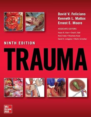
Trauma, Ninth Edition 9th Edition

Trauma, Ninth Edition 9th Edition

The world’s leading resource for to diagnosing and treating any injury―quickly, safely, and effectively

Doody’s Core Titles for 2023!

Unparalleled in its breadth and depth of expertly crafted content, 
Trauma takes you through the full range of injuries you are likely to encounter. With a full-color atlas of anatomic drawings and surgical approaches, this trusted classic provides thorough coverage of kinematics and the mechanisms of trauma injury, the epidemiology of trauma, injury prevention, the basics of trauma systems, triage, and transport, and more. It then reviews generalized approaches to the trauma patient, from pre-hospital care and managing shock, to emergency department thoracotomy and the management of infections; delivers a clear, organ-by-organ survey of treatment protocols; and shows how to handle specific challenges in trauma―including alcohol and drug abuse, and combat-related wounds―in addition to post-traumatic complications such as multiple organ failure.

  • 500 photos and illustrations
  • Color atlas
  • Numerous X-rays, CT scans, and algorithms
  • High-yield section on specific approaches to the trauma patient
  • A-to-Z overview of management of specific traumatic injuries
  • Detailed discussion of the management of complications

DOWNLOAD THIS MEDICAL BOOK 

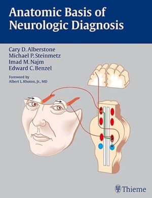
Anatomic Basis of Neurologic Diagnosis 1st Edition

Anatomic Basis of Neurologic Diagnosis 1st Edition

A visual guide to diagnosing neurologic disorders

2010 Benjamin Franklin Silver Award Winner!

Anatomic Basis of Neurologic Diagnosis is a lavishly illustrated book that places special emphasis on the paramount importance of signs and symptoms for the accurate diagnosis of neurologic disorders. It opens with a comprehensive review of neuroembryology, enabling readers to gain knowledge of normal nervous system development and related developmental disorders. The second section of the book comprises an easily accessible presentation of the anatomy of regional parts and to-the-point information on the cardinal manifestations of disease. Separate chapters in the third section of the book present the anatomy of different functional systems and provide practical approaches to diagnosing patients with system disorders. A final chapter covers the anatomy of the vascular system and cerebrospinal fluid.

Highlights:

  • Practical organization of chapters, according to regions and functional systems, reflects the clinician’s approach to patient care
  • Full-color illustrations provide an indispensable visual aid to learning and reviewing clinically relevant neurologic anatomy and pathways
  • Numerous tables summarize key points

Ideal for reading cover-to-cover, this book is essential for residents and students seeking to fully understand the complexity of clinical neuroanatomy. Seasoned clinicians will find the book a valuable refresher.

DOWNLOAD THIS MEDICAL BOOK

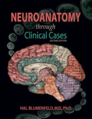
Neuroanatomy Through Clinical Cases, Second Edition 2nd Edition

Neuroanatomy Through Clinical Cases, Second Edition 2nd Edition

Neuroanatomy through Clinical Cases brings a pioneering interactive approach to the teaching of neuroanatomy, using over 100 actual clinical cases and high-quality radiologic images to bring the subject to life. The Second Edition is fully updated with the latest advances in the field, and includes several exciting new cases. This approach allows students to appreciate the clinical relevance of structural details as they are being learned, and to integrate knowledge of disparate functional systems, since a single lesion may affect several different neural structures and pathways. Most of the book comprises chapters that explain the major neuroanatomical systems. Each chapter first presents background material including an overview of relevant neuroanatomical structures and pathways, and a brief discussion of related clinical disorders. The second half of each chapter is devoted to clinical cases. The cases begin with a narrative of how the patient developed symptoms, and what deficits were found upon neurological examination. Boldface type highlights important symptoms and signs. A series of questions challenges the reader to deduce the neuroanatomical location of the patient s lesion, and the diagnosis. Discussion and answers follow, and an epilogue reveals the actual outcome. One of the book s most innovative features is the inclusion of CT and MRI scans that depict each patient s lesion. These radiographs help the reader develop skills in interpreting the same kinds of diagnostic images employed in clinical practice.

DOWNLOAD THIS MEDICAL BOOK

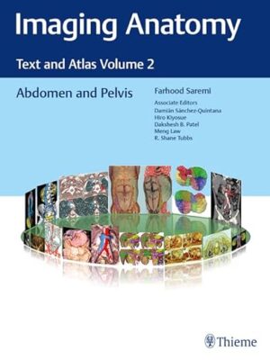
Imaging Anatomy: Text and Atlas Volume 2: Abdomen and Pelvis 1st Edition

Imaging Anatomy: Text and Atlas Volume 2: Abdomen and Pelvis 1st Edition

Unique anatomic atlas provides an indispensable virtual desk dissection experience

Normal imaging anatomy and variants, including diagnostic and surgical anatomy, are the cornerstones of radiologic knowledge. Imaging Anatomy: Text and Atlas Volume 2, Abdomen and Pelvis is the second in a series of four richly illustrated radiologic references edited by distinguished radiologist Farhood Saremi. The atlas is coedited by esteemed colleagues Damián Sánchez-Quintana, Hiro Kiyosue, Dakshesh B. Patel, Meng Law, and R. Shane Tubbs with contributions from an impressive cadre of international authors. Succinctly written text and superb images provide readers with a virtual, user-friendly dissection experience.

This exquisitely crafted atlas combines fundamental core anatomy principles with modern imaging and postprocessing methods to increase understanding of intricate anatomical features. Twenty-two concise chapters cover the abdominal wall, alimentary tract, liver, biliary system, pancreas, spleen, peritoneum, genitourinary system, pelvic floor, neurovasculature, and surface anatomy. Relevant anatomical components of the abdomen and pelvis are discussed, including musculature, arteries, veins, lymphatics, ducts, and innervation.

Key Highlights

  • High-quality cross-sectional multiplanar and volumetric color-coded CT, MRI, and angiography imaging techniques provide detailed insights on specific anatomy
  • Cross-sectional and topographic cadaveric views by internationally known anatomists coupled with more than 1,600 illustrations clearly elucidate difficult anatomical concepts
  • Consistently formatted chapters include an introduction, embryology, review of anatomy, discussion of anatomical variants, postsurgical anatomy, and congenital and acquired pathologies

This unique resource provides an excellent desk reference for differentiating normal versus pathologic anatomy. It is essential reading for medical students, radiology residents and veteran radiologists, internists, and general surgeons, as well as vascular and transplant surgeons.

DOWNLOAD THIS MEDICAL BOOK

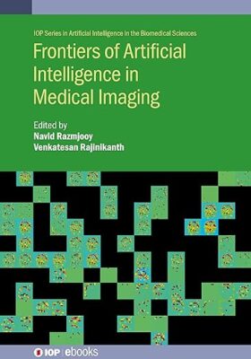
Frontiers of Artificial Intelligence in Medical Imaging

Frontiers of Artificial Intelligence in Medical Imaging

Frontiers of Artificial Intelligence in Medical Imaging considers the recent advancements happening in hospitals to diagnose various diseases accurately using AI supported detection procedures.

DOWNLOAD THIS MEDICAL BOOK

Fundamentals of Urine and Body Fluid Analysis 5th Edition

Fundamentals of Urine and Body Fluid Analysis 5th Edition

Learn how to accurately analyze urine and body fluids with Fundamentals of Urine and Body Fluid Analysis, 5th Edition. Known for its clear writing style, logical organization, and vivid full-color illustrations, this renowned textoffers the perfect level and depth of information for understanding the fundamental principles of urine and body fluids frequently encountered in the clinical laboratory.This includes the collection and analysis of urine, fecal specimens, vaginal secretions, and other body fluids such as cerebrospinal, synovial, seminal, amniotic, pleural, pericardial, and peritoneal fluids. Author Nancy Brunzel also shares her extensive knowledge and expertise in the field as she highlights key information and walks you through essential techniques and procedures ― showing you how to correlate data with your knowledge of basic anatomy and physiology in order to understand pathologic processes.

  • Study questions and case studies in each chapter reinforce comprehension and application, with an answer key located in the back of the book.
  • UNIQUE! Table of crystal images based on shape serves as a single, comprehensive guide to the identification of crystals in urine sediment.
  • UNIQUE! Image Gallery of Urine Sedimentprovides alternate views of sediment components to augment the numerous classic photomicrographs already present in the Microscopic Examination of Urine chapter.
  • UNIQUE! Quick Guides to urine and body fluid photomicrographs make it fast and easy to find a photo of a specific cell type or component of interest.
  • UNIQUETables with high quality polarizing microscopy photomicrographs demonstrate the differences in birefringent intensity of substances with and without a red compensator.
  • The most complete collection of high-quality, full-color images enables optimal identification of microscopic components in urine and other body fluids.
  • NEW! Fully updated content provides valuable information on the latest techniques and advances in the field.
  • NEW! Enhanced content, new tables, and new images facilitate the microscopic differentiation of monocytes, macrophages, and mesothelial cells in pleural, peritoneal, and pericardial fluids.
  • NEW! More than 250 photomicrographs of cells and other components in body fluid and urine sediment serve as a visual quick reference for identification during analysis.
  • NEW! Thumbprint images embedded in numerous tables enhance learning and serve as an invaluable resource when performing fluid analysis at the bench.

DOWNLOAD THIS MEDICAL BOOK 

Preclinical Immunology and Microbiology Review 2023: For USMLE Step 1 and COMLEX-USA Level 1 (USMLE Prep)

Preclinical Immunology and Microbiology Review 2023: For USMLE Step 1 and COMLEX-USA Level 1 (USMLE Prep)

The only official Kaplan Preclinical Immunology and Microbiology 2023 covers the comprehensive information you need to ace the exam and match into the residency of your choice.


  • Up-to-date: Updated annually by Kaplan’s all-star faculty

  • Integrated: Packed with clinical correlations and bridges between disciplines

  • Learner-efficient: Organized in outline format with high-yield summary boxes

  • Trusted: Used by thousands of students each year to succeed on USMLE Step 1


Looking for more prep? Our 
Preclinical Medicine Complete 7-Book Subject Review 2023 has this book, plus the rest of the 7-book series.

DOWNLOAD THIS MEDICAL BOOK

Manual of Childhood Infection: The Blue Book (Oxford Specialist Handbooks in Paediatrics) 4th Edition


Manual of Childhood Infection: The Blue Book (Oxford Specialist Handbooks in Paediatrics) 4th Edition

Infectious diseases remain a leading cause of child morbidity and mortality worldwide. Now in its fourth edition, Manual of Childhood Infections is a simple-to-use, evidence-based, and practical handbook on how to recognize, investigate, and manage both common and rare infectious diseases in children and babies. Endorsed by the Royal College of Paediatrics and Child Health and the European Society for Paediatric Infectious Diseases, this fully updated version of the established ‘Blue Book’ complements the Pan European initiatives and UK diploma courses to harmonise patient management and training in Paediatric Infectious Diseases (PID), making it essential reading for UK and European paediatricians.

Manual of Childhood Infections is divided into two alphabetized sections for easy access to information, covering key diagnosis and management features of infections alongside crucial points of epidemiology and clinical features. This fourth edition forms practical reading for practising paediatricians, featuring updates to all key chapters based on a literature review alongside new chapters focusing on emerging problems for Europe.

DOWNLOAD THIS MEDICAL BOOK

Forensic Pathology for Police, Death Investigators, Attorneys, and Forensic Scientists 2010th Edition

Forensic Pathology for Police, Death Investigators, Attorneys, and Forensic Scientists 2010th Edition

Forensic Pathology for Police, Death Investigators, Attorneys, and Forensic Scientists is a forensic pathology book specifically written for professionals who interact with forensic pathologists. The book includes sections that address various general topics which are not normally present in the typical forensic pathology text, such as descriptions of medical, pathology and forensic pathology training, basic anatomy and physiology, an overview of other forensic science disciplines, and autopsy performance. Forensic Pathology for Police, Death Investigators, Attorneys, and Forensic Scientists also covers classic topics in forensic pathology, including death investigation, death certification, postmortem changes, and the entire range of case types, ranging from natural deaths to drug-related deaths to various types of violent death. The text is written in easy-to-understand language, and is complemented by hundreds of high-quality photographs.

DOWNLOAD THIS MEDICAL BOOK

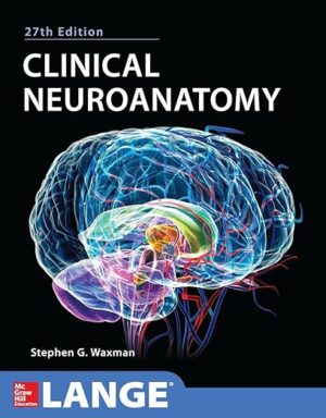
Clinical Neuroanatomy 27/E (Lange Medical Book) 27th Edition

Clinical Neuroanatomy 27/E (Lange Medical Book) 27th Edition

A streamlined, comprehensive synopsis of neuroanatomy and its functional and clinical applications

For more than seventy years, Clinical Neuroanatomy has been the best way for medical students, residents, trainees in health-related fields, and clinicians in practice to gain an understanding of neuroanatomy, its functional underpinnings, and its relationship to the clinic. Emphasizing the important concepts, facts, and structures, this full-color and engagingly written text includes clear, memorable tables and diagrams, and is state of the art in pathophysiology and diagnosis and treatment of neurological disorders.

Here’s why Clinical Neuroanatomy is essential for board review or as a clinical refresher:

  • More than 300 full-color illustrations
  • Clinical correlations help you interpret and remember essential neuroanatomic concepts in terms of function and clinical application
  • Numerous computed tomography (CT) and magnetic resonance images (MRIs) of the normal brain and spinal cord; functional magnetic resonance images that provide a noninvasive window on brain function; and neuroimaging studies that illustrate common pathological entities that affect the nervous system
  • Coverage of the latest advances in molecular and cellular biology in the context of neuroanatomy
  • A unique Introduction to Clinical Thinking section that puts neuroanatomy in a clinical perspective
  • Clear, easy-to-read tables that encapsulate important information
  • A complete practice exam to test your knowledge
  • Coverage of the basic structure and function of the brain, spinal cord, and peripheral nerves as well as clinical presentations of disease processes involving specific structures

DOWNLOAD THIS MEDICAL BOOK 

1 53 54 55 56 57 947