Atlas of Surgical Pathology Grossing (Atlas of Anatomic Pathology)

Atlas of Surgical Pathology Grossing (Atlas of Anatomic Pathology)

Written by expert pathologists, Atlas of Surgical Pathology Grossing is an excellent resource for pathologists, medical and pathology assistant students, residents, both in surgery and pathology, and pathology assistants.


DOWNLOAD THIS MEDICAL BOOK

Atlas of Intestinal Pathology: Volume 1: Neoplastic Diseases of the Intestines

Atlas of Intestinal Pathology: Volume 1: Neoplastic Diseases of the Intestines

Intestinal neoplasia comprises a large part of a surgical pathologist’s workload. Pathologists play a key role not only in the classification of malignancies but also in assisting screening programs, identifying incidental neoplasms, and guiding treatment by providing essential prognostic features for individual entities. A large variety of neoplasms affect the intestines, and there is ongoing discovery of new entities and prognostic features for known diseases. Pathologists and trainees should have a solid understanding of key morphologic features, pitfalls, and differential diagnoses. Importantly, pathologists should recognize and communicate features that help their clinical colleagues in making treatment decisions, with the ultimate goal of benefiting the patient first and foremost.
The first volume of the Atlas of Intestinal Pathology provides a comprehensive yet concise, primarily visual review of intestinal neoplasms.  It also serves as a useful resource primarily for pathologists and trainees in pathology by providing a concise yet comprehensive summary of morphology of intestinal neoplasia. Clinical practitioners and trainees also benefit from an understanding of the pathologic correlates to the diseases they manage.


DOWNLOAD THIS MEDICAL BOOK

Atlas of Genetic Diagnosis and Counseling 3rd ed

Atlas of Genetic Diagnosis and Counseling 3rd ed

Dr. Harold Chen shares his almost 50 years of clinical genetics practice in this new edition of a comprehensive pictorial atlas, featuring almost 290 genetic disorders, malformations, and malformation syndromes. The author provides a detailed outline for each disorder, describing its genetics, basic defects, clinical features, diagnostic tests, and counseling issues, including recurrence risk, prenatal diagnosis, and management. Numerous color photographs of prenatal ultrasounds, imagings, cytogenetics, and postmortem findings illustrate the clinical features of patients at different ages, patients with varying degrees of severity, and the optimal diagnostic strategies. The disorders cited are supplemented by case histories and diagnostic confirmation by cytogenetics, biochemical, and molecular techniques, when available.

Since the publication of the previous edition in 2012, the atlas has been widely accepted and used in light of rapid progress in genetic and gnomic information. In this new edition, additional genetic disorders are added, as well as extensive updates to the previous disorders with new illustrations, supplemented by case and family history, clinical features, and laboratory data, especially molecular confirmation if available. The atlas is written in outline format for ease of use.

Atlas of Genetic Diagnosis and Counseling, Third Edition is of great value to medical geneticists, genetic counselors, pediatricians, neonatologists, developmental pediatricians, perinatologists, obstetricians, neurologists, pathologists, and any physicians and health care professionals caring for handicapped children such as craniofacial surgeons, plastic surgeons, otolaryngologists, and orthopedists. It is the definitive volume for helping all physicians to understand and recognize genetic diseases and malform

ation syndromes and better evaluate, counsel, and manage affected patients.
 

DOWNLOAD THIS MEDICAL BOOK

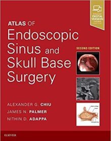
Atlas of Endoscopic Sinus and Skull Base Surgery 2nd Edition

Atlas of Endoscopic Sinus and Skull Base Surgery 2nd Edition

Gain a clear understanding of the entire spectrum of today’s rhinology and anterior skull base surgery with Atlas of Endoscopic Sinus and Skull Base Surgery, 2nd Edition. This thoroughly updated title increases your knowledge and skill regarding both basic or advanced procedures, taking you step by step through endoscopic approaches to chronic sinus disease, nasal polyps, pituitary tumors, cerebrospinal fluid leaks, sinonasal tumors, and more. All-new videos provide real-time guidance for procedures such as endoscopic anterior craniofacial resection, endoscopic approach and removal of orbital tumors, sphenoidotomy, and many more.


  • Covers the full range of modern rhinology and anterior skull base surgery, from septoplasty and sphenoethmoidectomy to extended frontal sinus procedures, endoscopic craniofacial resections and complex skull base reconstructions.
  • Clearly conveys the anatomy and detailed steps of each procedure with concise, step-by-step instructions; visual guidance features high-definition, intraoperative endoscopic photos paired with detailed, labeled anatomic illustrations.
  • Features all-new videos expertly narrated by Dr. Palmer and Dr. Chiu.
  • Includes new content on anterior skull base surgery that reflect new developments in the field.
  • Helps you provide optimal patient care before, during, and after surgery with detailed information on relevant anatomy and surgical indications, instrumentation, potential pitfalls, and post-operative considerations.
  • Expert Consult™ eBook version included with purchase. This enhanced eBook experience allows you to search all of the text, figures, and references from the book on a variety of devices.


DOWNLOAD THIS MEDICAL BOOK

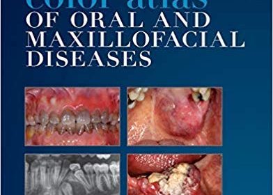
Color Atlas of Oral and Maxillofacial Diseases 1st Edition

Color Atlas of Oral and Maxillofacial Diseases 1st Edition

Introducing an essential new practical atlas for dental students and clinicians alike! The Color Atlas of Oral and Maxillofacial Diseases provides comprehensive, practical information on the most common oral and maxillofacial diseases and disorders. This new text uses a quick-access atlas format to help you easily look up clinical signs, diagnosis, and treatments. Nearly 750 high-quality images accompanied by brief narratives demonstrate exactly what clinical signs to look for – making an intervention as timely as possible. Written by four of the top dental authorities in the world, this concise resource is sure to become a clinical favorite. Expert Consult TM eBook version included with purchase. This enhanced eBook experience allows you to search all of the text, figures, and references from the book on a variety of devices


  • NEW! Quick-access atlas format makes it easy to look up clinical signs, diagnosis, and treatment of oral and maxillofacial diseases
  • NEW! Nearly 750 high-quality radiographs and color clinical photos facilitate the identification of lesions and diseases.
  • NEW! Comprehensive, focused coverage highlight diseases that may affect the oral and maxillofacial regions.
  • NEW! Full-color design and illustrations.
  • NEW! Logical organization reflects the sequence in which content is generally presented to predoctoral students.
  • NEW! Expert Consult TM eBook version included with purchase allows you to search all of the text, figures, and references from the book on a variety of devices


DOWNLOAD THIS MEDICAL BOOK


FOR MORE BOOKS VISIT EDOWNLOADS.ME

Oculoplastic Surgery Atlas: Eyelid and Lacrimal Disorders 2nd Edition

Oculoplastic Surgery Atlas: Eyelid and Lacrimal Disorders 2nd Edition

When the first edition of this atlas was published in 2002, it was well received throughout the oculoplastic and ophthalmology community for its simplicity and clinical focus.  Now, a decade later, surgical techniques have been updated to reflect current practice models and advances in technology. The field of oculoplastic surgery has grown and evolved to include all aspects of eyelid and facial plastic surgery and the literature must now reflect the advancements of this field. Oculoplastic Surgery Atlas: Eyelid and Lacrimal Disorders, 2nd edition

combines text and diagrams, plus surgical videos that enable readers to perform this surgery with the best possible instruction and preparation. Written for ophthalmology specialists and residents, this new edition presents many aspects of facial cosmetic surgery, including lacrimal anatomy, nasolacrimal duct disorders, canaliculitis, and canalicular involving eyelid laceration.


DOWNLOAD THIS MEDICAL BOOK HERE

Oculoplastic Surgery Atlas: Cosmetic Facial Surgery 2nd ed

Oculoplastic Surgery Atlas: Cosmetic Facial Surgery 2nd ed

When the first edition of this atlas was published in 2005, it was well received throughout the oculoplastic and ophthalmology community for its simplicity and clinical focus.  Now, a decade later, surgical techniques have been updated to reflect current practice models and advances in technology. The field of oculoplastic surgery has grown and evolved to include all aspects of facial plastic surgery and the literature must now reflect the advancements of this field. Oculoplastic Surgery Atlas: Cosmetic Facial Surgery, 2nd edition combines text and diagrams, plus surgical videos that enable readers to perform this surgery with the best possible instruction and preparation. Written for ophthalmology specialists and residents, this new edition presents many aspects of facial cosmetic surgery, including blepharoplasty, endoscopic forehead surgery, rhytidectomy, and  soft tissue and lip augmentation.

Review of the previous edition:

“The book offers insight into the possibilities and technology of modern aesthetic cosmetic surgery in the face region. … The book can be recommended for the aesthetic surgeon and for ophthalmologists. … The ophthalmologic plastic surgeon will find this book of interest especially in showing a lot of additional possibilities and dimensions in this field. The book will be an asset in every ophthalmic surgeons library.

DOWNLOAD THIS MEDICAL BOOK HERE

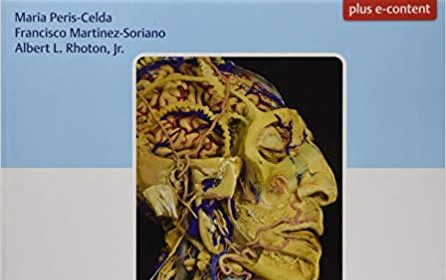
Rhoton’s Atlas of Head, Neck, and Brain: 2D and 3D Images 1st Edition

Rhoton’s Atlas of Head, Neck, and Brain: 2D and 3D Images 1st Edition

An internationally renowned and beloved author, educator, brain anatomist, and neurosurgeon, Professor Albert Rhoton has a special place in medical history. He was revered by students and colleagues and is regarded as one of the fathers of modern microscopic neurosurgery. A driving principle in his anatomy lab was the simple phrase, “Every Second.” This was embraced in his philosophy that every second of every day, a patient’s life was improved by a surgeon assisted by the anatomic knowledge his lab helped elucidate and distribute.

Rhoton’s Atlas of Head, Neck, and Brain is the visually exquisite crowning achievement of Dr. Rhoton’s brilliant career and unwavering dedication to the intertwined pursuits of surgical anatomy and neurosurgery. The atlas reflects the unparalleled contributions Dr. Rhoton made to the contemporary understanding of neurosurgical anatomy. Dr. Peris-Celda, with the collaboration of an impressive cadre of international multidisciplinary experts, worked closely under Dr. Rhoton’s tutelage on this project. This book is the culmination of 5 years of work and experience gleaned from more than 40 years of surgical anatomy research and exquisite dissection techniques performed in Dr. Rhoton’s laboratory.

Special Features

  • Each anatomic dissection meticulously labeled with English and Latin descriptors for easy cross referencing with other resources.
  • Multiple views of the most complex regions of the head, neck, and brain provide a deeper understanding of anatomy.
  • More than 600 anatomical images systematically organized in four major sections: Osteology of the Head and Neck; Face and Neck; Ear, Nose, Pharynx, Larynx, and Orbit; and Neuroanatomy and Cranial Base.
  • Superb 2D images presented in a large printed format to optimize the viewing experience.
  • 3D digital images fully realize the beauty of the dissections and enhance the learning process.
  • Specimens injected with colored silicone provide better visualization of arteries and veins.

Breathtakingly stunning, this atlas is certain to be a treasured reference for medical students, residents, and clinicians specializing in neurosurgery, facial plastic surgery, otolaryngology, maxillofacial surgery, and craniofacial surgery for many years to come.


DOWNLOAD THIS MEDICAL BOOK HERE

Atlas of Clinical Neurology 4th ed

Atlas of Clinical Neurology 4th ed

Like its preceding editions, this atlas is an indispensable guide to the field of neurology, featuring the most clinically essential images and figures. Chapters offer insight and research written by deeply practiced, knowledgeable neurologists that is supplemented with detailed imagery, tables, algorithms, and delineative drawings. Topics covered include developmental and genetic diseases, neuroendocrine disorders, critical care neurology, cerebrovascular disease, dementias, behavioral neurology, neuro-oncology, movement disorders, epilepsy, neuromuscular diseases, infectious diseases, neuroimmunology, neurotoxic disorders, and headache. The authors also delve into specific issues currently prevalent in neuroscientific research, including Alzheimer’s disease, dementia, Machado-Joseph disease, Huntington’s disease, and brain scanning with PET and fMRI. The Atlas of Clinical Neurology, 4th Edition serves as a comprehensive and premier visual resource for neurologists.


DOWNLOAD THIS MEDICAL BOOK PDF


FOR MORE BOOKS VISIT EDOWNLOADS.ME

Atlas on Narrow Band Imaging in Upper Aerodigestive Tract Lesions 1st ed

Atlas on Narrow Band Imaging in Upper Aerodigestive Tract Lesions 1st ed

This book covers all aspects of narrow band imaging (NBI), including research, general principles and diagnostic features in various ENT pathologies. In addition to highlighting the advantages of NBI over white light endoscopy for the detection of early cancers and lesions that are less than 1 cm in diameter, the book offers a step-by-step approach to help readers easily grasp various pathologies of the upper aerodigestive tract. It discusses both malignant and non-malignant aspects of NBI and provides extensive information on inflammatory lesions of the upper aerodigestive tract, including various clinical features of laryngopharyngeal reflux. Subsequent chapters cover the most difficult aspects of NBI in treated mucosa (post-chemo radiated), in which the potential recurrence of disease is a key question. It also addresses the limitations of NBI, including false positive and false negative cases.

Given its scope, the book will benefit otolaryngologists, oncosurgeons, radiation oncologists and oral medicine practitioners, as well as graduate students of ENT, radiotherapy, oral medicine and oncosurgery.

DOWNLOAD THIS MEDICAL BOOK HERE


FOR MORE BOOKS VISIT EDOWNLOADS.ME

1 2 3 4 5 19