Atlas of Essential Dermatopathology

[amazon template=image&asin=1447144708]

The book is not intended to be an all-encompassing atlas or textbook but rather a foundation of principles in dermatopathology, highlighting key elements in the field for trainees and will also serve as a basic resource for the pathologist in general practice. In addition to the sketches and minimal text, we envision accompanying high resolution histopathologic micrographs for ultimate correlation as well.

DOWNLOAD THIS BOOK FREE HERE

http://upsto.re/FjE3jCA

Dermatological Atlas of Indigenous People 1st ed. 2017 Edition

[amazon template=iframe image2&asin=3319594443]

About the Author

Atlas of Mitral Valve Repair

[amazon template=iframe image2&asin=0781746922]

This full-color atlas with accompanying video DVD provides a complete and practical “how-to” guide to planning and performing mitral valve repair surgery for mitral regurgitation. The book reviews the natural history of mitral regurgitation, the functional anatomy of the mitral valve, and the use of echocardiography in preoperative evaluation and surgical planning. Chapters describe and illustrate all techniques currently used for mitral valve repair and discuss results.

A bound-in DVD presents narrated video clips of six cases that show the application of specific techniques. Each case begins with preoperative echocardiograms demonstrating the mitral valve defect and proceeds through key surgical maneuvers.

DOWNLOAD THIS BOOK FREE HERE

http://upsto.re/FBXThBA

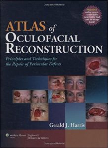
Atlas of Oculofacial Reconstruction: Principles and Techniques for the Repair of Periocular Defects 1 Har/Psc Edition

[amazon template=iframe image2&asin=0781796512]

This full-color atlas is a practical, step-by-step guide to the reconstruction of periocular defects following tumor excision or tissue-loss trauma. The book addresses the specific anatomic concerns in each oculofacial sector with tailored surgical principles and techniques designed to improve aesthetic outcomes.

Full-color illustrations with detailed explanatory legends depict each step of each surgical technique. Flap design and mobilization are shown directly on surgical photographs, rather than in idealized drawings. The clear, accessible writing style will appeal to ophthalmic and plastic surgeons, non-ophthalmic surgeons, and non-surgical ophthalmic specialists.

A companion Website will include an online image bank.

DOWNLOAD THIS BOOK FREE HERE

http://upsto.re/FBXBsYa

Atlas of Dermatology in Internal Medicine

[amazon template=iframe image2&asin=1461406870]

Atlas of Dermatology in Internal Medicine is the only concise text-atlas to cover the most common and most important cutaneous manifestations of systemic disease in children and adults.  It features more than 150 clinical photographs that are accompanied by format-driven, clinically focused text on the diagnosis and management of cutaneous manifestations of connective tissue, pulmonary, renal, GI, endocrine, malignant, infectious, and HIV disease.  There is also a separate chapter on skin diseases commonly seen in the ICU.  A special feature is its systematic coverage of clinically relevant dermatopathology.  The book is a helpful tool for physicians and trainees in internal medicine, family medicine, pediatrics, emergency medicine, and critical care medicine, as well as family, emergency, and critical care nurse practitioners.

DOWNLOAD THIS BOOK FREE HERE

http://upsto.re/FB74ms7

Surgical Atlas of Sports Orthopaedics and Sports Traumatology

[amazon template=iframe image2&asin=3662508540]

From the Back Cover

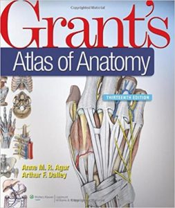
Grant’s Atlas of Anatomy, 13th Edition 13th Edition

[amazon template=iframe image2&asin=1608317560]

A cornerstone of gross anatomy since 1943, Grant’s Atlas of Anatomy reaches students worldwide with its realistic dissection illustrations, detailed surface anatomy photos, clinical images and comments, and quick-reference muscle tables. Renowned for its accuracy, pedagogy, and clinical relevance, this classic atlas boasts significant enhancements, including updated artwork, new conceptual diagrams, and vibrantly re-colored illustrations. Clinical material is clearly highlighted in blue text for easy identification.

DOWNLOAD THIS BOOK FREE HERE

http://upsto.re/FTu3JXQ

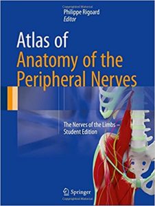
Atlas of Anatomy of the Peripheral Nerves: The Nerves of the Limbs – Student Edition 1st ed. 2017 Edition

[amazon template=iframe image2&asin=3319430882]

This innovative atlas focuses on peripheral nerves and provides a brand new approach compared to regular anatomy books. Using a modern 3D approach, it offers an alternative to conventional anatomical structures. It reviews all the anatomy and the morphology of these structures from an original point of view.
In these three-dimensional diagrams, as well as in the watercolor drawings enhanced with a 3D inlay, each type of nerve is depicted in a minute detail.
The atlas simplifies the anatomy and make it easy and understandable by allowing readers to develop a mental “real-time 3D GPS”.
The integration of MRI sections related to the drawings and the descriptions of the main nerve injuries provide medical students with a flexible but effective transition to the radiological interpretation and furthers the clinical learning process.
After a detailed evaluation of the morphofunctional anatomy of the peripheral nerves, the authors present a collection of relevant data on neuromuscular transmission, both from classical and recent literature, ranging from the central and peripheral nervous system to the effector muscle. This information offers a basis for understanding the physiology, the pathology, and the repair prospects of peripheral nerves from a purely theoretical point of view.
The book is divided into three main parts:
– Fundamental notions: from immunohistochemistry to limb innervation
– The upper limb: the brachial plexus and related peripheral nerves
– The lower limb: the lumbosacral plexus and related peripheral nerves
This atlas also features 261 outstanding full-colour 2D and 3D illustrations. Each picture has been designed in 2D and 3D with a combination of the original editor’s personal drawings/paintings and 3D-modeling tools.
This book is a valuable resource for anyone studying medicine, anaesthesiology, neurosurgery, spine surgery, pain, radiology or rheumatology and is also of high interest to the whole medical community in general.

DOWNLOAD THIS BOOK FREE HERE

http://upsto.re/FTQb4eV

Atlas of Neuromuscular Diseases: A Practical Guideline 2nd ed

[amazon template=iframe image2&asin=370911604X]

This atlas presents a comprehensive outline of neuromuscular diseases, written by respected American and European authors. It discusses all aspects of neuromuscular disorders including cranial and spinal nerves, motor neuron diseases, nerve plexus, peripheral nerves, mono- and polyneuropathies, entrapment syndromes, neuromuscular junctions, and muscle disease. Each chapter is structured into the following sections: anatomy, symptoms, signs, pathogenesis, diagnosis and differential diagnosis, therapy and prognosis. The diagnostic tools in neuromuscular disease are explained and practical guidelines are offered on how to advance from symptoms to syndromes. The therapeutic options for each disease are also described. In this new edition, the structure of the chapters has been reorganized and chapters on principles of peripheral nerves, nerve pain, nerve surgery and rehabilitation have been added. The current trend of increased use of imaging techniques such as US and MRI in the diagnosis and follow-up of neuromuscular disorders is also reflected.

DOWNLOAD THIS BOOK FREE HERE
http://upsto.re/FJAF7HW

Surgical Atlas of Perforator Flaps: A Microsurgical Dissection Technique

[amazon template=iframe image2&asin=9401798338]\

The aims of this Atlas are to introduce the clinical applications of perforator flaps in plastic surgery and skin replacement. After a brief introduction of the concept and surgical rationale of perforator flaps, the book presents 24 different flaps from donor sites including upper limb, lower limb and the trunk. It shows the surgical dissection techniques of each flap step by step, from vascular anatomy, flap design and elevation to harvest and transfer. The book is composed of operative pictures, drawings and concise interpretation. Written by a group of micro surgeons and hand surgeons working in the fields of orthopedic, trauma, hand, plastic and reconstructive surgery, this Atlas offers a valuable reference work for medical researchers and doctors in microsurgery, orthopedic surgery and plastic and reconstructive surgery.

DOWNLOAD THIS BOOK FREE HERE

http://upsto.re/FJAppUg

1 11 12 13 14 15 19