Rhinoplasty: An Anatomical and Clinical Atlas 1st ed. 2018 Edition

[amazon_link asins=’3319673130′ template=’ProductAd’ store=’aishabano-20′ marketplace=’US’ link_id=’2f3cbee7-292c-11e8-abf8-8b719f87a8c4′]

In this atlas, sequential anatomical dissections are presented which show each component of the nose in unprecedented meticulous detail.  Anatomical photographs are often paired with anatomical drawings and even intraoperative clinical photographs to illustrate each part of the nose.

Rhinoplasty: An Anatomical and Clinical Atlas, provides an in-depth understanding of nasal anatomy and a wide variety of operative techniques. In rhinoplasty surgery, the surgeon must understand the tight linkage between surface aesthetics, underlying anatomy, and selection of operative techniques. The underlying anatomy is only revealed to a limited degree at the time of surgery and the surgeon must then adapt the operative plan to fit the actual anatomy observed in the operating room to achieve the patient’s desired aesthetic result. Ultimately, the goal  of this atlas is to allow the surgeon to see the operative techniques in both cadavers and clinical cases which represents the best possible learning approach.

DOWNLOAD THIS BOOK FREE HERE

https://upsto.re/3F6UgH8

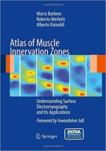
Atlas of Muscle Innervation Zones: Understanding Surface Electromyography and Its Applications

Invasive electromyography is a well-established diagnostic tool that has been used for decades by neurologists. Recently, new and alternative devices have increasingly become available that permit diagnosis without the use of needles. This developing area of science and the new tools have not, however, been sufficiently investigated in academic training. Consequently a gap exists between what science is making possible and the competence acquired during graduate studies. This handy volume has the aim of filling this gap by providing the information required by medical practitioners in rehabilitation, sports, and occupational health as well as by rehabilitation therapists, ergonomists, and sport coaches. The techniques that are presented and explained will help in monitoring and recording changes, evaluating the effectiveness of treatments and training, evaluating work stations, and preventing and documenting the evolution of occupational disorders of the neuromuscular system.

DOWNLOAD THIS BOOK FREE HERE
https://upsto.re/3AnmhtY

Human Chromosome Atlas: Introduction to diagnostics of structural aberrations 1st ed

This atlas presents the results of cases of structural chromosome aberrations based on the currently available methods of analysis for different types of abnormality. It particularly focuses on which spectrum should be chosen when combining the different techniques to achieve the best method of diagnosis in individual cases, for example direct preparation of cells and mitoses, short or long-time cell culture, fluorescence in situ hybridization (FISH), analysis of interphases, microarray or DNA sequencing. Generally it has to be taken into account, that the development of new and improved investigation methods is forthcoming. Thus, by improvement of diagnostic possibilities new fields of investigation arise, and special groups of patients with cytogenetic analyses can be re-analysed under new research questions.

DOWNLOAD THIS BOOK FREE HERE

https://upsto.re/3EYxpmy

Atlas of Lacrimal Drainage Disorders 1st ed. 2018 Edition

About the Author

Atlas of Head and Neck Endocrine Disorders: Special Focus on Imaging and Imaging-Guided Procedures 1st ed

From the Back Cover

Normal and Abnormal Fetal Face Atlas: Ultrasonographic Features 1st ed

This atlas offers a guide to studying the fetal face by means of ultrasound analysis. It describes key phases in cranial-facial development, allowing the reader to learn the related semiology from its most simple iteration to the most complex one.
The overall examination of a newborn’s face offers a rich source of information and can guide the general examination. The same applies in the context of fetal ultrasound examination. The analytical study of the fetal face not only makes it possible to screen for anomalies related to the face itself, but also yields valuable insights into the brain, the limbs, and the heart. In addition, it allows ultra-sonographers to unravel the puzzle of fetal dimorphism.
Written in a pedagogical style, the book guides walks the reader through the diagnostic reasoning process step by step.
The authors are pioneers in this field and teach in various university and master’s degree ultrasound programs. Their aim is to share with readers their diagnostic approaches and their knowledge and passion for 2D and 3D ultrasound techniques. Each chapter includes algorithms, biometry curves, and simple guidelines that allow users to go “from sign to syndrome”. The first chapter, which focuses on innovative embryology adapted to the needs of ultra-sonographers, was written by Gérard Couly, a maxilla-facial surgeon and the founding father of the specialty
DOWNLOAD THIS BOOK FREE HERE

Atlas of Head and Neck Endocrine Disorders: Special Focus on Imaging and Imaging-Guided Procedures 1st ed

This atlas draws on a multidisciplinary approach to provide a comprehensive overview on endocrine disorders of the head and neck, with particular emphasis on the role of imaging and image-guided procedures. The first section discusses the basic characteristics of the imaging methods and other techniques used for evaluation and diagnosis. The remainder of the book focuses on application of these methods in thyroid, parathyroid, and other endocrine disorders of the head and neck. The coverage is wide ranging, encompassing Graves’ disease, toxic multinodular goiter, toxic adenoma, thyroiditis, non-toxic goiter, benign nodules, and the different forms of thyroid carcinoma, as well as parathyroid adenoma, hyperplasia, and carcinoma and paragangliomas. Informative, high-quality images are provided by international experts in endocrine disorders, including endocrinologists, pathologists, radiologists, nuclear medicine physicians, and surgeons, who also discuss sample cases and provide syntheses of the relevant scientific literature.

DOWNLOAD THIS BOOK FREE HERE
https://upsto.re/FoQ573p

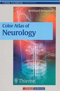
Color Atlas Of Neurology

Neurology—made visible Every practitioner in modern medicine is confronted daily with neurologic symptoms, diagnoses, and clinical problems. Yet there is scarcely any other medical specialty that is so fraught with complexities and abstractions. This pocket atlas is designed to provide a better, easier-to-understand visual guide on what the reader needs to know about neurology. In a unique way, neurology is made “visible” in the truest sense of the word. Coverage includes: – The basic principles of neuroanatomy and neurophysiology (structure of the CNS, peripheral nerves, stimulus transmission, nerve conduction velocity, etc.) – Diagnostic methods and procedures (clinical examinations, electrophysiologic techniques, imaging studies, etc.) – Neurologic disorders including their clinical manifestations, pathogenesis, and principles of treatment These topics and more are covered in elaborately drawn, meticulously labeled illustrations. The effective concept of placing the illustrations opposite the descriptive text for a particular subject has created word-and-picture units that combine maximum teaching impact with an optimum density of information. Neurologic relationships can be grasped literally “at a glance.” This pocket atlas is intended for medical students, physicians, and other medical professionals (nurses, physical therapists, occupational therapists, speech therapists) who could profit from a visual guide to neurology.

DOWNLOAD THIS BOOK FREE HERE

https://upsto.re/FYwtjBo

Atlas of Essential Dermatopathology

[amazon template=iframe image2&asin=1447144708]

The book is not intended to be an all-encompassing atlas or textbook but rather a foundation of principles in dermatopathology, highlighting key elements in the field for trainees and will also serve as a basic resource for the pathologist in general practice. In addition to the sketches and minimal text, we envision accompanying high resolution histopathologic micrographs for ultimate correlation as well.

DOWNLOAD THIS BOOK FREE HERE

https://upsto.re/FLHaJRU

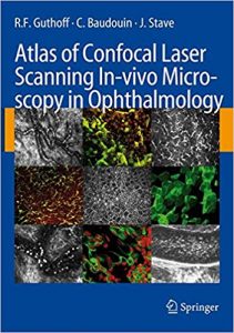
Atlas of Confocal Laser Scanning In-vivo Microscopy in Ophthalmology 2006th Edition

[amazon template=iframe image2&asin=3540327053]

This lavishly illustrated atlas demonstrates normal in-vivo anatomy of the cornea, limbus, and conjunctiva; quantifies various cellular structures using cell-density calculations; and establishes correlations between novel optical sections of various diseases of the ocular surface and clinical findings. It also describes early signs of different eye diseases and supports the reader in diagnostic and therapeutic management.

DOWNLOAD THIS BOOK FREE HERE

https://upsto.re/FLSvaHC

1 8 9 10 11 12 19