Imaging of the Pancreas: Acute and Chronic Pancreatitis (Medical Radiology)
[amazon template=iframe image2&asin=3642055494]

Using numerous high-quality illustrations, this volume assesses strengths and limitations of techniques for the imaging of pancreatitis. Ultrasound, computed tomography, magnetic resonance imaging and interventional imaging are considered separately in the settings of acute and chronic pancreatitis, with an additional section on imaging of complications. The significance of the imaging findings for clinical and therapeutic decision making is clearly explained, and protocols are provided to help obtain the best possible images.

With the aid of numerous high-quality illustrations, this volume explains the strengths and limitations of the different techniques employed in the imaging of pancreatitis. Ultrasound, computed tomography, magnetic resonance imaging and interventional imaging are each considered separately in the settings of acute and chronic pancreatitis. A further section is devoted to imaging of the complications of these conditions. Throughout, care has been taken to ensure that the reader will achieve a sound understanding of how the imaging findings derive from the pathophysiology of the disease processes. The significance of the imaging findings for clinical and therapeutic decision making is clearly explained, and protocols are provided that will assist in obtaining the best possible images.

DOWNLOAD THIS BOOK FREE HERE

http://upsto.re/FZkMGXF

Respiratory Outcomes in Preterm Infants: From Infancy through Adulthood (Respiratory Medicine) 1st ed. 2017 Edition

[amazon template=iframe image2&asin=3319488341]

Atlas of Airway Surgery: A Step-by-Step Guide Using an Animal Model 1st ed. 2017 Edition

[amazon template=iframe image2&asin=3319497383]

Angelo Ghidini is a member of the Head and Neck Department at the University Hospital of Modena, Italy, where he has been Co-Director and Professor of Endoscopic Surgery of the Paranasal Sinuses since 2003. His other responsibilities have included teaching activities relating to phonosurgery and speech therapy rehabilitation (ENT Department, Bufalini Hospital, Cesena, Italy) and cadaver endoscopic dissection of the paranasal sinuses and skull base (Laboratoire d’Anatomie de Lyon, France). Dr. Ghidini is a member of most scientific societies in the field. In recent years his activity has focused especially on the management of pediatric and neonatal airways and congenital laryngotracheal malformations.

Francesco Mattioli currently works in the Otolaryngology Department at Modena General Hospital. After his residency in this discipline, he worked at the Pediatric Surgery Department of the Hôpital Robert Debrè in Paris, the Catholic Hospital of North Kingnakopp, Kenya and the Hôpital St. Jean de Dieu in Tanguità, Benin. He has authored more than 30 publications in peer-reviewed journals.
Sergio Bottero is  Head of Otolaryngology in the Division of Pediatric Otolaryngology, Bambino Gesù Hospital, Rome, Italy. The Division of Pediatric Otolaryngology at the hospital is one of the leading centers in Paediatric Otolaryngology in Italy, annually performing about 3000 inpatient medical/surgical procedures, 1000 minor surgery outpatient procedures, and 6000 outpatient diagnostic tests and receiving some 35,000 specialized clinic visits. Dr. Bottero is the author of many articles in peer-reviewed international journals.
Professor Livio Presutti has been Director of the Department of Head and Neck Surgery (ENT, ocular surgery, maxillofacial surgery, plastic surgery, and neurosurgery) of Modena General Hospital and Professor of Surgery at the University of Reggio Emilia since 2003. He is co-founder of the International Working Group on Endoscopic Middle Ear Surgery and head of a multidisciplinary team (including radiologists, oncologists, pathologists and radiotherapists) which each week deliberates on the optimal therapeutic approach in cases of head and neck cancer. He has more than 30 years of experience in endoscope-assisted paranasal sinus surgery, microscope-assisted otoneurosurgery and laryngeal surgery and pioneered endoscope-assisted middle ear surgery and cerebellopontine angle surgery, as well as the use of new devices such as the Tullio and Diodi laser and piezoelectric surgery. He has published more than 100 articles in national and international journals.

DOWNLOAD THIS BOOK FREE HERE

http://upsto.re/F2Aw2kk

Nosokomiale Pneumonie (German Edition)

[amazon template=iframe image2&asin=3662498200]

Dieses Buch bietet differenzierte Diagnostik und Therapie – was, wann, warum – zur sofortigen sicheren Anwendung entsprechend der neuesten Studien und Leitlinien aller wichtigen deutschen, europäischen und amerikanischen Fachgesellschaften. Zudem umfasst es die Darstellung der aktuellen Hygienekonzepte zur Prävention, die angesichts der Verbreitung multiresistenter Erreger immer wichtiger erscheinen.

DOWNLOAD THIS BOOK FREE HERE

http://upsto.re/FC8vHcD

Atlas of Diffuse Lung Diseases: A Multidisciplinary Approach 1st ed. 2017 Edition

[amazon template=iframe image2&asin=3319427504]

This atlas is designed as an easy-to-use reference guide that identifies and illustrates the key patterns of diffuse lung diseases observed on high-resolution computed tomography (HRCT) and then documents in more detail the characteristics and appearances of the individual diseases, grouped on the basis of their prevalent pattern. A further feature of the book is its interdisciplinary nature: contributions from experts in various specialties are tightly interwoven throughout and many pathologic correlations are included. Less experienced readers will find that this atlas, with its wealth of figures and helpful color coding, steers them towards correct interpretation when confronted by the multiplicity and complexity of these diseases; those who are already experts, on the other hand, will benefit from the detailed coverage of individual diseases, which will deepen their understanding. At the end of the book, a graphically appealing and practice-oriented illustrated glossary with tips and tricks offers a further highly effective educational tool. Given the clinical spectrum of diseases covered, this atlas will prove invaluable for a wide range of healthcare workers, especially radiologists, pulmonologists, and pathologists.

DOWNLOAD THIS BOOK FREE HERE

http://upsto.re/FCUFuuz

Chest Sonography 4th ed. 2017 Edition

[amazon template=iframe image2&asin=3319440713]

This book, widely regarded as the standard work in the field, presents the state of the art in chest sonography with the aid of a wealth of excellent illustrations. It provides the reader with concise, easy-to-assimilate information on all aspects of the use of the modality, including indications, investigative techniques, diagnostic decision making, and imaging artifacts and pitfalls. Numerous tips and tricks and potential sources of diagnostic error are highlighted to aid in daily clinical practice. This fourth edition has been extensively revised to take full account of the latest techniques, study results, and meta-analyses, with inclusion of important additional illustrative material. An entirely new chapter is devoted to interstitial syndrome, and detailed guidance is provided on contrast-enhanced sonography, ultrasound techniques for evaluation of the mediastinum, and the role of chest sonography in emergency medicine. As the value of the technique continues to grow, readers will find Chest Sonography to be a superb up-to-date resource and guide.

DOWNLOAD THIS BOOK FREE HERE

http://upsto.re/F3xc87f

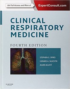
Clinical Respiratory Medicine, 4e 4th Edition

[amazon template=iframe image2&asin=1455707929]

2013 BMA Medical Book Awards Highly Commended in Internal Medicine!

Clinical Respiratory Medicine provides practical guidance to help you more effectively diagnose and manage the full range of pulmonary disorders, including those seen in today’s most challenging patient populations. Now with over 400 brand-new review questions and 25 videos available online, this medical reference book delivers all of the answers you need to ensure the best outcomes.

  • Better manage and treat patients with pulmonary disease with complete clinical coverage of the critical information relevant to your everyday practice, presented in a templated, user-friendly format.
  • Find critical information quickly with the help of diagnostic algorithms.


  • Test your knowledge of respiratory medicine with the help of 400 brand-new review questions.
  • Watch and learn. Over 25 videos of practical procedures are available online at www.expertconsult.com.
  • Thoroughly understand the needs and recognize co-morbidities of particular patient populationsthrough entirely new chapters on lung structure, echocardiography, and obesity and its effects.
  • Access the latest research and advancements in lung cancer, benign tumors, and the importance of pulmonary physiology in understanding lung function and the disease processes that occur.

Take a practical approach to the diagnosis and management of patients with respiratory disorders using this ideal source for reference in clinical practice

DOWNLOAD THIS BOOK FREE HERE

http://upsto.re/FQ4ZTJa

Handbook of Tuberculosis 1st ed. 2017 Edition

[amazon template=iframe image2&asin=3319262718]

This concise, clinically focused handbook offers a complete overview of tuberculosis and reviews the latest guidelines, treatment options, clinical trials and management of this disease. Handbook of Tuberculosis is a well-rounded book written by renowned researchers at the Johns Hopkins Center for Tuberculosis Research. The easily accessible text offers infectious disease specialists, pulmonologists, and other healthcare workers an excellent quick reference tool, with full color tables and figures enhancing the text further.

Tuberculosis is a life-threatening lung infection that affects people of all ages. It is the second biggest cause of death from infectious diseases and is the most common cause of death amongst patients with HIV/AIDS. Education on the recognition of tuberculosis and the subsequent treatment of the disease is empirical to the prevention of wide-spread resistance. There is a lack of knowledge on how to treat coinfected and comorbid patients, this handbook offers the latest medical guidance and treatment options to equip physicians to manage such patients.

DOWNLOAD THIS BOOK FREE HERE

http://upsto.re/Ay7bmok

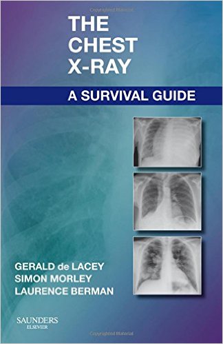
The Chest X-Ray: A Survival Guide, 1e 1st Edition

[amazon template=iframe image2&asin=0702030465]

Featuring a practical, clinical approach – and written in a quick-access style – this portable, economical reference helps you build a strong foundation in chest x-ray interpretation. Three radiologists with years of clinical and teaching experience present fundamental principles and key anatomical concepts…walk you through examples of classic chest x-ray features that provide subtle evidence of abnormality…and explore a variety of problems and dilemmas common to everyday clinical practice. High-quality drawings and digital chest x-rays – combined with secrets from the radiologists’ toolbox, helpful differential diagnoses, handy checklists, and key references – deliver all the assistance you need to enhance your interpretation skills.

  • Provides a strong foundation of essential knowledge for an informed, systematic approach to accurate chest x-ray interpretation.
  • Features the work of three radiologists who offer you the benefit of their many years of clinical and teaching experience.
  • Emphasizes common errors and misdiagnoses to help ensure correct image readings.
  • Presents step-by-step guidance in a bulleted, quick-access format, in short chapters focused on clinical problems, to make it easy to master the information that you need to know.
  • Makes difficult anatomic concepts easier to grasp by pairing radiographs with color line drawings.
  • Explains the nomenclature special to the field through a glossary of important terms.
  • Highlights the most important concepts in diagnosis/interpretation via Key Points in each chapter.

DOWNLOAD THIS BOOK FREE HERE

http://upsto.re/ArpMEsQ

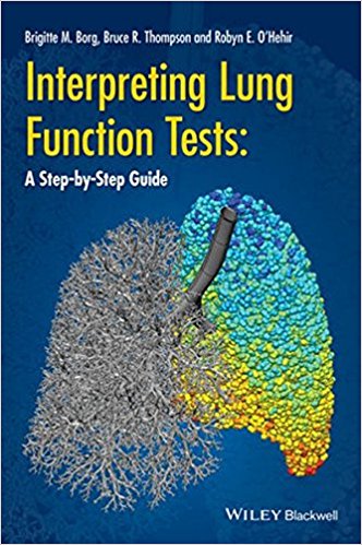
Interpreting Lung Function Tests: A Step-by Step Guide 1st Edition

Lung function assessment is the central pillar of modern respiratory diagnosis, providing invaluable information to assist in clinical decision making and management strategies.Interpreting Lung Function Tests: A Step-by Step Guide is a practical “how-to” training manual, which provides the reader with the necessary skills to interpret lung function test results, and to write a concise and informative report on the outcome.

Interpreting Lung Function Tests: A Step-by Step Guide

  • provides unique guidance on the reporting of pulmonary function tests, including illustrative cases and sample reports.
  • utilizes the many references available on interpretation of lung function and provides a teaching/reference tool for report writing of lung function results routinely performed in clinical practice.
  • provides the reader with the skill to interpret and write a concise, yet informative report
  • provides examples of results and written reports (with commentary where necessary as further explanation).
  • focuses primarily on tests performed as part of routine clinical testing: spirometry, static lung volumes, gas transfer, bronchial provocation tests, and maximal respiratory pressures.

Interpreting Lung Function Tests: A Step-by Step Guideis a superb new resource to educate medical students, junior doctors, family physicians, as well as advanced trainee physicians specializing in respiratory medicine, respiratory scientists, and respiratory physicians

DOWNLOAD THIS BOOK FREE HERE

http://upsto.re/AiDjugR

1 11 12 13 14 15 18