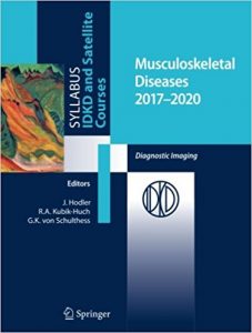
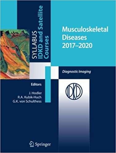
Musculoskeletal Diseases 2017-2020: Diagnostic Imaging 1st ed. 2017 Edition

[amazon template=iframe image2&asin=3319540173]

This book focuses on with musculoskeletal imaging: during the last few years, there have been considerable advances in this area, driven by clinical as well as technological developments. The authors, internationally renowned experts in their field, have contributed chapters that are disease-oriented and cover all relevant imaging modalities, including magnetic resonance imaging, computed tomography, and positron emission tomography. As a result, this book offers a comprehensive review of the state of the art in musculoskeletal imaging. It is a valuable resource for general radiologists, radiology residents, rheumatologists, orthopaedic surgeons, and clinicians from other specialities wishing to update their knowledge in this discipline.

About the Author

Juerg Hodler is Professor of Radiology and chairman of the Dept. of Radiology at the University Hospital of Zurich, Switzerland. He started his career in 1990 at the University of California, San Diego where he was research fellow in Osteoradiology. Subsequently he completed a Master of Business Administration (MBA) at Heriot-Watt University, Edinburgh, Scotland. From 1992 to 2001 he has been chief of section at the Orthopedic University Hospital of Zurich, in 2001 he became chief at the Department of Radiology at the Orthopedic University Hospital of Zurich and in 2007 deputy medical director at the Orthopedic University Hospital Balgrist, Zurich. In 2010, he became a professor at the University of Zurich and was elected head of the Department of Radiology, University Hospital Zurich. He has peer-reviewed over 200 original papers (focusing on MR imaging of joints, MR-anatomical correlation, asymptomatic MR findings, clinical application of MR sequences) and written 28 book chapters and four books (including co-editing). He is course co-director of the International Diagnostic Course in Davos IDKD.

Rahel Kubik-Huch earned her medical degree and doctorate from the University of Zurich in 1991 and her titular professorship for diagnostic radiology in 2005. She completed her residency in diagnostic radiology at the University Hospital of Zürich, Switzerland, where she subsequently worked as senior physician. Since 2002 she has worked at the Kantonsspital Baden, Baden, Switzerland, where she was appointed head of the Institute of Radiology and director of the Dept. of Medical Services in 2005. In 2010, she also became a member of the executive board of the hospital. Her specialist focus is magnetic resonance tomography (especially breast, abdominal and genitourinary imaging) and breast imaging (all modalities, including breast MRI and interventions under imaging guidance). She has published numerous research articles in peer-reviewed journals, contributed to several book chapters and received a number of scientific awards, including the Swiss Society of Radiology Anniversary Award (“Jubiläumspreis”). She is a course co-director of the International Diagnostic Course Davos (IDKD), course advisor for the Breast Imaging Satellite Course, Pearl of the IDKD and a former organizer of the Advanced Breast and Pelvis Imaging Course at the ESMRMB where she has remained a member of the faculty.

DOWNLOAD THIS BOOK FREE HERE

http://upsto.re/Azn8uWh


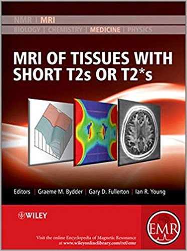
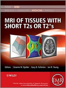
MRI of Tissues with Short T2s or T2*s 1st Edition

[amazon template=iframe image2&asin=0470688351]

The content of this volume has been added to eMagRes (formerly Encyclopedia of Magnetic Resonance) – the ultimate online resource for NMR and MRI.

Up to now MRI could not be used clinically for imaging fine structures of bones or muscles. Since the late 1990s however, the scene has changed dramatically. In particular, Graeme Bydder and his many collaborators have demonstrated the possibility – and importance – of imaging structures in the body that were previously regarded as being “MR Invisible”. The images obtained with a variety of these newly developed methods exhibit complex contrast, resulting in a new quality of images for a wide range of new applications.

This Handbook is designed to enable the radiology community to begin their assessment of how best to exploit these new capabilities. It is organised in four major sections – the first of which, after an Introduction, deals with the basic science underlying the rest of the contents of the Handbook. The second, larger, section describes the techniques which are used in recovering the short T2 and T2* data from which the images are reconstructed. The third and fourth sections present a range of applications of the methods described earlier. The third section deals with pre-clinical uses and studies, while the final section describes a range of clinical applications. It is this last section that will surely have the biggest impact on the development in the next few years as the huge promise of Short T2 and T2* Imaging will be exploited to the benefit of patients.

In many instances, the authors of an article are the only research group who have published on the topic they describe. This demonstrates that this Handbook presents a range of methods and applications with a huge potential for future developments.

About EMR Handbooks / eMagRes Handbooks 

The Encyclopedia of Magnetic Resonance (up to 2012) and eMagRes (from 2013 onward) publish a wide range of online articles on all aspects of magnetic resonance in physics, chemistry, biology and medicine. The existence of this large number of articles, written by experts in various fields, is enabling the publication of a series of EMR Handbooks / eMagRes Handbooks on specific areas of NMR and MRI. The chapters of each of these handbooks will comprise a carefully chosen selection of articles from eMagRes. In consultation with the eMagRes Editorial Board, the EMR Handbooks / eMagRes Handbooks  are coherently planned in advance by specially-selected Editors, and new articles are written (together with updates of some already existing articles) to give appropriate complete coverage. The handbooks are intended to be of value and interest to research students, postdoctoral fellows and other researchers learning about the scientific area in question and undertaking relevant experiments, whether in academia or industry.

DOWNLOAD THIS BOOK FREE HERE
http://upsto.re/A8UVe59

Recognizing Child Abuse in Radiology 1st ed. 2017 Edition

[amazon template=iframe image2&asin=3319443224]

This book is both a personal review of experience in the diagnosis of child abuse and its differential diagnosis, intended to instruct beginners, and a reliable source of information for the more experienced health care provider. Emphasis is placed on normal findings that simulate abuse, true and false diagnoses of metabolic bone disease as the cause of fractures, abusive head trauma, and the problem of zealous abuse-denying “experts.” Plain radiography is emphasized, with appropriate mention of, ultrasound, CT, MRI, – in detecting childhood abuse is clearly explained, and individual chapters focus on neuroradiological evaluation and the radiological assessment of abdominal, thoracic, and subcutaneous abnormalities. Guidance is provided on how to exclude a range of alternative diagnoses, on the documentation of concerns over potential abuse, and on the needs of the clinician in relation to the radiologist. Recognizing Child Abuse in Radiology will be of value for radiologists in training and practice, for health care workers, and for other interested professionals.

About the Author

Alan E. Oestreich, MD, is a pediatric radiologist since 1980 at the Cincinnati Children’s Hospital Medical Center in Cincinnati, Ohio, and is Emeritus Professor of radiology at the University of Cincinnati College of Medicine. From 1972 – 1979 he was pediatric radiologist at the University of Missouri-Columbia. He a Fellow of the American College of Radiology, Honorary Member of the European Society of Paediatric Radiology, emeritus member of the Society for Pediatric Radiology, and past chairman of the Radiology Section of the National Medical Association. He is a member of the Child Abuse Team at Cincinnati Children’s Hospital. He has written and lectured extensively on the subject of radiology of child abuse. Among his previous books are An Atlas of Pediatric Orthopedic Radiology, with co-author Alvin Crawford, MD. He received his MD from the Johns Hopkins Medical School in 1965.

Marguerite M. Caré, MD is an assistant professor in the Department of Radiology, Division of Neuroradiology, at Cincinnati Children’s Hospital Medical Center. Caré joined the CCHMC faculty after completing a pediatric radiology fellowship in 1998 and a pediatric neuroradiology fellowship in 2000 at Cincinnati Children’s Hospital Medical Center. She has been the radiology representative to the multidisciplinary Child Abuse Team at the CCHMC Mayerson Center since 1998 and has recently become the radiology representation to the CCHMC Trauma Surgical Program. Caré has a major interest in the neuroimaging findings in child abuse and has presented at both local and national meetings on this topic. She also has research interests in Tuberous Sclerosis with her neurology colleagues. Caré serves on the CCHMC Radiology Department education committee with a primary role in third year medical student education for the department.

DOWNLOAD THIS BOOK FREE HERE
http://upsto.re/A87fj56

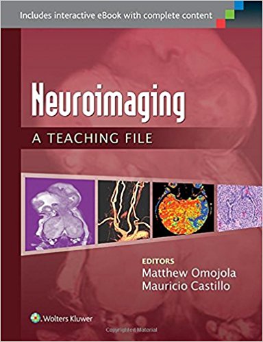
Neuroimaging: A Teaching File (LWW Teaching File Series) First Edition

[amazon template=iframe image2&asin=1451173288]

With 100 illustrated cases in print and an eBook containing the 100 print cases plus another200 cases, this volume in the Teaching File Series gives you ready access to actual cases culled from the teaching files of major medical centers.  Neuroradiology: A Teaching File covers a wide variety of CNS diseases that are presented as unknowns with coverage of both brain and head and neck cases.  Each case features two to four images and follows the Teaching File template:  clinical history, findings, differential diagnosis, diagnosis, discussion, questions, key issues for the report, what the clinician needs to know, and answers. It’s the next-best thing to a personal consultation with teaching hospital experts!

Key Features

  • Clear, concise discussions mimic those performed on a daily basis between residents and faculty members.
  • Cases are now randomized, to better prepare you for challenges faced in an everyday clinical setting.
  • Both brain and head and neck cases are highlighted by color perfusion, PET, and functional/MR spectroscopy images. 
  • Common neuroimaging cases are featured, including both normal anatomy and the use of advanced imaging in differential diagnosis.
  • Collaboration with clinical services and pathology provides an interdisciplinary perspective whenever possible.
  • Dozens of cases feature full-color images.
  • New softcover format for the entire series.

Now with the print edition, enjoy the bundled interactive eBook edition, offering tablet, smartphone, or online access to:

  • 300  cases, including 200 eBook only cases
  • Complete content with enhanced navigation.
  • Powerful search tools and smart navigation cross-links that pull results from content in the book, your notes, and even the web.
  • Cross-linked pages, references, and more for easy navigation.
  • Highlighting tool for easier reference of key content throughout the text.
  • Ability to take and share notes with friends and colleagues.
  • Quick reference tabbing to save your favorite content for future use.

DOWNLOAD THIS BOOK FREE HERE
http://upsto.re/A8qAtx8

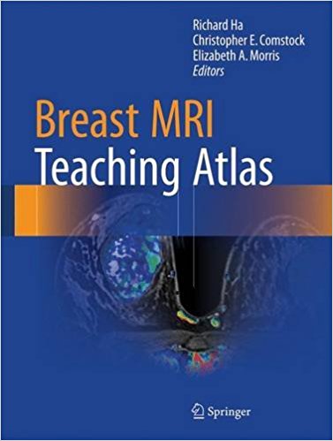
Breast MRI Teaching Atlas 1st ed. 2017 Edition

[amazon template=iframe image2&asin=1493964070]

This atlas serves as a basic introduction to breast MRI. Organized by case, it emphasizes pertinent breast MRI findings and common indications for breast MRI. Topics include breast MRI basics, benign findings, breast malignancy, high-risk conditions, interesting cases, and breast implants. Brief teaching points accompany each case and highlight the importance of the findings. Breast MRI Teaching Atlas is an ideal resource for diagnostic radiologists, residents, fellows, and clinicians involved in the care of breast cancer patients, including surgeons, oncologists, and obstetricians/gynecologists.

DOWNLOAD THIS BOOK FREE HERE

http://upsto.re/A8qFPpM

Oncologic Imaging: Spine and Spinal Cord Tumors 1st ed. 2017 Edition

[amazon template=iframe image2&asin=9812876995]

This book is a detailed guide to image interpretation in patients with spine and spinal cord tumors that will enable clinicians and residents to improve their diagnostic abilities. The book opens by introducing basic concepts in the imaging of spinal tumors, namely the compartmental approach and the histologic basis for the different imaging appearances. These concepts are explained with the aid of representative cases and schematic illustrations. The second part of the book represents a “training step” in which various spinal tumors are described in detail, focusing on the imaging findings. Representative cases are presented with radiographic, CT, and MR images; in addition, schematic illustrations and pathologic or operative images are included in selected cases. The third part is a “practice step” describing tips for correct imaging diagnosis. Individual chapters in this part focus on incidence-, age-, location-, and imaging pattern-based approaches to bone, extradural, intradural extramedullary, intramedullary, and pediatric spinal tumors. The presented cases will enhance the reader’s understanding of the different tumor patterns and assist in solving diagnostic problems that may be encountered in daily practice.

DOWNLOAD THIS BOOK FREE HERE

http://upsto.re/A8q4eCh

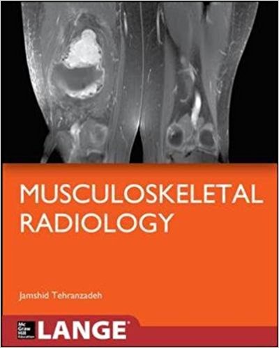
Basic Musculoskeletal Imaging (Lange Medical Books) 1st Edition

[amazon template=iframe image2&asin=007178702X]

A complete introductory text to musculoskeletal imaging

Basic Musculoskeletal Imaging is an engagingly written, comprehensive textbook that addresses the fundamental principles and techniques of general diagnostic and advanced musculoskeletal imaging. In order to be as clinically relevant as possible, the text focuses on the conditions and procedures most often encountered in real-world practice, such as:

  • Upper and lower extremity trauma
  • Axial skeletal trauma
  • Arthritis and infection
  • Tumors
  • Metabolic bone diseases
  • Bone infarct and osteochondrosis
  • Shoulder, knee, spine, elbow, wrist, hip, and ankle MRI

You will also find authoritative coverage of:

  • Signs in musculoskeletal imaging
  • The key concepts of using different modalities in musculoskeletal imaging
  • Current advances in musculoskeletal scintigraphy

The book is enhanced by superb figures and illustrations, including a four-page full-color insert; “Pearls” that summarize must-know information; and an outstanding introduction to musculoskeletal ultrasound by international experts from France and Brazil.

DOWNLOAD THIS BOOK FREE HERE
http://upsto.re/A8xouUC

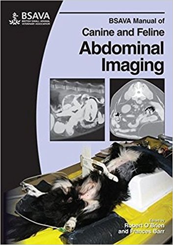
BSAVA Manual of Canine and Feline Abdominal Imaging 1st Edition

[amazon template=iframe image2&asin=190531910X]

The BSAVA Manual of Canine and Feline Abdominal Imaging is the third Manual in the diagnostic imaging series. The first section of the Manual outlines the approach to abdominal imaging, with chapters dedicated to radiography and ultrasonography. The remainder of the Manual is devoted to the individual body systems, including the liver and gallbladder, the kidneys and ureters, and the small intestine. Extensively illustrated throughout.

DOWNLOAD THIS BOOK FREE HERE
http://upsto.re/A8jAqk6

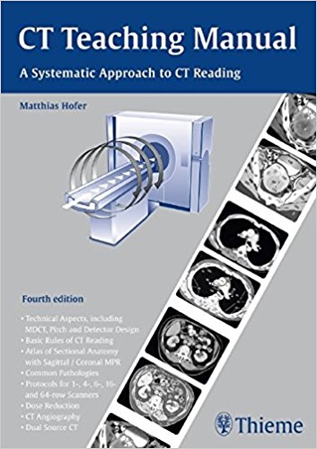
CT Teaching Manual: A Systematic Approach to CT Reading 4th Edition

[amazon template=iframe image2&asin=3131243546]

Ideal for residents starting in radiology and radiologic technologists,
this concise manual is the perfect introduction to the physics and practice of
CT and the interpretation of basic CT images. Designed as a systematic learning
tool, it introduces the use of CT scanners for all organs, and includes
positioning, use of contrast media, representative CT scans of normal and
pathological findings, explanatory drawings with keyed anatomic structures, and
an overview of the most important measurement data. Finally, self-assessment
quizzes – including answers – at the end of each chapter help the reader monitor
progress and evaluate knowledge gained. New in this fourth edition: – Updated
examination protocols for multidetector CT and CT angiography – An extended
chapter on dual source CT – A new chapter on contrast injector

DOWNLOAD THIS BOOK FREE HERE
http://upsto.re/A88sGBr

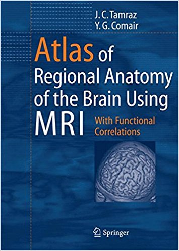
Atlas of Regional Anatomy of the Brain Using MRI: With Functional Correlations

[amazon template=iframe image2&asin=3540278761]

A unique review of the essential topographical anatomy of the brain from an MRI perspective, correlating high-quality anatomical plates with high-resolution MRI images. The book includes a historical review of brain mapping and an analysis of the essential reference planes used. It provides a detailed review of the sulcal and the gyral anatomy of the human cortex, guiding readers through an interpretation of the individual brain atlas provided by high-resolution MRI. The relationship between brain structure and function is approached in a topographical fashion with an analysis of the necessary imaging methodology and displayed anatomy. An extensive coronal atlas rounds off the book.

From the Back Cover

The volume provides a unique review of the essential topographical anatomy of the brain from an MRI perspective, correlating high-quality anatomical plates with the corresponding high-resolution MRI images. The book includes a historical review of brain mapping and an analysis of the essential reference planes used for the study of the human brain. Subsequent chapters provide a detailed review of the sulcal and the gyral anatomy of the human cortex, guiding the reader through an interpretation of the individual brain atlas provided by high-resolution MRI. The relationship between brain structure and function is approached in a topographical fashion with analysis of the necessary imaging methodology and displayed anatomy. The central, perisylvian, mesial temporal and occipital areas receive special attention. Imaging of the core brain structures is included. An extensive coronal atlas concludes the book. Neuroscientists, neuroradiologists, neurologists, neurosurgeons and students of human behavior should find this book useful guiding them to a better understanding of the localization of brain function.

DOWNLOAD THIS BOOK FREE HERE
http://upsto.re/A88Fnb9

1 42 43 44 45 46 60