Atlas of PET/MR Imaging in Oncology 2013th Edition
[amazon template=iframe image2&asin=3642312918]

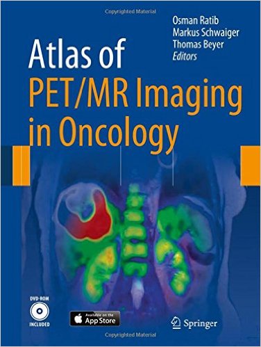
This new project on PET-MR imaging in oncology includes digital interactive software matching the cases in the book. The interactive version of the atlas is based on the latest web standard, HTML5, ensuring compatibility with any computer operating system as well as a dedicated version for Apple iPad.

The book opens with an introduction to the principles of hybrid imaging that pays particular attention to PET/MR imaging and standard PET/MR acquisition protocols. A wide range of illustrated clinical case reports are then presented. Each case study includes a short clinical history, findings, and teaching points, followed by illustrations, legends, and comments.

The multimedia version of the book includes dynamic movies that allow the reader to browse through series of rotating 3D images (MIP or volume rendered), display blending between PET and MR, and dynamic visualization of 3D image volumes. The movies can be played either continuously or sequentially for better exploration of sets of images.

The editors of this state-of-the-art publication are key opinion leaders in the field of multimodality imaging. Professor Osman Ratib (Geneva) and Professor Markus Schwaiger (Munich) were the first in Europe to initiate the clinical adoption of PET/MR imaging. Professor Thomas Beyer (Zurich) is an internationally renowned pioneering physicist in the field of hybrid imaging. Individual clinical cases presented in this book are co-authored by leading international radiologists and nuclear physicians experts in the use of PET and MRI.

DOWNLOAD THIS BOOK FREE HERE

http://upsto.re/AFuhgZf

Atlas of Emergency Medicine 4th Edition 4th Edition

[amazon template=iframe image2&asin=0071797254]

The most complete and trusted visual compendium of emergency medicine ― extensively updated with more than 1500 full-color illustrations.

Now in its fourth edition, The Atlas of Emergency Medicine remains the best guide to visual diagnosis of acute medical problems encountered in emergency practice. Packed with more than 1500 images, 500 new to this edition, it is the most comprehensive source of high quality emergency medicine images available, and enhanced with clear clinical information on medical emergencies.

Organized by organ-system/special populations/general issues and then by problem, The Atlas of Emergency Medicine is your “look quick, act fast” reference designed to help assess, diagnose, and treat patients efficiently. It covers basic and subtle diagnosis of a broad spectrum of typical and atypical conditions.

The book’s format is ideally suited to visual learning. One to four images per topic show you exactly what to look for. Accompanying the images is succinct “need-to-know” information for each clinical problem, including management options and clinical pearls. All treatment and diagnostic testing information and guidelines are fully updated.

NEW to this edition is the inclusion of more than 50 video clips that highlight key visual topics in emergency medicine.

DOWNLOAD THIS BOOK FREE HERE
http://upsto.re/AFcGcH7

Atlas of Emergency Medicine Procedures 1st ed. 2016 Edition

[amazon template=iframe image2&asin=1493925067]

This full-color atlas is a step-by-step, visual guide to the most common procedures in emergency medicine. Procedures are described on a single page, or two-page spreads, so that the physician can quickly access and review the procedure at hand. The atlas contains more than 600 diagnostic algorithms, schematic diagrams and photographic illustrations to highlight the breadth and depth of emergency medicine. Topics are logically arranged by anatomic location or by type of procedure and all procedures are based on the most current and evidence-based practices known.

DOWNLOAD THIS BOOK FREE HERE

http://upsto.re/AFBeN9A

Zollinger’s Atlas of Surgical Operations, 9th Edition 9th Edition

[amazon template=iframe image2&asin=0071602267]

The classic step-by-step atlas of general surgery procedures – now in color and updated with the latest laparoscopic operations

A Doody’s Core Title for 2011!

1615 color-highlighted illustrations

“…a beautifully illustrated atlas of modern surgical procedures….While in other atlases line drawings may simplify and operative photographs may add realism, the illustrations in this atlas allow the surgeon to visualize both the anatomy and the operation. Beyond this, the softly colored illustrations are aesthetically pleasing and invite reading of the accompanying narrative….In sum, while the ninth edition of Zollinger’s Atlas of Surgical Operations differs from the early editions in many ways, it nonetheless evokes the enduring qualities of its predecessors and will guide yet another generation of surgeons.”–JAMA

For more than half-a-century, Zollinger’s Atlas of Surgical Operations has been the gold-standard reference for learning how to perform the most common surgical procedures using safe, well-established techniques. The ninth edition continues this tradition of excellence with the addition of color illustrations and coverage of more than 230 procedures, including many of the most important laparoscopic operations.

Following the proven effective design of previous editions, each procedure is fully explained on two pages. The right page contains beautifully rendered line drawings with color highlights that depict every important action a surgeon must consider while performing the operation. The facing page includes consistently formatted coverage of indications, preoperative preparation, anesthesia, position, operative preparation, incision and exposure, procedure, closure, and postoperative care.

Features

  • Coverage of gastrointestinal, hepatobiliary, pancreatic, vascular, gynecologic, and additional procedures, including hernia repair, vascular access, breast procedures, sentinel lymph node biopsy, thyroidectomy, and many more
  • New: coverage of the latest laparoscopic procedures, including right and left colectomy, distal pancreatectomy with splenic preservation, right and left adrenalectomy, bariatric roux-en-Y gastric bypass and adjustable gastric banding, and more

Time-tested, updated, and enhanced by color, Zollinger’s remains the ultimate teaching atlas of surgery.

DOWNLOAD THIS BOOK FREE HERE
http://upsto.re/AFvRBf3

Atlas of Suturing Techniques: Approaches to Surgical Wound, Laceration, and Cosmetic Repair 1st Edition

[amazon template=iframe image2&asin=0071836578]

The ultimate visual compendium of optimal suturing and wound repair practice, covering 75 must-know techniques

Atlas of Suturing Techniques: Approaches to Surgical Wound, Laceration, and Wound Repair presents the building blocks of suturing and wound repair in an at-a-glance, economical atlas format. In its pages, the author shares his vast expertise and it is his intention to help physicians across many specialties to improve outcomes for patients requiring repair of any wound. Starting with chapters on principles of wound management and wound repair, the atlas then moves on to equipment, positioning, and other key principles. The next section covers 75 suture techniques, providing detailed step-by-step instructions adjacent to drawings and photos of the techniques. Finally, the last section covers each area of anatomy, guiding you in what suture to use when, how to modify certain sutures in specific locations, and even how to prepare and execute skin grafts.

Atlas of Suturing Techniques: Approaches to Surgical Wound, Laceration, and Wound Repair goes beyond the scope of other texts, which often limit their focus to either general operative principles or the specific use of flaps in particular reconstructive areas, such as facial and nasal reconstruction. While these texts provide insufficient coverage of actual suture placement techniques, this one-stop resource excels at providing a clear, easy-to-follow atlas of suturing―one that enables you to prevent scars and other negative patient outcomes. Unfortunately, even the best designed repair can be undone by less-than-optimal suturing techniques.

  • Reviews the fundamental principles of suturing and wound repair in an accessible, affordable Atlas format
  • Covers 75 suture techniques and provides unique, step-by-step instructions with precise drawings and photos of the techniques
  • Includes 80 videos
  • Helpful anatomical orientation in last section specifies what suture to use in a wide range of clinical scenarios

DOWNLOAD THIS BOOK FREE HERE
http://upsto.re/AFvKJLg

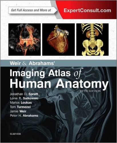
Weir & Abrahams’ Imaging Atlas of Human Anatomy, 5e 5th Edition

[amazon template=iframe image2&asin=0723438269]

Imaging is ever more integral to anatomy education and throughout modern medicine. Building on the success of previous editions, this fully revised fifth edition provides a superb foundation for understanding applied human anatomy, offering a complete view of the structures and relationships within the body using the very latest imaging techniques.

It is ideally suited to the needs of medical students, as well as radiologists, radiographers and surgeons in training. It will also prove invaluable to the range of other students and professionals who require a clear, accurate, view of anatomy in current practice.

Fully revised legends and labels and over 80% new images – featuring the latest imaging techniques and modalities as seen in clinical practice

Covers the full variety of relevant modern imaging – including cross-sectional views in CT and MRI, angiography, ultrasound, fetal anatomy, plain film anatomy, nuclear medicine imaging and more – with better resolution to ensure the clearest anatomical views

Unique new summaries of the most common, clinically important anatomical variants for each body region – reflects the fact that around 20% of human bodies have at least one clinically significant variant

New orientation drawings – to help you understand the different views and the 3D anatomy of 2D images, as well as the conventions between cross-sectional modalities

Now a more compete learning package than ever before, with superb new BONUS electronic enhancements embedded within the accompanying eBook, including:

Labelled image ‘stacks’ – that allow you to review cross-sectional imaging as if using an imaging workstation

Labelled image ‘slide-lines’ – showing features in a full range of body radiographs to enhance understanding of anatomy in this essential modality

Self-test image ‘slideshows’ with multi-tier labelling – to aid learning and cater for beginner to more advanced experience levels

Labelled ultrasound videos – bring images to life, reflecting this increasingly clinically practiced technique

Questions and answers accompany each chapter – to test your understanding and aid exam preparation

34 pathology tutorials – based around nine key concepts and illustrated with hundreds of additional pathology images, to further develop your memory of anatomical structures and lead you through the essential relationships between normal and abnormal anatomy

DOWNLOAD THIS BOOK FREE HERE
http://upsto.re/Avo9Ldm

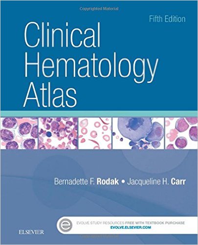
Clinical Hematology Atlas, 5e 5th Edition

[amazon template=iframe image2&asin=0323322492]

Ensure you are accurately identifying cells at the microscope with Clinical Hematology Atlas, 5th Edition. An excellent companion to Rodak’s Hematology: Clinical Principles & Applications, this award-winning atlas offers complete coverage of the basics of hematologic morphology, including examination of the peripheral blood smear, basic maturation of the blood cell lines, and information on a variety of clinical disorders. Nearly 500 photomicrographs, schematic diagrams, and electron micrographs vividly illustrate hematology from normal cell maturation to the development of various pathologies so you can be sure you’re making accurate conclusions in the lab.

Schematic diagrams, photomicrographs, and electron micrographs are found in every chapter to visually enhance understanding of hematologic cellular morphology.

Smaller trim size, concise text, and spiral binding make it easy to reference the atlas in the laboratory.

Chapter on normal newborn peripheral blood morphology covers the normal cells found in neonatal blood.

Chapter on body fluids illustrates the other fluids found in the body besides blood, using images from cytocentrifuged specimens.

The most common cytochemical stains, along with a summary chart for interpretation, are featured in the leukemia chapters to help classify both malignant and benign leukoproliferative disorders.

Chapter featuring morphologic changes after myeloid hematopoietic growth factors is included in the text.

Morphologic abnormalities are covered in the chapters on erythrocytes and leukocytes, along with a description of each cell, in a schematic fashion.

User resources on the Evolve companion site feature review questions and summary tables to further enhance their learning experience.

NEW! Appendix with comparison tables of commonly confused cells includes lymphocytes versus neutrophilic myelocytes and monocytes versus reactive lymphoctyes to help users see the subtle differences between them.

NEW! Glossary of hematologic terms at the end of the book provides a quick reference to easily look up definitions.

DOWNLOAD THIS BOOK FREE HERE
http://upsto.re/AvGdNAa

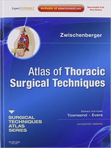
Atlas of Thoracic Surgical Techniques: (A Volume in the Surgical Techniques Atlas Series) (Expert Consult – Online and Print), 1e

[amazon template=iframe image2&asin=141604017X]

Atlas of Thoracic Surgical Techniques, a title in the Surgical Techniques Atlas Series edited by Drs. Townsend and Evers, presents state-of-the-art updates on the full range of thoracic surgical procedures performed today. Dr. Joseph B. Zwischenberger, along with esteemed international contributors, offers you expert advice on a variety of thoracic techniques, including lung-volume reduction surgery, video-assisted thoracoscopic surgery, and laparoscopic approaches to many procedures to help you expand your repertoire and hone your clinical skills. Color illustrations enhance the detailed instructions, and Expert Consult functionality gives you easy access to the full text online.

Includes convenient access to the full-text online at expertconsult.com.

Offers step-by-step guidance on a variety of thoracic surgical techniques, giving you more options for the challenges you face.

Discusses the hottest topics in thoracic surgery, including lung-volume reduction surgery, video-assisted thoracoscopic surgery, and laparoscopic approaches to many procedures.

Presents more than 200 full-color illustrations and step-by-step intraoperative photographs for expert visual guidance.

Discusses pearls and pitfalls to help you avoid complications.

Uses a consistent, easy-to-follow chapter format that includes clinical anatomy, pre-operative considerations, operative steps, and post-operative care to make reference easy.

Visually master a wide range of operative techniques, with authoritative guidance

DOWNLOAD THIS BOOK FREE HERE
http://upsto.re/AsuWTrJ

Atlas of Upper Gastrointestinal and Hepato-Pancreato-Biliary Surgery 2nd ed. 2016 Edition

[amazon template=iframe image2&asin=3662465450]

The first edition of this book was judged by many to be the best available atlas of upper gastrointestinal and hepato-pancreato-biliary surgery. The second edition builds upon this success by covering all of the significant intervening developments, including both new procedures and adaptations of established techniques. The atlas opens with an introductory section on the basic principles of operative surgery. All of the important surgical techniques are then presented in a series of sections devoted to the esophagus, stomach, and duodenum, the liver, the biliary system, portal hypertension, the pancreas, and the spleen. General, oncologic, and transplantation surgical procedures are described step by step with the aid of more than 900 superb illustrations drawn by a team of artists with extensive experience. The contributors were selected on the basis of their extensive experience in the procedures discussed and most are established educators.

DOWNLOAD THIS BOOK FREE HERE
http://upsto.re/AQenRNW


1 16 17 18 19