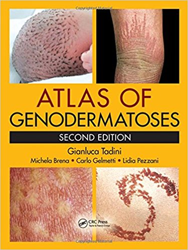
Atlas of Genodermatoses, Second Edition 2nd Edition
[amazon template=iframe image2&asin=1466598352]

Diagnosing a genetic skin disease can sometimes be a difficult task for a dermatologist. This is especially true for genodermatosesgenerally considered rare diseases seldom seen by practicing clinicians. As a result, professionals often have little experience with their diagnosis.

The Atlas of Genodermatoses presents a unique collection of such cases gathered patiently over the course of the years by the authors. It provides an unsurpassed overview of the major genodermatoses encountered in practice, even if only on rare occasions.

This book discusses almost 200 inherited diseases of the skin, hair, and nails. The entry for each disease includes its epidemiology, laboratory findings, genetics, pathogenesis, cutaneous and extracutaneous findings, differential diagnosis, disease course, complications, and follow-up and therapy, where appropriate.

In addition to being a clinical primer, this atlas is also a work of scientific research. The new edition rewrites the classification of some diseases, adds some newly described conditions, and updates established information with the latest molecular genetic studies and references. Specialists in both dermatology and pediatrics should find the atlas an invaluable frontline resource in the clinic.

DOWNLOAD THIS BOOK FREE HERE

http://upsto.re/Ar5Frpy

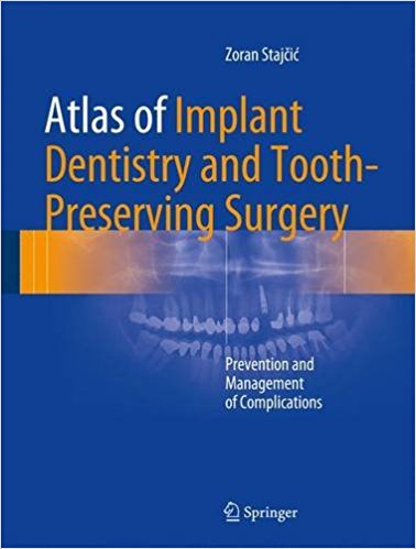
Atlas of Implant Dentistry and Tooth-Preserving Surgery: Prevention and Management of Complications 1st ed. 2017 Edition

This atlas is unique in comparing the two disciplines of dental implant surgery and tooth-preserving surgery with respect to common procedures, problems, and failures and in providing excellent guidance on the prevention and management of complications. The etiology of a wide variety of implant-related and non-implant-related complications and failures is carefully explained. Since many complications have their roots in oral and periodontal surgical maneuvers, these maneuvers are themselves discussed and extensively illustrated. The most frequently used tooth preservation procedures are also fully described, with emphasis on correct surgical technique as a means to avoid complications. The use of these procedures is constantly weighed against the effects of tooth removal and insertion of dental implants. The text is complemented by the inclusion of a substantial number of helpful references. While the atlas is intended primarily for dentists involved in outpatient implant dentistry and oral surgery, oral and maxillofacial surgeons will also find the descriptions of innovative techniques or maneuvers to be of interest, including those relating to the selection of incision and flap design and the sinus floor elevation techniques.

DOWNLOAD THIS BOOK FREE HERE

http://upsto.re/ApxRub7

Atlas of Peripheral Blood: The Primary Diagnostic Tool 1st Edition

[amazon template=iframe image2&asin=0781777801]

This book will be a full-color atlas of both benign and neoplastic proliferations in the blood. The book will illustrate the morphologic features on peripheral blood smears of the various disorders and the text will focus on diagnostic criteria, differential diagnosis and modern classification terminology. The book may be used as a stand-alone resource and should be useful to trainees and clinicians that routinely review peripheral blood smears. The book will be accompanied by an on-line image bank containing numerous additional full-color images of the various disorders.

DOWNLOAD THIS BOOK FREE HERE

http://upsto.re/AxHMipV

Atlas of Infectious Disease Pathology (Atlas of Anatomic Pathology) 1st ed. 2017 Edition

[amazon template=iframe image2&asin=3319547011]

Infectious diseases may be encountered in nearly every aspect of pathology. This atlas provides an informative reference for the identification of the common and esoteric pathogens, presenting in a wide array of specimen types. The focus of the presented images is on the hematoxylin and eosin-stained appearances of these infections and highlight common special stains that can be used to aid in the diagnosis of the infectious agent.  Where appropriate, commentary regarding additional testing such as immunohistochemistry and molecular-based methods is supplied.

The Atlas of Infectious Disease Pathology is organized primarily by pathogen type followed by discussion of the various manifestations that may occur in individual organ systems. The reader will be provided with a comprehensive overview of the histopathology of the majority of infectious diseases encountered in general and subspecialty practice alike.

DOWNLOAD THIS BOOK FREE HERE

http://upsto.re/AxDYxJS

Atlas of Elastosonography: Clinical Applications with Imaging Correlations 1st ed. 2017 Edition

[amazon template=iframe image2&asin=331944199X]

With the aid of an extensive series of high-quality images, this atlas describes all the potential applications of elastosonography in routine clinical practice. After a brief introduction on technical and physical aspects, the diagnostic benefits of elastosonography in a range of settings are illustrated with the aid of numerous representative cases. The coverage encompasses pathologies of the liver, spleen, pancreas, kidney, breast, thyroid, testis, musculoskeletal system, and vascular system. In addition, helpful correlations with imaging appearances on ultrasound and computed tomography are included. Elastosonography is a powerful, relatively new diagnostic technique that assesses the elasticity of tissues as an indicator of malignancy. Readers will find this book to be an excellent aid to use and interpretation of the technique.

About the Author

Dirk-André Clevert, MD, is Associate Professor of Radiology and Section Chief of the Interdisciplinary Ultrasound Center at Munich University Hospital Grosshadern, which is part of one of the largest medical complexes in Germany. He has headed the Interdisciplinary Ultrasound Center since 2004 and performs all interventional ultrasound investigations at the center, including contrast-enhanced ultrasound. Dr. Clevert has published more than 115 articles in national and international medical journals and is a member of the advisory boards of several scientific journals. His research interests span the fields of contrast-enhanced sonography and interventional and vascular ultrasound.

Mirko D’Onofrio, MD, has worked as an Assistant Professor in the Department of Radiology at Verona University Hospital since 2004. He is an associate of SIRM (Italian Society of Medical Radiology), SIUMB (Italian Society of Ultrasound in Medicine and Biology), and EFSUMB (European Federation of Societies for Ultrasound in Medicine and Biology). Dr. D’Onofrio is a member of ESGAR (European Society of Gastrointestinal and Abdominal Imaging) and has been a fellow of ESGAR since 2009. He is on the editorial board of the World Journal of Gastroenterology.

Emilio Quaia is Associate Professor of Radiology in the Faculty of Medicine of the University of Trieste. He has been principal investigator in several clinical trials. He is the author of 105 scientific papers in different areas.

DOWNLOAD THIS BOOK FREE HERE

http://upsto.re/A8j3Nhh


Surgical Anatomy and Techniques to the Spine Book + Image bank CD-ROM

[amazon template=iframe image2&asin=1416003134]

Written by three neurosurgeons and an orthopedic surgeon, this comprehensive reference’s multidisciplinary approach makes it relevant and informative to all surgeons regardless of their specialty or level of surgical experience. After thoroughly reviewing the anatomy and approaches to the spine, the authors lead you through a full range of state-of-the-art techniques in spine surgery, including those procedures commonly performed for degenerative disease, trauma, scoliosis, tumor, and infection. Abundant full-color illustrations, three-dimensional images and cadaver dissection photos walk you through both simple and complex spinal surgical procedures. The bonus CD-ROM contains all the images from the book, including some of the most sought-after anatomic photographs and radiographic images in neurosurgery, brought together for the first time uniquely for this volume.

  • Multiple-viewpoint, full-color photographs and illustrations provide a thorough presentation of procedures and relevant anatomy.
  • Concise, atlas-style format makes techniques easy to understand and reproduce.
  • The Bonus CD-ROM includes all the images from the book in an easily searchable format for use in electronic presentations.
  • Multidisciplinary coverage makes the book relevant and informative to all spine surgeons.
  • Facilitates diagnosis and complication avoidance with clinical pearls and hints..

DOWNLOAD THIS BOOK FREE HERE

http://upsto.re/A8Krers

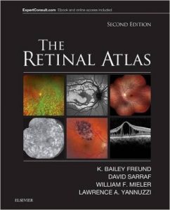
The Retinal Atlas, 2e 2nd Edition

[amazon template=iframe image2&asin=0323287921]

With more than 5,000 images, a unique page layout, and comprehensive illustrations of the entire spectrum of vitreous, retina, and macula disorders, The Retinal Atlas, 2nd Edition, is an indispensable reference for retina specialists and comprehensive ophthalmologists as well as residents and fellows in training. For this edition, an expanded author team made up of Drs. K. Bailey Freund, David Sarraf, William F. Mieler, and Lawrence A. Yannuzzi, each an expert in retinal research and imaging, provide definitive up-to-date perspectives in this rapidly advancing field. This award-winning title has been thoroughly updated with new images with multimodal illustrations, new coverage and insight into key topics, and new disorders and classifications, while retaining the innovative page layout that has made it the most useful and most complete atlas of its kind.

    • Provides a complete visual guide to advanced retinal imaging and diagnosis of the full spectrum of retinal diseases, including early and later stages of disease.
    • Enhances understanding by presenting comparison imaging modalities, composite layouts, high-power views, panoramic disease visuals, and selected magnified areas to hone in on key findings and disease patterns.
    • Features color coding for different imaging techniques, as well as user-friendly arrows, labels, and magnified images that point to key lesions and intricacies.

  • Expert Consult™ eBook version included with purchase. This enhanced eBook experience allows you to search all of the text, figures, and references from the book on a variety of devices.
  • Covers all current retinal imaging methods including: optical coherence tomography (OCT), indocyanine green angiography, fluorescein angiography, and fundus autofluorescence.
  • Depicts and explains expanding OCT uses, including spectral domain and en face OCT, and evolving retinal imaging modalities such as ultra-wide-field fundus photography, angiography and autofluorescence.
  • Presents a select team of experts, all of whom are true international leaders in retinal imaging, and have assisted in contributing to the diverse library of common and rare case illustrations.

DOWNLOAD THIS BOOK FREE HERE

http://upsto.re/AckB82J

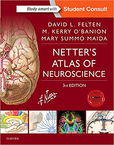
Netter’s Atlas of Neuroscience, 3e (Netter Basic Science) 3rd Edition

[amazon template=iframe image2&asin=0323265111]

Ideal for students of neuroscience and neuroanatomy, the new edition of Netter’s Atlas of Neuroscience combines the didactic well-loved illustrations of Dr. Frank Netter with succinct text and clinical points, providing a highly visual, clinically oriented guide to the most important topics in this subject. The logically organized content presents neuroscience from three perspectives: an overview of the nervous system, regional neuroscience, and systemic neuroscience, enabling you to review complex neural structures and systems from different contexts.

You may also be interested in:

A companion set of flash cards, Netter’s Neuroscience Flash Cards, 3rd Edition, to which the textbook is cross-referenced.

  • Coverage of both regional and systemic neurosciences allows you to learn structure and function in different and important contexts.
  • Combines the precision and beauty of Netter and Netter-style illustrations to highlight key neuroanatomical concepts and clinical correlations.
  • Reflects the current understanding of the neural components and supportive tissue, regions, and systems of the brain, spinal cord, and periphery.
  • Uniquely informative drawings provide a quick and memorable overview of anatomy, function, and clinical relevance.
  • Succinct and useful format utilizes tables and short text to offer easily accessible “at-a-glance” information.
  • Provides an overview of the basic features of the spinal cord, brain, and peripheral nervous system, the vasculature, meninges and cerebrospinal fluid, and basic development.
  • Integrates the peripheral and central aspects of the nervous system.
  • Bridges neuroanatomy and neurology through the use of correlative radiographs.
  • Highlights cross-sectional brain stem anatomy and side-by-side comparisons of horizontal sections, CTs and MRIs.
  • Features video of radiograph sequences and 3D reconstructions to enhance your understanding of the nervous system.

  • Student Consult eBook version included with purchase. This enhanced eBook experience includes access — on a variety of devices — to the complete text, 14 videos, and images from the book.
  • Expanded coverage of cellular and molecular neuroscience provides essential guidance on signaling, transcription factors, stem cells, evoked potentials, neuronal and glial function, and a number of molecular breakthroughs for a better understanding of normal and pathologic conditions of the nervous system.
  • Micrographs, radiologic imaging, and stained cross sections supplement illustrations for a comprehensive visual understanding.
  • Increased clinical points — from sleep disorders and inflammation in the CNS to the biology of seizures and the mechanisms of Alzheimer’s — offer concise insights that bridge basic neuroscience and clinical application.

DOWNLOAD THIS BOOK FREE HERE

http://upsto.re/AcB593c

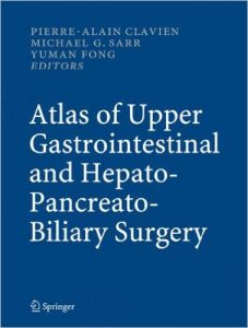
Atlas of Upper Gastrointestinal and Hepato-Pancreato-Biliary Surgery 1st Edition

[amazon template=iframe image2&asin=3540200045]

Atlas of Upper Gastrointestinal and Hepato-Pancreato-Biliary Surgery covers in detail the technical aspects of all important procedures of the upper abdomen (esophagus, duodenum, stomach, liver, biliary system, pancreas, portal hypertension and spleen) including general, oncologic and transplantation surgery. Each chapter has been written by an expert renowned in his or her field and been reviewed by a second expert. All of the approximately 900 illustrations have been drawn by the same team of three artists of notable experience. The liver section follows the internationally accepted terminology of the Brisbane 2000 conference of the IHPBA. In this atlas experts of upper abdominal surgery share their experience and show how they do it – step by step.

DOWNLOAD THIS BOOK FREE HERE

http://upsto.re/AtyMdci

1 15 16 17 18 19